RBCCV 44205-1424 DOI: 10.5935/1678-9741.20120099
Minimally invasive aortic valve replacement:
an alternative to the conventional technique
Troca valvar aórtica minimamente invasiva: uma alternativa à técnica convencional
Jeronimo Antonio Fortunato Júnior
1, Alexandre Gabelha Fernandes
2, Jeferson Roberto Sesca
2,
Rogério Paludo
3, Maria Evangelista Paz
4, Luciana Paludo
5, Marcelo Luiz Pereira
6, Amélia Araujo
71. Brazilian Red Cross Hospital – (branch of Paraná) Positivo University, Cardiac surgeon, Master of Surgery, Professor of Cardiovascular and Thoracic Surgery at Positivo University. Demonstration of an alternative technique to conventional cardiac surgery.
2. Brazilian Red Cross Hospital – (branch of Paraná) Positivo University; Heart Surgery Resident.
3. Brazilian Red Cross Hospital – (branch of Paraná) Positivo University; Perfusionist.
4. Brazilian Red Cross Hospital – (branch of Paraná) Positivo University; Surgical instrumentator.
5. Brazilian Red Cross Hospital – (branch of Paraná) Positivo University; Anesthesiologist.
6. Brazilian Red Cross Hospital – (branch of Paraná) Positivo University; Assistant surgeon.
7. Brazilian Red Cross Hospital – (branch of Paraná) Positivo University; Cardiologist. Responsible for the pre- and postoperative follow-up period.
Work performed at the Brazilian Red Cross Hospital Brazilian (branch of Paraná) / Positivo University, Curitiba, Paraná, Brazil.
Correspondence Address: Jeronimo Antonio Fortunato Jr.
50 Amaury GG Matei Street, Santo Inácio - Curitiba, Paraná Brazil – Zip code: 82010-620.
E-mail: [email protected]
Article received on May 9th, 2012 Article accepted on October 3rd, 2012
Abstract
Objectives: To demonstrate the use of minimally invasive surgery for aortic valve replacement and compare its results with the traditional method.
Methods: Between 2006 and 2011 sixty patients underwent surgery on aortic valve, after written consent, 40 of them by minimally invasive technique with right anterior minithoracotomy access (Group 1/G1) and 20 by median sternotomy (Group 2/G2). Compare the operating times and postoperative evolution intra-hospital.
Results: The average times of bypass and aortic cross-clamp in G1 were, respectively, 142.7 ± 59.5 min and 88.6 ± 31.5 min and, in G2, 98.1 ± 39.1 min and 67.7 ± 26.2 min (P
< 0.05), a difference in medians of 39 minutes in bypass time and 23 minutes in aortic cross-clamp were observed in favor
of conventional technique. The blood loss by the thoracic drains was signiicantly lower in the Group: minimally invasive 605.1 ± 679.5 ml (G1) versus 1617 ± 1390 ml (G2) (P < 0.05).The average time of ICU and hospital stay were shorter in G1: 2.3 ± 1.8 and 5.5 ± 5.4 days versus 5.1 ± 3.6 and 10 ± 5.1 in G2 (P < 0.05), respectively. Vasoactive drug use was also less post-operative at 12.8% in minimally invasive group G1 versus 45% in G2.
Conclusion: Aortic valve replacement through minimally invasive techniques, although intraoperative times larger, did not demonstrate to affect postoperative results in this case proved to be better when compared to the traditional approach.
Rev Bras Cir Cardiovasc 2012;27(4):570-82 Fortunato Júnior JA, et al. - Minimally invasive aortic valve replacement:
an alternative to the conventional technique
INTRODUCTION
The minimally invasive cardiac surgery (MICS) has increased in popularity over the past 15 years. The small incisions have been associated with good aesthetic result and less surgical trauma, consequently less pain and rapid postoperative recovery. For a while, these same arguments will not attract the attention of the physician population. This concept has been changing with the wider dissemination of the technique and best results in recent
reports. The beneits of minimally invasive incisions are supported primarily with conirmation of reduction of
hospital costs without harming the achieved results with median sternotomy [1-3].
Also in recent years, using access alternative, the percutaneous or transapical aortic valve and endovascular devices was developed, including aortic stenting and even annular ring reducers for mitral valve and devices for occlusion of interatrial or interventricular defects [4 - 8].
Nevertheless, the median sternotomy is still the traditional access to surgical treatment of heart disease because it allows excellent control of all cardiac structures and asserts itself as a safe technique with low morbimortality.
Our goal is to demonstrate the use of minimally invasive surgery for the treatment of aortic valve and compare its results with the conventional technique (median sternotomy).
METHODS
This is a retrospective study and gathered all patients undergoing aortic valve surgery in the period from 2006 to 2011. Sixty patients underwent valve surgery, 40 of them by the minimally invasive technique and preferably with access the via right anterolateral thoracotomy (Group 1/G1) and 20 by median sternotomy (Group 2/G2). The preoperative clinical characteristics are described in Table 1. In selecting the data in Table 1, the presentation of the predominant aortic valve dysfunction was chosen: stenosis
or insuficiency that deined the clinical and surgical
indication, either by transvalvular gradient or aortic regurgitation on echocardiography.
Exclusion criteria for a minimally invasive procedure included: reoperation, need for concomitant CABG or patients who opted for the conventional technique. All patients in G1 signed authorization for the alternative procedure.
Resumo
Objetivo: Demonstrar o uso da cirurgia minimamente invasiva para tratamento da valva aórtica e comparar seus resultados com o método tradicional.
Métodos: Entre 2006 e 2011, 60 pacientes foram submetidos à cirurgia na valva aórtica, após consentimento escrito, destes 40 pela técnica minimamente invasiva com acesso por minitoracotomia ântero-lateral direita (Grupo 1/G1)e 20 por esternotomia mediana (Grupo 2/G2). Comparamos os tempos operatórios e a evolução pós-operatória intra-hospitalar.
Resultados: Os tempos médios de circulação extracorpórea (CEC) e pinçamento aórtico no G1 foram, respectivamente, 142,7 ± 59,5 min e 88,6 ± 31,5 min e, no G2, 98,1 ± 39,1 min e 67,7 ± 26,2 min (P<0,05), uma diferença nas medianas de 39 minutos no tempo de CEC e 23 minutos no pinçamento aórtico foram observados a favor da técnica convencional. A perda sanguínea pelos drenos torácicos foi signiicativamente menor no grupo minimamente invasivo: 605,1 ± 679,5 ml (G1) versus 1617 ± 1390 ml (G2) (P<0,05). Os tempos médios de internamento em UTI e hospitalar foram menores em G1: 2,3 ± 1,8 dias e 5,5 ± 5,4 dias versus 5,1 ± 3,6 dias e 10 ± 5,1 dias em G2 (P<0,05), respectivamente. O uso de drogas vasoativas no pós-operatório também foi menor no grupo minimamente invasivo 12,8% em G1 versus 45% em G2.
Conclusão: Troca valvar aórtica com o uso de técnicas minimamente invasivas, apesar de demonstrar maiores tempos intraoperatórios, não afeta os resultados pós-operatórios, que nesta casuística mostraram-se melhores quando comparado ao método tradicional.
Descritores: Procedimentos cirúrgicos minimamente invasivos. Valva aórtica/cirurgia. Doenças das valvas cardíacas. Abbreviations, acronyms and symbols
CVA Cerebrovascular accident
BIS Bispectral index
MICS Minimally invasive cardiac surgery
ECC Extracorporeal circulation
G1 Group 1
G2 Group 2
HTK Histidine-tryptophan-ketoglutarate
Min Minutes
CVP Central venous pressure
TEE Transesophageal echocardiography
Echocardiographic evaluation, coronariography and carotid artery Doppler were performed in all patients, while peripheral vascular Doppler and abdominal aorta only in patients undergoing cardiopulmonary bypass (CPB) and Peripheral circulation (G1).
All patients in this series underwent speciic protocol
for anesthesia, used systematically in the institution, with the intention of immediate extubation in the operating room. The technique used a device for continuous electroencephalogram analysis (BIS®), calculating the bispectral index to assess the depth of anesthesia and its
supericialization at the end of the surgery. Ramifentanil®
and propofol® were used. Patients with the following characteristics were extubated in the operating room: BIS above 60, level of responsive awareness, adequate pulmonary ventilation and hemodynamic stability in average time of 15 to 30 minutes (min) after skin suture.
In G1, a right minithoracotomy was performed (± 5 cm) on the 2nd or 3rd right intercostal space or upper J-shaped ministernotomy. The peripheral CPB was performed by the femoral vessels [9,10] to all G1 procedures which were performed with the aid of chest videoscopy.
In peripheral CPB, a manometer was used with negative pressure for vacuum-assisted venous drainage. The arterial femoral cannulation (17 French) and venous kiys (21 French) especially designed for peripheral CPB, were used in all these cases (DLP®, Medtronic Inc., Minneapolis, USA).
Intermittent cold blood cardioplegia was performed
in aortic root or the coronary ostia, in the irst 20 cases
of G1 and G2. In the last 20 of G1, histidine-tryptophan-ketoglutarate (HTK) or commercially known as Custodiol® in infusion of 20 ml/kg body weight was used
in a single dose.
Transesophageal echocardiography (TEE) was performed in all patients in G1, both for introduction of arterial and venous cannulae as monitoring and
conirmation of the surgical outcome.
The instruments used in G1 involving a 5mm or 10mm diameter thoracoscope according to the need of the visual
ield and angle lens of 30°. The instruments (ESTECH® Inc, California, USA) speciically designed for cardiac
surgery included: chest retractor, scissors, knot pushers, aortic clamp (Chitwood®), and needle holders. Other instruments such as forceps, electrocautery, video cameras and light source were the same ones used in conventional laparoscopy.
In G2, a median sternotomy was performed with CPB and cannulation of the aorta and right atrium, both by the conventional technique. Transthoracic clamping and intermittent blood cardioplegia were performed in all patients.
Surgical technique for minimally invasive access
1. In all cases from G1, an orotracheal intubation with Carlens® or Portecs® cannulae was performed, for occlusion of the right lung during surgery. In cases of ministernotomy, where right pleuron was not openned, this occlusion was not necessary.
2. After cannula insertion, and effective right unilateral occlusion was guaranteed and also maintaining oxygenation with a single lung.
3. Central vessel puncture, jugular or subclavian infusion of drugs and monitoring of central venous pressure (CVP). The punction was preferable on the right side, as pneumothorax as a complication, which was not diagnosed on the left side could be very serious and prevent occlusion of the right lung.
4. We used as a routine a protocol for immediate extubation in the operating room. The combination of Propofol® and Ramifentanil® was used and the depth of
anesthesia was assessed by bispectral index (BIS).
5. Transthoracic deibrillation pads were placed in the
left, anterior and posterior thoracic region.
Table 1. Preoperative clinical characteristics of surgical groups
Variables Male sex Age
Ejection fraction Hypertension Diabetes
Preoperative atrial ibrillation Predominant valvular dysfunction Failure
Stenosis
Minimally invasive access 30/75%
52.4±15.1 60.5±9.3 27/67.5% 2/5% 5/12.5%
19/47.5% 21/52.5%
Median sternotomy 15/75% 58.6±14.3 55.4±11.8 16/80%
3/15% 3/15%
6/30.0% 14/70%
P-value Ns Ns Ns Ns Ns Ns
6. CPB was set up in a conventional manner, testing the vacuum system through a negative pressure gauge connected to the oxygenator venous reservoir. This test
was done during the circuit illing and removal of bubbles.
Variations from 40 to 100 mmHg were used to allow adequate venous drainage.
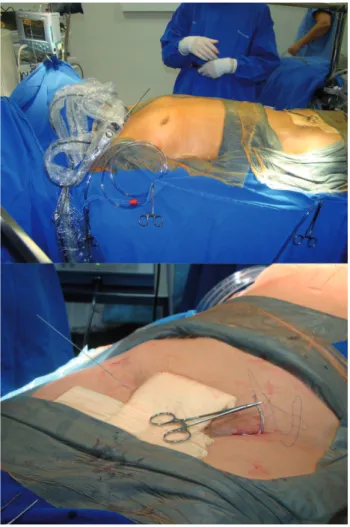
7. We dissected the left femoral artery and punctured the right femoral vein, even before heparinization (Figure
1). The CPB tubes were directed to the surgical ield,
positioned under the lower limbs.
8. The right anterolateral minithoracotomy was used in
patients with severe aortic insuficiency or stenosis with small or moderate calciication. We performed a right
sternal incision extending laterally with 5 cm in length; the intercostal space was incised until we could observe the right mammary artery.
The 2nd intercostal space was accessed in short patients with small chest on chest radiograph and the 3rd intercostal space in the other patients (Figure 2).
9. Upper J-shaped ministernotomy was used marking the 3rd intercostal space and performing an incision of 5 cm, which began at this point, following the cranial direction. Sternotomy was performed from the sternal furcula to the 3rd intercostal space to the right, trying not to affect the right mammary artery. This last access was used
in all patients with severe valvular and ring calciication
(Figure 3).
10. Video-assisted thoracoscopy was used in all patients, being introduced in the 2nd intercostal space, laterally to the thoracic incision, either by anterolateral mini-thoracotomy or ministernotomy. This display option
has expanded the visual ield and helped visualization and
cannulation of the right coronary ostium in cases of ostial cardioplegia, the observation of the left ventricle in cases of distension of this cavity (cardioplegia in the aortic root) and in the visualization and cleaning the interior of the left ventricle in search of debris or calcium emboli (Figure 4).
11. A ESTECH® retractor thoracic with a 4 cm single metal blade was used for exposure of the cavity in both alternative techniques. The "Finochietto" pediatric retractor was used as a good option in some cases, but the size of the short blades prevented from more routine use.
12. We followed the dissection and the identiication
of the pericardium. The pericardium was opened on the
ascending aorta from the pericardial delection to the right
atrium. Exposure points were used to keep the pericardium open and pulled the chest wall.
13. After heparinization, cannulation of the femoral vessels was performed, primarily through the right femoral vein, once punctured; we introduced a rigid metal tab that
progressed to the right atrium, conirmed by TEE. Dilators
were introduced sequentially to dilate the vessel until the cannula, with occlusive dilator, was introduced to the right atrium, again with the need to ensure its position with
TEE. After the venous cannula was positioned, we ixed it
to the skin and connected it to the CPB venous tube. 14. The same procedure was done with the arterial cannulation, only in this case, the progression of the cannula reached its maximum length in the abdominal aorta. Being connected to the arterial segment in the CPB tube, permeability and wrist were tested.
15. A 2 cm incision was performed in the 2nd intercostal space in the anterior axillary line for the placement of Chitwood® transthoracic clamp in patients undergoing anterolateral minithoracotomy, the videoscopy helped aortic clamping performed laterally along the
pericardial delection. The transthoracic clamping in tge
ministernotomy was performed by thoracic incision with conventional tweezers (DeBakey®) (Figure 5A).
Fig. 1 - Position of the patient, the surgical ield and peripheral access for ECC. A: panoramic view of the surgical ield, B: 2
cm incision in the left inguinal region for blood exposure and percutaneous puncture of the right femoral vein
Rev Bras Cir Cardiovasc 2012;27(4):570-82 Fortunato Júnior JA, et al. - Minimally invasive aortic valve replacement:
Fig. 3 – Upper J Mini sternotomy, from the 3rd intercostal space to the sternal notch. A: exposure with retractor and exposure of the ascending aorta after opening the pericardium. Trocar for videoscopy positioned laterally to the right by counterinsicion B:
transthoracic clamping, with observation of the surgical ield and start of cardioplegic infusion in the aortic root with rigid cannula,
C: observation of left ventricular distention during cardioplegia in the aortic root; D: transverse aortotomy, E: cardioplegia performed in the coronary ostia, F: metallic prosthesis implanted
Fig. 2 - Right anterolateral minithoracotomy and use of Finochietto or ESTECH® pediatric retractor. A: panoramic view, B:
lateral aortic clamping with Chitwood®, C: aortic exposure via minithoracotomy, D: Thoracoscopic visualization with calciic
16. In this moment, the CPB began. The need for higher or lower drainage was oriented by the surgeon requesting variations in vacuum pressure, assessing the complete emptying of the right atrium.
17. Before the transthoracic clamping, we made a pouch in the aortic root for the introduction of the cardioplegia cannula, which was also used at the end of the procedure to remove air from the left cavities. This same cannula was
withdrawn always on CPB and low low, to reduce the risk
of aortic dissection.
18. Hypothermic blood cardioplegia 4/1 were measured every 15 minutes and the CPB maintained between 28 and 30 degrees. In cases which HTK solution was used (Custodiol®), only one infusion (20 ml/kg) was made in the aortic root to perform the entire procedure, the coronary ostia were cannulated in case of predominant aortic
insuficiency or where we can notice distention of the left
ventricle by videoscopy. In such cases, it was extremely important to maintain 28º because HTK solution maintains its maximum effect [12].
19. At this time, we opened the heart cavity through transverse aortotomy. Only wires were used for exposure of the aortic valve: two equidistant polyester sutures in
the anterolateral and anteromedial proximal aorta sides (Figure 5B).
20. No vacuum was alternatively used to drain the left ventricle, for this purpose we used an aspiration cannula introduced by aortotomy into the left ventricle and, after
placement of the prosthetic valve through the lealets.
21. We continued with the aortic valve replacement in all cases using the conventional method.
22. After completion of primary surgical time, we tried to be very careful for maximum removal of air from
the heart cavities, also guided by TEE. The irst step was
to conduct the maximum Trendelenburg position. The cardioplegia cannula, attached to the aortic root, was enough to suck all the residual air in the left ventricle. In
this moment, the TEE conirmed the complete elimination
of air from the heart chambers, before we could remove CPB. Periods of interruption of CPB with constant suction of aortic root helped deaeration.
23. Pacemaker wires (2) were placed in the right ventricle which was still on CPB, with the heart drained.
24. After review of hemostasis, protamine solution began (1/1) by continuous infusion. Before completing the heparin reversal, we withdrew the venous cannula. Since
Fig. 4 – Observation of the heart by videothoracoscopy. A: visualization of the ostium and right coronary lealet, B: observation
of the left ventricle; C: visualization of the left ventricle in search of emboli or calcium debris
Fig. 5 - Aortic clamping in minimally invasive procedures. A: placement of Chitwood® transthoracic clamp in patients undergoing anterolateral thoracotomy, the videoscopy assisted in clamping performed laterally to the aorta near the pericardial recess between the aorta and pulmonary artery, B: transverse aortotomy with equidistant points, placed on the anterior exposure to the aorta, C: mini sternotomy transthoracic clamping was performed by thoracic incision with conventional tweezers (DeBakey®), and cardioplegia in the aortic root
Rev Bras Cir Cardiovasc 2012;27(4):570-82 Fortunato Júnior JA, et al. - Minimally invasive aortic valve replacement:
we had used percutaneous, only local compression was performed.
25. After reversing the anticoagulation, a 4-0 prolene "U" pouch was made in the artery around the femoral cannula for occlusion after its removal.
26. A chest tube was enough to make drainage, and was placed in the subxiphoid position in the ministernotomy or the 5th intercostal space with anterior axillary line in cases of anterolateral minithoracotomy.
27. After all sutures were done; we had anesthesia
supericialization according to the anesthetic protocol. Patients
with the following characteristics were extubated in the operating room: BIS above 60, level of responsive awareness, adequate pulmonary ventilation and hemodynamic stability in average time of 15 to 30 minutes after skin suture.
Statistical Analysis
Continuous data were expressed as mean ± standard deviation and categories evaluated in frequencies and percentages. To compare continuous variables, t-test or Fisher's exact test were used. The P-value <0.05 was
considered statistically signiicant.
RESULTS
The surgeries performed by median sternotomy were prior to the experience of the surgical team with minimally invasive procedures in aortic valve (13 patients) and were also performed in some cases requiring intervention besides valve replacement: 1) three cases of concomitant revascularization of the left anterior descending artery; 2) three cases of valvular reoperation and 3) a patient who opted for the open procedure.
In G1, aortic valve replacement was performed in all patients (40 cases). We decided to implant the metallic prosthesis in 33 cases (St. Jude Medical System®). Seven
patients aged over 70 years received bioprosthetic implant (Braile Biomedica®). Upper J Ministernotomy to the right was performed on ten patients because they presented
severe valve calciication and dilation of the ascending
aorta, in other cases 75% (30/40 cases), we performed a right anterolateral minithoracotomy through the 2nd (5/30
cases) or 3rd intercostal space (25/30 cases) (Figure 6). Among the 20 patients from G2, 7 received biological prostheses (Braile Biomedica®) and thirteen metallic prostheses (St. Jude Medical System®), median sternotomy was performed in all patients.
The mean CPB and aortic clamping in G1 were respectively: 142.7 ± 59.5 min and 88.6 ± 31.5 min and in G2, 98.1 ± 39.1 and 67.7 ± 26, 2 (P <0.05), a difference in the medians of 39 min on CPB and 23 min in aortic clamping was observed in favor of the conventional technique. In our service, we systematically use immediate extubation attempt, when the patient is still in the operating room. Almost all patients from G1 group were extubated immediately after the surgery, 92.5% of them, and only 75% were extubated in G2 (Table 2).
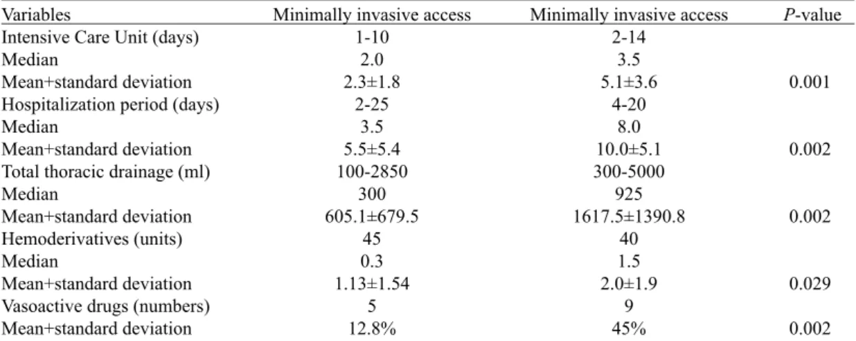
The total blood loss through chest tubes was signiicantly
lower in the minimally invasive group: 605.1 ± 679.5 ml (G1) versus 1617 ± 1390 mL (G2) (P <0.05). Mean time of hospitalization in the intensive care unit (ICU) and hospital were lower in G1: 2.3 ± 1.8 and 5.5 ± 5.4 days versus 5.1 ± 3.6 and 10 ± 5.1 G2 (P <0.05), respectively. The use of vasoactive drugs in the postoperative period was also lower in the minimally invasive group, 12.8% in G1 versus 45% in G2 (Table 3).
Two (5%) patients died in the group undergoing minimally invasive procedure and one (5%) in the
median sternotomy group, without statistical signiicance.
Postoperative complications were observed in both groups
and showed no signiicant difference being reported in
Table 4.
Table 2. Surgical times
Variables
ECC time (minutes) Median
Mean+standard deviation Aortic clamping (minutes) Median
Mean+standard deviation Extubation in the operating room Valve type
Biological Metallic
Minimally invasive access
127.0 142.7±59.5 80.0 88.6±31.5 37/92.5% 7/17.5% 33/82.5% Median sternotomy 88.0 98.1±39.1 57.0 67.7±26.2 15/75.0% 7/35.0% 13/65% P-value 0.004 0.012 0.031 Ns Ns Ns= not signiicant
Table 3. Postoperative variables
Variables
Intensive Care Unit (days) Median
Mean+standard deviation Hospitalization period (days) Median
Mean+standard deviation Total thoracic drainage (ml) Median
Mean+standard deviation Hemoderivatives (units) Median
Mean+standard deviation Vasoactive drugs (numbers) Mean+standard deviation
Minimally invasive access 1-10 2.0 2.3±1.8 2-25 3.5 5.5±5.4 100-2850 300 605.1±679.5 45 0.3 1.13±1.54 5 12.8%
Minimally invasive access 2-14 3.5 5.1±3.6 4-20 8.0 10.0±5.1 300-5000 925 1617.5±1390.8 40 1.5 2.0±1.9 9 45% P-value 0.001 0.002 0.002 0.029 0.002
Table 4. Postoperative complications
Variables Mortality
Neurological events New atrial ibrillation Renal failure respiratory failure Pleural effusion Surgical wound infection Reoperation for bleeding Dissection of the ascending aorta Conversion to sternotomy
Minimally invasive access 2/5% 3/7.5% 3/7.5% 2/5% 2/5% 2/5% 0/0% 3/7.5% 3/7.5% 2/5%
Minimally invasive access 1/5% 1/5% 2/10% 2/10% 3/15% 1/5% 1/5% 1/5% 2/10% __ P-value Ns Ns Ns Ns Ns Ns Ns Ns Ns Ns Ns = not signiicant
DISCUSSION
The concept of minimally invasive heart surgery incisions occurred in the mid-nineties. In the beginning, smaller incisions to access the mitral, aortic and coronary valves were introduced, such as the upper and lower hemi-sternotomy with transection of the sternum and the lateral thoracotomy [11,12]. Left thoracotomy for single revascularization of anterior descending and right artery
to give access to the mitral valve or coronary artery were also used. The right anterolateral thoracotomy had been used in the past with preference for mitral disease, but it was discontinued due to the best results with median thoracotomy or sternotomy [13-15].
Except for myocardial revascularization without CPB, minimally invasive surgery, mainly under the aortic valve, was once considered dangerous due to the high mortality rate when compared to the conventional
Rev Bras Cir Cardiovasc 2012;27(4):570-82 Fortunato Júnior JA, et al. - Troca valvar aórtica minimamente invasiva:
technique. Bridgewater et al. [16] demonstrated a 43% mortality in minimally invasive surgery compared with 7% in the conventional surgery for the treatment of aortic valve. Even when other centers showed more encouraging results, still it would not attract the attention of cardiac surgeons in the world [17,18].
Currently, minimally invasive cardiac surgery has shown its best results when using the aid of videothoracoscopy. Besides these video equipment, the extrathoracic access to CPB was implemented, the so-called "port-access technology", an innovative technique for vascular access and peripheral aortic endoclamping [9,18]. The inclusion of transthoracic clamping did not change the idea of the technique. Brinkman et al. [19] presented the favorable experience of using port-access surgical treatment of aortic
valve using transthoracic clamp with lexible Cosgrove®.
Since 1995 multicenter studies have been presented to demonstrate the effectiveness of this new method. Galloway et al. [10] in 1999, gathered data from 121 centers and included 1063 patients who underwent minimally invasive techniques with results similar to those of the conventional surgery, with the advantage of less aggression and pain, and use of hemoderivatives, in addition to hospital discharge and return to daily activities much earlier. In 2009, Dr. Galloway reported their data from a decade of experience with the method [2]. Grossi et al. [20] and Greco et al. [21] in 2002 and Mishra et al. [22] in 2005, reported highly favorable experiences of video-assisted techniques.
Speciically in the aortic valve, Tabata et al. [23]
presented their experience with 1005 patients undergoing minimally invasive technique to treat aortic diseases, including from simple valve replacement procedures and also procedures in the ascending aorta, aortic root and reoperation, with excellent postoperative results. Cunningham et al. [24] in 2011, reported 101 patients after a short learning curve and results similar to those found with the conventional technique, when the aortic valve was treated by minithoracotomy.
In Brazil, Jatene et al., in 1997, Souto et al., in 2000, and Salerno et al., also in 2000, reported their initial experience with video-assisted surgery, but still in the vicinity of the heart. Mulinari et al. [25] in 1997 presented their experiences with ministernotomy under direct vision, including, among others, also with aortic valve procedures and concluded that the ministernotomy is a safe access and is associated with low morbidity. Dias et al. [26] in 2001 reported their positive experience with ministernotomy in relation to the aortic valve treatment.
Other national authors also have used the
ministernotomy to treat aortic valve and deined the
alternative technique as comparable to the traditional procedure. In this cases series, CPB was used in a conventional manner and was not vacuum-assisted since
the cannulation was transthoracic occupying the same
surgical ield. This surgical option required larger chest incisions, reducing the aesthetic beneit and the expected
reduction of postoperative discomfort [27-29].
Only in 2005 after the beginning of our experience [30-32] and Poffo et al. studies, in 2006 [33], a new phase of minimally invasive cardiac surgery started in our environment, including video-assisted surgery, the intracardiac procedures through peripheral CPB, vacuum assistance and minithoracotomy.
The right anterolateral minithoracotomy was the most used in this series, performed on the 2nd or 3rd intercostal space with variable incision between 4 and 7 cm and the aid of video-assisted surgery allowed adequate visualization of the aortic valve, making it possible to exchange them. Gersak et al. [34] in 2003 were the pioneers in aortic valve replacement performed under complete indirect vision, in other words, by video-assisted surgery. In order to do that,a 3cm submammary incision was used close to the 3rd intercostal space, which allowed perpendicular view of the aortic valve and prevented any aid by direct vision. Plass et al. [35] used the anterolateral minithoracotomy in most
of their cases and the best intercostal space was deined by
CT three-dimensional analysis.
The videosurgery is most used in atrioventricular disorders and assists in several surgical intrathoracic attitudes. Although they were not emphasized in the minimally invasive procedures for the treatment of aortic valve, we observed in our study, that the use of videoscopy
expands the visual ield. The visualization of the right coronary ostium is dificult even in large incisions, as the
right coronary cusp. The cleaning of the left ventricle can be performed more safely when using an indirect vision-assisted procedure, including in conventional surgery. All these procedures can be implemented with the aid of videosurgery. The video-assisted procedure also helps in the transthoracic clamping in right minithoracotomy because it allows excellent visualization of the aorta, pulmonary artery and left atrium, decreasing the risk of injury to these structures, as it is observed when treating diseases of the atrioventricular valves [19].
In most of our patients (30/40 cases), we used the access via a 5cm right anterolateral minithoracotomy in the 3rd intercostal space. In ten (25%) patients with aortic disease, the access was performed in J hemisternotomy. We chose this access in cases where the ascending aorta
was dilated or when the aortic valve was too calciied,
since with this technique direct vision facilitates the aortic clamping and handling the compromised valve. Other accesses as in inverted T, H or L hemisternotomy to the left have also been suggested by some authors, but they are
associated with greater trauma, minor aesthetic beneits
The CPB time and myocardial anoxia were longer in G1, similar to those found in the literature, but without
sacriicing the beneits of the technique [19,39]. Other
authors have shown that, with more experience with the method, these times become almost similar to those of the sternotomy [23,40,41]. In our series, the minimally invasive access group demonstrated CPB and aortic clamping times, respectively: 142.7 ± 59.5 min and 88.6 ± 31.5 min. These data were comparable to those presented by Plass et al. [35] in an article published about the subject in 2009, were longer than those with median sternotomy, but similar in relation to morbimortality [42,43].
The times of postoperative outcome reduced in the
minimally invasive group in this series were also conirmed
by other authors [44]. These authors suggest that less contact with the chest cavity maintains the expandability and lung function, facilitating earlier extubation and a postoperative recovery to be faster. This fact was
also identiied when we demonstrated the high rate of
immediate extubation in our G1 (92.5%). The maintenance of lung function, the increased thoracic stability associated with reduced postoperative pain are probably responsible for shorter hospital times, when compared to the times of recovery after median sternotomy [45,46].
The need for inotropic support was greater in G2 (42%) and only 12% in cases of minimally invasive procedures. This fact is rarely discussed in articles presented in the literature, but it was reported by Moustafa et al. [46] comparing 0% versus 50% of inotropes used only in cases of conventional sternotomy. Szwerc et al. [47] compared the partial and total sternotomy in aortic valve surgery and also observed a reduction in the use of inotropes in the alternative procedure.
Surgical bleeding, especially during the postoperative period was reduced in G1 compared to the conventional approach (P <0.05). The use of hemoderivatives was also lower in our series. These elements are much emphasized by several authors who report, in addition to shorter hospital times, reduced blood loss and need for hemoderivatives in cases of minimally invasive surgery [11,48,49].
Reoperation for bleeding was low and similar in both groups analyzed (7.5% vs. 5%). Vanoverbeke et al. [41] showed 7.5% reexploration by bleeding in the minimally invasive group with no difference when comparing with the conventional technique and Brinkman et al. [19] in 2010, 8.1% of reoperations for bleeding in patients undergoing port-access procedures.
The use of access via femoral artery, considered as a complicating factor in minimally invasive surgery was not associated with major complications in our series. Only 1 patient in G1 showed complications at the site of arterial cannulation, which underwent reexploration. The femoral cannulation (extrathoracic) facilitates the use of smaller incisions, because it does not occupy space in the surgical
ield. Comments about additional costs, among cannulas
and instruments, have been challenged by many authors,
who conirmed the reduction of total hospital costs when
using the minimal accesses [44,45,50].
The conversion to sternotomy occurred in two (5%) cases of G1, both by dissection in the ascending aorta that impossible to be treated by the minimal incisions. A third case of dissection was successfully corrected by minithoracotomy. It was noted that when we use the
minithoracotomy, we ind greater dificulty in correcting
minor bleeding in the aortic suture, sometimes progressing
to larger dissections. Relecting on these complications,
a complementary care was used by our team with aortic
clamping at low pressures, the aortotomy rafia in two
planes, the cardioplegia cannula removal and control of bleeding (even if minimal) always on CPB and at low pressures. A meta-analysis published in 2009 [48] included 4856 patients undergoing aortic valve replacement procedures for minimally invasive or conventional procedures, referring 3% conversion to sternotomy. In this same report there was no difference in mortality, although CPB and aortic clamping times were longer.
Three (7.5%) patients developed cerebrovascular
accident (CVA) in G1 and 1 (5%) in G2, with no signiicant
difference between groups, and both direct relationship
was observed with complication of severe calciication
of the aortic valve suggest with no relationship with air embolism. Only one patient from G1 progressed to permanent sequel. Modi et al. [51] also reported a 2.6% of CVA in 12 years of use of minimally invasive surgery.
Other minor complications, such as atrial ibrillation,
pleural effusion, and others, were similar in both groups. Regarding mortality and postoperative complications, we found that morbimortality was the same in both groups. A 2009 report published by the European Association of Cardiothoracic Surgery, Scarci et al. [49] reviewed
115 articles and conirmed that minimally invasive
procedures do not increase the risk of death or other major complications, and it depends on the patient's preference or the experience of the surgical team using this method.
Six patients in the open surgery group (sternotomy) had concomitant procedures. In the three cases of associated revascularization of the anterior descending coronary artery, due to the minimal increase in surgical time, we
do not consider it as an inluencing factor in perioperative
outcomes. The reoperations could be seen as a bias in this study, but the small number of cases did not apparently change the outcome. Many international authors have also used smaller incisions in aortic valve reoperations, but we chose not to use it in this early experience [23]. The main emphasis of this work was to demonstrate the feasibility of this method and its similarity in postoperative results, especially in relation to morbimortality.
Rev Bras Cir Cardiovasc 2012;27(4):570-82 Fortunato Júnior JA, et al. - Minimally invasive aortic valve replacement:
CONCLUSION
The major advantages of the minimally invasive technique were observed due to a minimal surgical trauma, in less postoperative pain and reduced blood loss; as a result we had less use of hemoderivatives and shorter periods of postoperative recovery, statistically lower when compared to those found for the conventional technique.
In this sample we could demonstrated that the minimally invasive technique can be used safely and effectively in cases of aortic valve surgery without changing the results already found for the median sternotomy.
The access by ministernotomy in cases of severely
calciied aortic stenosis is a good option to the right
minithoracotomy technique, thus keeping the idea of smaller incisions.
REFERENCES
1. Gersak B. Sostaric M, Kalisnik JM, Blumauer R. The preferable use of port access surgical technique for right and left atrial procedures. Heart Surg Forum. 2005;8(5):E354-63.
2. Galloway AC, Schwartz CF, Ribakove GH, Crooke GA, Gogoladze G, Ursomanno P, et al. A decade of minimally invasive mitral repair: long-term outcomes. Ann Thorac Surg. 2009;88(4):1180-4.
3. Modi P, Hassan A, Chitwood WR Jr. Minimally invasive mitral valve surgery: a systematic review and meta-analysis. Eur J Cardiothorac Surg. 2008;34(5):943-52.
4. Rossi RI, Cardoso CO, Machado PR, Francois LG, Horowitz ES, Sarmento-Leite R. Transcatheter closure of atrial septal defect with Amplatzer device in children aged less than 10 years old: immediate and late follow-up. Catheter Cardiovasc Interv. 2008;71(2):231-6.
5. Chamié F, Chamié D, Ramos S, Tress JC, Victer R. Fechamento percutâneo das comunicações interatriais complexas. Rev Bras Cardiol Invas. 2006;14(1):47-55.
6. Gaia DF, Palma JH, Ferreira CBND, Souza JAM, Agreli G, Gimenes MV, et al. Implante transcateter de valva aórtica: resultados atuais do desenvolvimento e implante de uma nova prótese brasileira. Rev Bras Cir Cardiovasc 2011;26(3):338-47.
7. Gaia DF, Palma JH, Ferreira CB, Souza JA, Agreli G, Guilhen JC, et al. Transapical aortic valve implantation: results of a Brazilian prosthesis. Rev Bras Cir Cardiovasc. 2010;25(3):293-302.
8. Leon MB, Smith CR, Mack M, Miller DC, Moses JW, Svensson LG, et al; PARTNER Trial Investigators. Transcatheter aortic-valve implantation for aortic stenosis in patients who cannot undergo surgery. N Engl J Med. 2010;363(17):1597-607.
9. Baldwin JC. Editorial (con) re minimally invasive port-access mitral valve surgery. J Thorac Cardiovasc Surg. 1998;115(3):563-4.
10. Galloway AC, Shemin RJ, Glower DD, Boyer JH Jr, Groh MA, Kuntz RE, et al. First report of the Port Access International Registry. Ann Thorac Surg. 1999;67(1):51-6.
11. Cosgrove DM 3rd, Sabik JF, Navia JL. Minimally invasive valve operations. Ann Thorac Surg. 1998;65(6):1535-8.
12. Cosgrove DM 3rd, Sabik JF. Minimally invasive approach to aortic valve operations. Ann Thorac Surg. 1996;62(2):596-7.
13. Grossi, EA, Galloway AC, Ribakove GH, Zakow PK, Derivaux CC, Baumann FG, et al. Impact of minimally invasive valvular heart surgery: a case-control study. Ann Thorac Surg. 2001;71(3):807-10.
14. Calafiore AM, Giammarco GD, Teodori G, Bosco G, D'Annunzio E, Barsotti A, et al. Left anterior descending coronary artery grafting via left anterior small thoracotomy without cardiopulmonary bypass. Ann Thorac Surg. 1996;61(6):1658-63.
15. Lisboa LAF. Minitoracotomia para revascularização do miocárdio com artéria torácica interna em lesão isolada proximal na artéria coronária interventricular anterior ou na artéria coronária direita: estudo prospectivo de 120 pacientes [Tese de Doutorado]. São Paulo:Faculdade de Medicina da Universidade de São Paulo;1999.
16. Bridgewater B, Steyn RS, Ray S, Hooper T. Minimally invasive aortic valve replacement through a transverse sternotomy: a word of caution. Heart. 1998;79(6):605-7.
17. Navia JL, Cosgrove DM 3rd. Minimally invasive mitral valve operations. Ann Thorac Surg. 1996;62(5):1542-4.
19. Brinkman WT, Hoffman W, Dewey TM, Culica D, Prince SL, Herbert MA, et al. Aortic valve replacement surgery: comparison of outcomes in matched sternotomy and PORT ACCESS groups. Ann Thorac Surg. 2010;90(1):131-5.
20. Grossi EA, Galloway AC, LaPietra A, Ribakove GH, Ursomanno P, Delianides J, et al. Minimally invasive mitral valve surgery: a 6-year experience with 714 patients. Ann Thorac Surg. 2002;74(3):660-3.
21. Greco E, Barriuso C, Castro MA, Fita G, Pomar JL. Port-access cardiac surgery: from a learning process to the standard. Heart Surg Forum. 2002;5(2):145-9.
22. Mishra YK, Khanna SN, Wasir H, Sharma KK, Mehta Y, Trehan N. Port-access approach for cardiac surgical procedures: our experience in 776 patients. Ind Heart J. 2005;57(6):688-93.
23. Tabata M, Umakanthan R, Cohn LH, Bolman RM 3rd, Shekar PS, Chen FY, et al. Early and late outcomes of 1000 minimally invasive aortic valve operations. Eur J Cardiothorac Surg. 2008;33(4):537-41.
24. Cunningham MJ, Berberian CE, Starnes VA. Is transthoracic minimally invasive aortic valve replacement too time-consuming for the busy cardiac surgeon? Innovations. 2011;6(1):10-4.
25. Mullinari LA, Tyszka AL, Costa FDA, Carvalho RG, Silva Jr. AZ, Giublin R, et al. Miniesternotomia: um acesso seguro para a cirurgia cardíaca. Rev Bras Cir Cardiovasc. 1997;12(4):335-9.
26. Dias AR, Dias RR, Gaiotto F, O. Júnior JL, Cerqueira FMCN, Grinberg M, et al. Miniesternotomia no tratamento de lesões da valva aórtica. Arq Bras Cardiol. 2001;77(3):221-4.
27. Castilho F, Arnoni AS, Arnoni RT, Rivera JA, Almeida AFS, Abdulmassih Neto C, et al. Miniesternotomia e mini-incisão: experiência inicial do Instituto Dante Pazzanese de Cardiologia. Rev Bras Cir Cardiovasc. 2000;15(1):39-43.
28. Dias RR, Sobral MLP, Avelar Junior SF, Santos GG, Lima MAVB, Haddad V, et al. Cirurgia da valva aórtica: estudo prospectivo e randomizado da miniesternotomia versus cirurgia convencional. Rev Bras Cir Cardiovasc. 1999;14(2):98-104.
29. Tyszka AL, Watanabe R, Cabral MMC, Cason AM, Hayashi EK, Nogueira GA, et al. Acesso minimamente invasivo para troca da valva aórtica: resultados operatórios imediatos comparativos com a técnica tradicional. Rev Bras Cir Cardiovasc. 2004;19(1):34-41.
30. Fortunato Jr JA, Branco Filho AA, Granzotto PCN, Moreira LMS, Martins ALM, Pereira ML, et al. Videotoracoscopia para fechamento de fístula coronário-pulmonar: relato de caso. Rev Bras Cir Cardiovasc. 2010;25(1):109-11.
31. Fortunato Jr JA, Branco Filho AD, Branco A, Martins ALM,
Pereira M. Reoperação de valva mitral totalmente endoscópica: relato de caso. Rev Bras Cir Cardiovasc. 2008;23(3):411-4.
32. Fortunato Jr JA, Branco Filho AA, Branco A, Martins ALM, Pereira ML, Ferraz JGG, et al. Padronização da técnica para cirurgia cardíaca videoassistida: experiência inicial. Rev Bras Cir Cardiovasc. 2008;23(2):183-9.
33. Poffo R, Pope RB, Selbach RA, Mokross CA, Fukuti F, Silva Júnior I, et al. Cirurgia cardíaca videoassistida: resultados de um projeto pioneiro no Brasil Rev Bras Cir Cardiovasc. 2009;24(3):318-26.
34. Gersak B, Sostaric M, Kalisnik JM. Endoscopic aortic valve replacement. Heart Surg Forum. 2003;6(6):E197-9.
35. Plass A, Scheffel H, Alkadhi H, Kaufmann P, Genoni M, Falk V, et al. Aortic valve replacement through a minimally invasive approach: preoperative planning, surgical technique, and outcome. Ann Thorac Surg. 2009;88(6):1851-6.
36. Suenaga E, Suda H, Katayama Y, Sato M, Yamada N. Limited upper sternotomy for minimally invasive aortic valve replacement. Kyobu Geka. 2000;53(12):1028-31.
37. Nair RU, Sharpe DA. Limited lower sternotomy for minimally invasive mitral valve replacement. Ann Thorac Surg. 1998;65(1):273-4.
38. Gundry SR, Shattuck OH, Razzouk AJ, del Rio MJ, Sardari FF, Bailey LL. Facile minimally invasive cardiac surgery via ministernotomy. Ann Thorac Surg. 1998;65(4):1100-4.
39. Cooley DA. Minimally invasive valve surgery versus the conventional approach. Ann Thorac Surg. 1998;66(3):1101-5.
40. Aris A, Cámara ML, Montiel J, Delgado LJ, Galán J, Litvan H. Ministernotomy versus median sternotomy for aortic valve replacement: a prospective, randomized study. Ann Thorac Surg. 1999;67(6):1583-7.
41. Vanoverbeke H, Van Belleghem Y, Francois K, Caes F, Bové T, Van Nooten G. Operative outcome of minimal access aortic valve replacement versus standard procedure. Acta Chir Belg. 2004;104(4):440-4.
42. Bakir I, Casselman FP, Wellens F, Jeanmart H, De Geest R, Degrieck I, et al. Minimally invasive versus standard approach aortic valve replacement: a study in 506 patients. Ann Thorac Surg. 2006;81(5):1599-604.
43. Corbi P, Rahmati M, Donal E, Lanquetot H, Jayle C, Menu P, et al. Prospective comparison of minimally invasive and standard techniques for aortic valve replacement: initial experience in the irst hundred patients. J Card Surg 2003;18(2):133-9.
44. Cohn LH, Adams DH, Couper GS, Bichell DP, Rosborough DM, Sears SP, et al. Minimally invasive cardiac valve surgery improves patient satisfaction while reducing costs of cardiac valve replacement and repair. Ann Surg. 1997;226(4):421-6.
Rev Bras Cir Cardiovasc 2012;27(4):570-82 Fortunato Júnior JA, et al. - Minimally invasive aortic valve replacement:
45. Doll N, Borger MA, Hain J, Bucerius J, Walther T, Gummert JF, et al. Minimal access aortic valve replacement: effects on morbidity and resource utilization. Ann Thorac Surg. 2002;74(4):S1318-22.
46. Moustafa MF, Benckart DH, Wiechmann RJ, Savage Minimally Invasive Aortic Valve Surgery Asian Cardiovasc Thorac Ann. 2007;472:15-6.
47. Szwerc MF, Benckart DH, Wiechmann RJ, Savage EB, Szydlowski GW, Magovern GJ Jr, et al. Partial versus full sternotomy for aortic valve replacement. Ann Thorac Surg. 1999;68(6):2209-13.
48. Brown ML, McKellar SH, Sundt TM, Schaff HV. Ministernotomy versus conventional sternotomy for aortic
valve replacement: a systematic review and meta-analysis. J Thorac Cardiovasc Surg. 2009;137(3):670-9.
49. Scarci M, Young C, Fallouh H. Is ministernotomy superior to conventional approach for aortic valve replacement? Interact Cardiovasc Thorac Surg. 2009;9(2):314-7.
50. Christiansen S, Stypmann J, Tjan TD, Wichter T, Van Aken H, Scheld HH, et al. Minimally-invasive versus conventional aortic valve replacement: perioperative course and mid-term results. Eur J Cardiothorac Surg. 1999;16(6):647-52.