RBCCV 44205-1477 DOI: 10.5935/1678-9741.20130051
Minimally invasive redo mitral valve surgery
without aortic crossclamp
Reoperação da valva mitral minimamente invasiva sem pinçamento da aorta
Rodrigo Milani
1, MD, PhD; Paulo Roberto Slud Brofman
1, MD, PhD; Sergio Oliveira
1; Luiz Patrial
Neto
1; Matheus Rosa
1; Victor Hugo Lima
1; Luis Fernando Binder
1; Aline Sanches
11Pontifícia Universidade Católica do Paraná (PUCPR), Curitiba, PR, Brazil.
Work carried out at Pontifícia Universidade Católica do Paraná (PUCPR), Curitiba, PR, Brazil.
Correspondence address: Rodrigo Milani
Pontifícia Universidade Católica do Paraná
Rua Cezar Correia de Souza, 54 - Santa Felicidade, Curitiba, PR, Brazil – Zip code: 82015-220.
E-mail: rodrigo.milani@sbccv.org.br
Article received on January 10th, 2012
Article approved on December 28th, 2012 Abstract
Introduction: Reoperations of the mitral valve have a
higher rate of complications when compared with the irst surgery. With the ield of video-assisted techniques for the irst
surgery of mitral valve became routine, reoperation cases began to arouse interest for this less invasive procedures.
Objective: To assess the results and the technical dificulties in 10 patients undergoing minimally invasive redo mitral valve surgery.
Method: Cardiopulmonary bypass was installed through a cannula placed in the femoral vessels and right internal jugu -lar vein, conducted in 28 degrees of temperature in ventricu
-lar ibrillation. A right lateral thoracotomy with 5 to 6 cm in
the third or fourth intercostal space was done, pericardium was displaced only at the point of atriotomy. The aorta was not clamped.
Results: Ten patients with mean age of 56.9 ± 10.5 years, four
were in atrial ibrilation rhythm and six in sinusal. Average time between irst operation and reoperations was 11 ± 3.43 years. The mean EuroSCORE group was 8.3 ± 1.82. The mean ventric-ular ibrillation and cardiopulmonary bypass was respectively 70.9 ± 17.66 min and 109.4 ± 25.37 min. The average length of stay was 7.6 ± 1.5 days. There were no deaths in this series.
Conclusion: Mitral valve reoperation can be performed
through less invasive techniques with good immediate results,
low morbidity and mortality. However, this type of surgery re
-quires a longer duration of cardiopulmonary bypass, especially
in cases where the patient already has prosthesis. The presence
of a minimal aortic insuficiency also makes this procedure
tech-nically more challenging.
Descriptors: Minimally invasive surgical procedures, methods. Mitral valve, surgery. Surgical procedures minimally invasive. Vi
-deo-Assisted Surgery. Cardiac surgical procedures.
Resumo
Introdução: Reoperações da valva mitral apresentam maior
índice de complicações quando comparadas com a primeira
cirurgia. Com o domínio das técnicas videoassistidas para as primeiras cirurgias da valva mitral, os casos de reoperações passaram a despertar interesse para esses procedimentos me -nos invasivos.
Objetivo: Analisar os resultados e as diiculdades técnicas da retroca valvar mitral minimamente invasiva em 10 pacientes.
Métodos: A circulação extracorpórea foi instalada por meio
Objective
The goal of this study is to describe the immediate results of tem patients who underwent video-assisted minimally invasive mitral valve reoperation, under hypothermic
ventricular ibrillation and without aortic clamping.
METHODS
Tem patients who had had previous mitral valve surgery were submitted to reoperation. As previously described [3], conventional induction of anesthesia was used. Conventional orotracheal cannula was used for mechanical ventilation hence one-lung ventilation was not needed. Patients were placed in dorsal decubitus with right side elevation at 15 degrees. Cardiopulmonary bypass was established with dissection of the left femoral artery and vein, followed by the insertion of arterial cannula designed for peripheral access into the femoral artery and the positioning of long venous cannula in the right atrium close to the superior vena cava, which was inserted via the femoral vein. Another venous cannula was inserted in the right internal jugular vein via puncture.
Thorocotomy was performed in the right fourth intercostal space, beginning right below the nipple and extending 4 to 6 cm laterally. The 6.5 mm 30° optics was inserted into the right pleura through the same space as the main incision, via access located 2 to 3 cm posterolateral to the main access. Cardiopulmonary bypass had begun and lungs had stopped when the intercostal space was opened and optics was inserted.
Pericardium was opened at the level of the left atrium and adhesions in that area were removed. A pair of temporary pacing wires was placed in the right ventricle and connected INTRODUCTION
The use of sternotomy for mitral valve surgeries has led to an increase in long-term survival and acceptable rates of morbidity and mortality, which is why it is the treatment of choice in cases of severe mitral valve disease. Alternatives to sternotomy have been put forward in order to reduce morbidity and mortality, provide faster recovery, and offer better aesthetic results. These alternatives include: partial sternotomy, mini right anterolateral thoracotomy performed under direct vision or video-assisted as well as robotic mitral valve surgery. These procedures can be performed either using longer common instruments coupled with small retractors or with the help of complex, expensive instruments. Myocardial protection can be achieved through the use of endovascular clamp, aortic clamping with the placement of the clamp via parallel access, or through surgeries performed without aortic clamping and with the patient under hypothermia with
ventricular ibrillation.
Cardiopulmonary bypass cannot be avoided in intracardiac procedures. However, total trauma caused by surgery can be minimized by a smaller incision. Using an alternative access to the sternotomy results in the preservation of chest wall integrity, which in turn leads to improved lung function, less pain, shorter hospital stay, and faster return to normal routine. In an attempt to simplify this kind of procedure, avoiding aortic clamping and the consequent ischemia, some authors have published studies [1-3] showing the possibility of performing this procedure associated with hypothermia
and ventricular ibrillation, and without aortic occlusion. A hypothermic, decompressed heart, with a constant low of
oxygenated blood, is the key to myocardial protection; in other words, the heart is never ischemic and is always empty.
Abbreviations, acronyms & symbols
CPB Cardiopulmonary bypass ICU Intensive care unit CO2 Carbon dioxide
Resultados: Foram avaliados 10 pacientes com idade média
de 56,9±10,5 anos. Quatro encontravam-se em ritmo de ibrila-ção atrial e 6 em ritmo sinusal. O tempo médio entre a primeira operação e a reoperações foi de 11 ± 3,43 anos. O EuroSCORE médio do grupo foi de 8,3 ± 1,82. O tempo médio de ibrilação ventricular e de circulação extracorpórea foi respectivamente
70,9 ± 17,66 min e 109,4 ± 25,37 min. O tempo médio de interna-mento foi de 7,6 ± 1,5 dias. Não houve óbitos nessa série.
Conclusão: A reoperação da valva mitral pode ser feita por meio de técnicas menos invasivas com bons resultados imediatos
e baixa morbimortalidade. Entretanto, esse tipo de cirurgia requer maior tempo de circulação extracorpórea, especialmente nos casos em que o paciente já tenha uma prótese. A presença de uma mínima insuiciência aórtica também torna esse procedimento tecnicamente mais desaiador.
Descritores: Cirurgia Vídeo-Assistida. Doenças das valvas
cardíacas. Procedimentos cirúrgicos minimamente invasivos.
to a ibrillator. Patients were cooled down to 28°C and a
catheter with CO2 at 4 L/min was placed in the cavity. A small incision, less than 1 cm long, was made approximately 3 cm above the main incision in order to place the left atrial retractor. The opening of the left atrium was performed in the same way as in conventional surgeries. Prolene 4-0 sutures were applied to the edges of the left atrium to facilitate exposure. A customized retractor, which was designed
speciically for this kind of surgery, was inserted into the left
atrium (Figure 1).
The most technically challenging part of the procedure is the removal of previously implanted prosthesis. The removal is done in a similar way to conventional surgeries; however, the distance between the valve and the edge of the incision as well as the equipment used, which is usually quite delicate
for this part of the surgery, make it dificult to remove the
prosthesis.
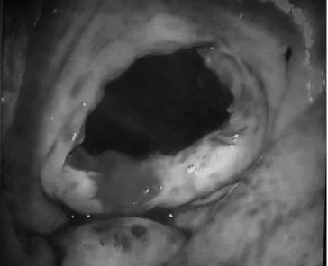
The remaining steps of the surgery were performed routinely, with the passage of stitches through the valve ring done in a similar way to conventional surgeries. Both visibility of the ring and lowering of the prosthesis to its position were very good (Figure 2).
A long appropriate instrument is required for placing the
knots to afix the prosthesis. Once the prosthetic valve was
placed (Figure 3), a left ventricle aspirator was inserted via the left atrium and through the prosthesis and atriorrhaphy
was done as usual. Deibrillation was performed externally.
After the cannulas were removed, heparin was completely reversed and the right pleural cavity was drained. The intercostal space used for access was closed with 2-0 ethibond suture and the skin was closed with intradermal suture.
• This study was approved by the Ethics Committee, protocol 0003761/10 on October 17th, 2010.
RESULTS
A total of ten patients, four males and six females, were evaluated. Patient ages ranged from 46 to 76 years-old, the mean being 56.9±10.5 years. Two patients (20%) were diabetic, six (60%) had hypertension, and four (40%) had history of smoking. Four patients (40%) were in functional
class II, ive (50%) were in functional class III, and one (10%)
was in functional class IV. Electrocardiogram at rest showed
sinus rhythm in six patients (60%) and atrial ibrillation in
four patients (40%). Four patients (40%) had normal ejection
fraction, ive patients (50%) had moderate left ventricular
dysfunction, and one patient (10%) had severe ventricular dysfunction. Five patients (50%) had undergone previous
mitral valve repair; the other ive (50%), mitral valve Fig. 2 – This technique provides good visibility
Fig. 1 - Retractor developed speciically for this type of operation introduced into the left atrium
replacement with biological prosthesis. The diagnosis which led to a new surgical procedure was: stenosis in two patients
(20%), insuficiency in three patients (30%), and double dysfunction in ive patients (50%). Mean length of time between the irst and second surgeries was 11±3.43 years,
ranging from 7 to 17 years. EuroSCORE ranged from 7 to 12, with a mean of 8.3±1.82.
Mechanical prostheses were implanted in eight patients (80%) and biological prostheses were implanted in the other two patients (20%). Size of the prosthesis ranged from 25 to 29 mm, with seven patients (70%) receiving a 27 mm prosthesis.
Mean time of ventricular ibrillation was 70.9±17.66
minutes, ranging from 45 to 100 minutes. Cardiopulmonary bypass time ranged from 66 to 150 minutes, with a mean of 109.4±25.37 minutes. After surgery, patients were taken to the ICU, where mean time of mechanical ventilation was 6.4±3.2 hours, ranging from zero to 12 hours. Length of stay in the ICU ranged from 2 to 3 nights, the mean being 2.5± 0.52 nights. Six patients (60%) had low output syndrome after removal of cardiopulmonary bypass, requiring inotropic support. None of the patients in this series needed the reoperation due to bleeding nor did they have stroke, acute myocardial infarction, or acute renal failure. One patient (10%) developed pneumonia in the postoperative period. There were no deaths in this study.
At hospital discharge, ive patients (50%) had sinus rhythm and ive patients (50%) had atrial ibrillation. Mean
length of hospital stay was 7.6±1.5 days, ranging from 5 to 10 days.
DISCUSSION
Since the mid-1990s, the medical community has shown growing interest in mini-incisions for valve replacement [4-8]. The right anterolateral thoracotomy seems to be an interesting access for mitral valve surgeries as it allows for direct vision of the line where left atriotomy is usually performed, good exposure of the valve, and minimum initial discomfort for the surgeon. In addition, aesthetic result of this incision is superior to others, since, in women, the breast hides the incision [9].
In the last decade, the use of video in cardiac surgeries has become commonplace. Some studies [10-15] have demonstrated that, in video-assisted surgeries, incisions can
be smaller, visualization of the surgical ield is improved, and
long-term results are the same as those of conventional mitral valve surgery. Furthermore, there is substantial reduction in trauma, mechanical ventilation as well as hospital stay, so the patient can return to routine activities faster than in conventional surgeries, where the patient cannot drive for 45 days as opposed to 10 to 15 days in minimally invasive surgeries.
Dogan et al. [11] published an article comparing conventional and minimally invasive surgeries. Forty patients were randomly assigned to two groups, Group I being comprised of patients undergoing minimally invasive surgery and Group II of patients undergoing conventional surgery. Cardiopulmonary bypass and aortic clamping times were slightly longer in Group I; however, there were no
statistically signiicant differences.
In Group I, none of the patients needed to be converted to conventional surgery and length of both mechanical ventilation and hospital stay was shorter, but again there
were no statistically signiicant differences. There was also
no difference between the two groups in terms of drainage by chest drains and pulmonary function. There were no major complications or deaths reported in this series; however, the authors stated that the biggest challenge in Group I was myocardial protection, as they faced a series of problems
with endovascular aortic clamping in addition to dificulties
using conventional occlusion forceps. The authors concluded
that despite the lack of statistically signiicant data in
favor of minimally invasive surgeries, they proved to be
just as effective as conventional surgeries, which justiies
performing the less invasive procedure.
In a similar article, Holzhey et al. [16] published a study comparing conventional and minimally invasive surgeries in patients over 70 years-old. Like other studies [17-19], the authors stated that duration of cardiopulmonary bypass, aortic clamping as well as the surgery itself was longer. There were no differences in terms of major complications and mortality in 5 to 8 years of follow-up. In the immediate postoperative period, the conventional surgery group showed greater incidence of arrhythmia and pacemaker implant; once
again, the difference was not statistically signiicant. The
conclusions reached were the same as the ones from other authors: minimally invasive surgeries are at the very least equal to conventional surgeries and the biggest challenge lies in myocardial protection methods.
Ventricular ibrillation associated with hypothermia has
been used as myocardial protection in coronary surgeries for many years, based on the fact that a decompressed heart in
ventricular ibrillation associated with hypothermia at 28°C
consumes roughly the same amount of energy as a heart in cardiac arrest induced by cardioplegia [3].
On the other hand, in valve surgeries, this kind of heart protection technique has always caused certain concern related to the incidence of stroke, since the cavities are open and the aorta is not clamped.
In a 2008 article, Umakanthan et al. [20] demonstrated
how safe ventricular ibrillation associated with hypothermia
underwent the whole procedure without aortic clamping,
under hypothermia and ventricular ibrillation, and with
continuous CO2 instillation. The average EuroSCORE of the group was above 8, indicating a group of high risk patients. Yet, there were neither deaths nor strokes observed in this series. The biggest challenge faced in this study was the removal of the mitral prosthesis that had been implanted in
the irst surgery, which led to patients in this condition being under ventricular ibrillation for a longer period of time. Six
patients developed low output syndrome after removal of cardiopulmonary bypass. Consequently, in the last cases, it took longer to start the reheating in order to avoid exposing a
heart under ibrillation to higher temperatures. The effect of
doing that was somewhat positive making it easier to remove
the CPB. Presence of mild aortic insuficiency hinders the
surgical procedure; therefore, in cases where there is slight
to mild insuficiency, the procedure should not be performed.
CONCLUSION
Minimally invasive mitral valve reoperations under
hypothermia associated with ventricular ibrillation without
aortic clamping can be safely performed, with good immediate results, low incidence of complications, and no need for ample dissections of the heart. However, longer length of time on cardiopulmonary bypass is required, especially in patients who have previously implanted prosthesis. This type of procedure should not be performed in this manner if there
is signiicant aortic insuficiency.
showed incidence of mortality in 30 days at 3%, low output syndrome at 4%, and stroke at 3%, which are equivalent to the results reported by other authors who used myocardial protection with aortic clamping [10,11,16]. The authors attributed the low incidence of stroke to: arterial pressure being kept above 30 mmHg during cardiopulmonary bypass, insertion of cannula for continuous aspiration into the left ventricle, and continuous CO2 instillation in the cavity throughout the procedure. The authors concluded this type of cardiac management is safe, easily reproduced, and it makes minimally invasive surgery simple.
After this study, minimally invasive mitral valve surgery, which had been limited to patients who had had no previous surgical intervention, started to be performed in patients undergoing reoperations as well.
In patients who have undergone previous surgeries, the heart
tends to have a large amount of adhesions, which are dificult
to remove through minimally invasive methods, making the aortic clamping almost impossible to achieve. That, as well as inconsistencies in the occlusion of the aorta via endovascular techniques severely limits the indication of less invasive procedures to patients undergoing reoperations. Aortic clamping
is unnecessary when the patient is under ventricular ibrillation;
therefore, it is possible to remove only the adhesions on top of the left atrium where the incision is made.
In 2010, Ricci et al. [21] presented a study in which 241 patients who had undergone previous mitral valve or other cardiac surgeries were evaluated. The authors started by describing the risks of performing a resternotomy due to adhesions, which could lead to serious injuries, especially to the right ventricle and innominate vein. In the results, the authors mentioned the need to convert the surgery to sternotomy in two patients, one due to aortic dissection; the other, left ventricular perforation. Average length of stay in intensive care was 24 hours, average time of mechanical ventilation was 12 hours, and average blood drainage in 24
hours was 450 ml. Ventricular ibrillation was used in six
of the patients who underwent surgery; in the remaining patients, endovascular clamping was used. Furthermore, the
authors reported a stroke rate of 5.8%, none in the ibrillation
group, and overall mortality of 4.9%. They concluded that the minimally invasive mitral valve reoperation is safe
and its beneits are: very low incidence of surgical wound
infection, short length of intensive care unit and hospital stay, good aesthetic results, and faster return to routine activities.
A report was published in 2008 by Fortunato et al.[22], describing the case of a patient who underwent endoscopic mitral valve surgery reoperation. The authors performed a comissurotomy in a valve that had been repaired 12 years earlier. They reached the conclusion that the procedure was feasible and effective.
In the present study, 10 patients who underwent minimally invasive mitral valve reoperation were evaluated. All patients
Authors' roles & responsibilities
RM Main author PRSB Coauthor SO Coauthor LPN Coauthor MR Coauthor VHL Coauthor LFB Coauthor AS Coauthor
REFERENCES
1. Akins CW. Noncardioplegic myocardial preservation for coronary revascularization. J Thorac Cardiovasc Surg. 1984;88(2):174-81.
2. Greene PS, Cameron DE, Grifiths EM, DiNatale JM, Gardner TJ. Does hypothermic ibrillatory arrest improve myocardial
valve surgery via left thoracotomy: experience with forty cases. J Thorac Cardiovas Surg. 2004;127(4):1026-31.
14. Poffo R, Bonim M, Selbach RA, Pillati M. Troca valvar mitral minimamente invasiva videoassistida. Rev Bras Cir Cardiovasc. 2007;22(4):491-4.
15. Fortunato Jr JA, Branco Filho AA, Branco A, Martins ALM, Pereira ML, Ferraz JGG, et al. Padronização da técnica para cirurgia cardíaca videoassistida: experiência inicial. Rev Bras Cir Cardiovasc. 2008;23(2):183-9.
16. Holzhey DM, Shi W, Borger MA, Seeburger J, Garbade J, Pfannmüller B, et al. Minimally invasive versus sternotomy approach for mitral valve surgery in patients greater than 70 years old: a propensity-matched comparison. Ann Thorac Surg. 2011;91(2):401-5.
17. Modi P, Hassan A, Chitwood WR Jr. Minimally invasive mitral valve surgery: a systematic review and meta-analysis. Eur J Cardiothorac Surg. 2008;34(5):943-52.
18. Chitwood WR Jr, Elbeery JR, Morgan JF. Minimally invasive mitral valve repair using transthoracic aortic occlusion. Ann Thorac Surg. 1997;63(5):1477-9.
19. Dagenais F. Minimally invasive mitral valve surgery: evolution, techniques and outcomes. Future Cardiol. 2008;4(6):609-16.
20. Umakanthan R, Leacche M, Petracek MR, Kumar S, Solenkova NV, Kaiser CA, et al. Safety of minimally invasive mitral valve surgery without aortic cross-clamp. Ann Thorac Surg. 2008;85(5):1544-9.
21. Ricci D, Pellegrini C, Aiello M, Alloni A, Cattadori B, D’Armini AM, et al. Port-access surgery as elective approach for mitral valve operation in re-do procedures. Eur J Cardiothorac Surg. 2010;37(4):920-5.
22. Fortunato Júnior JA, Branco Filho AD, Branco A, Martins ALM, Pereira M. Reoperação de valva mitral totalmente endoscópica: relato de caso. Rev Bras Cir Cardiovasc. 2008;23(3):411-4. 3. Milani RM. Análise dos resultados imediatos da operação para
revascularização do miocárdio sem pinçamento total da aorta [Dissertação de Mestrado]. Curitiba: Universidade Federal do Paraná; 2000.
4. Cosgrove DM 3rd, Sabik JF. Minimally invasive approach for aortic valve operations. Ann Thorac Surg. 1996;62(2):596-7.
5. Cosgrove DM 3rd, Sabik JF, Navia JL. Minimally invasive valve operations. Ann Thorac Surg. 1998;65(6):1535-8.
6. Koenertz W, Waldenberger F, Schmutzler M, Rilter J, Liu J. Minimal access valve surgery through superior partial sternotomy: a preliminary study. J Heart Valve Dis. 1996;5(6):638-40.
7. Navia JL, Cosgrove DM 3rd. Minimally invasive mitral operations. Ann Thorac Surg. 1996;62(5):1542-4.
8. Navia JL, Cosgrove DM. Minimally invasive mitral valve operations. Ann Thorac Surg. 1996;62(5):1542-4.
9. Mishra YK, Malhotra R, Mehta Y, Sharma KK, Kasliwal RR, Trehan N. Minimally invasive mitral valve surgery through right anterolateral minithoracotomy. Ann Thorac Surg. 1999;68(4):1520-4.
10. Grossi EA, Galloway AC, LaPietra A, Ribakove GH, Ursomanno P, Delianides J, et al. Minimally mitral valve surgery: 6-year experience with 714 patients. Ann Thorac Surg. 2002;74(3):660-3.
11. Dogan S, Aybek T, Risteski PS, Detho F, Rapp A, Wimmer-Greinecker G, et al. Minimally invasive port access versus conventional mitral valve surgery: prospective randomized study. Ann Thorac Surg. 2005;79(2):492-8.
12. Rodriguez E, Nifong LW, Chu MW, Wood W, Vos PW, Chitwood
WR. Robotic mitral valve repair for anterior lealet and bilealeat
prolapse. Ann Thorac Surg. 2008;85(2):438-44.