Periodontics
Carolina Barrera de Azambuja Juliano Cavagni
Marcius Comparsi Wagner Eduardo José Gaio
Cassiano Kuchenbecker Rösing
Department of Periodontology, School of Dentistry, Univ Federal do Rio Grande do Sul - UFRGS, Porto Alegre, RS, Brazil.
Corresponding Author: Juliano Cavagni
E-mail: [email protected]
Correlation analysis of alveolar bone
loss in buccal/palatal and proximal
surfaces in rats
Abstract: The aim was to correlate alveolar bone loss in the buccal/pala-tal and the mesial/disbuccal/pala-tal surfaces of upper molars in rats. Thirty-three, 60-day-old, male Wistar rats were divided in two groups, one treated with alcohol and the other not treated with alcohol. All rats received silk ligatures on the right upper second molars for 4 weeks. The rats were then euthanized and their maxillae were split and deleshed with sodium hypochlorite (9%). The cemento-enamel junction (CEJ) was stained with 1% methylene blue and the alveolar bone loss in the buccal/palatal sur-faces was measured linearly in 5 points on standardized digital photo-graphs. Measurement of the proximal sites was performed by sectioning the hemimaxillae, restaining the CEJ and measuring the alveolar bone loss linearly in 3 points. A calibrated and blinded examiner performed all the measurements. Intraclass Correlation Coeficient revealed values of 0.96 and 0.89 for buccal/lingual and proximal surfaces, respectively. The Pearson Correlation Coeficient (r) between measurements in buccal/ palatal and proximal surfaces was 0.35 and 0.05 for the group treated with alcohol, with and without ligatures, respectively. The best corre-lations between buccal/palatal and proximal surfaces were observed in animals not treated with alcohol, in sites both with and without liga-tures (r = 0.59 and 0.65, respectively). A positive correlation was found between alveolar bone loss in buccal/palatal and proximal surfaces. The correlation is stronger in animals that were not treated with alcohol, in sites without ligatures. Areas with and without ligature-induced peri-odontal destruction allow detection of alveolar bone loss in buccal/pala-tal and proximal surfaces.
Descriptors: Alveolar Bone Loss; Animals; Periodontitis.
Introduction
Periodontitis is a disease characterized by periodontal tissue destruc-tion caused by the host response against bacteria and their toxins. The absence of treatment can lead to the loss of the tooth affected by the disease.1
In the study of the pathogenesis of periodontitis, there are strong limi-tations in the use of models involving humans for logistic and ethical rea-sons. Because of these limitations, animal models have been widely used in literature. Among the experimental models for destructive periodontal disease study, monkeys, dogs and small animals have been used. Rats
Declaration of Interests: The authors certify that they have no commercial or associative interest that represents a conflict of interest in connection with the manuscript.
Submitted: May 08, 2012
are one of the most frequently used models, since they share similarities with humans. These similari-ties apply to periodontal anatomy, development and compositions of bioilms, histopathology and immu-nobiology of periodontal lesions.2
Following the experimental periods, it is impor-tant to quantify the destruction, in order to under-stand the effect of the factor studied in the experi-mental development of the disease. Animal models make it possible to make different morphological measurements, which of course is not possible with human models. These measurements enable metri-cal quantiication of the result and of the inlamma-tory process. For this purpose, different methods for measuring alveolar bone loss in rats have been pro-posed in the literature, such as histometric,3
mor-phometric4-6 conventional radiographic
measure-ments7 and computed tomography.8,9
Different studies have sought to demonstrate the ability to measure periodontal destruction by dif-ferent methods and effectively quantify the effect caused by the experimental model of periodontal disease. Fernandes et al.10 compared the
histomet-ric and morphomethistomet-ric methods and found that both are capable of detecting alveolar bone loss in rats. Li and Amar11 likewise compared the different
meth-ods—morphometric isolated, histometric and mor-phometric associated and micro-computed tomog-raphy—in 100 male rats, and their indings showed that all the methods are accurate for quantiication of alveolar bone loss.
In addition to the ability to detect the destruc-tion, there is the issue, raised in the literature, of the best site and the best way to measure the observed effects. For example, there are studies comparing the morphometric method with area versus linear measurements.12-14 However, these two systems
al-low evaluation of alveolar bone loss in lingual/ palatal and buccal surfaces only. To the best of our knowledge, studies evaluating proximal bone loss are inexistent in the literature to date.
Thus, the aim of this study was to correlate al-veolar bone loss in buccal/palatal and proximal sur-faces of upper molars in Wistar rats.
Methodology
Sample
The data used in the present study included a subset of the project entitled “Effect of the chemi-cal dependence of 15% ethanol on ligature-induced alveolar bone loss in Wistar rats.” Thirty-three male, 60-day-old Wistar rats were used, 15 not treated and 18 treated with alcohol. A randomized, blind and controlled animal model study was per-formed. The project compared alcohol treated ani-mals during the whole study period with aniani-mals not exposed to alcohol. It was a methodological study that compared intragroup measurements of alveolar bone loss, without taking alcohol exposure speciically into consideration. All rats received 4-0 silk ligatures (Ethicon, Johnson & Johnson, São Paulo, Brazil) for induction of alveolar bone loss around the upper right second molars. The left side remained as intragroup control. In all, 66 hemimax-illae were used (33 with ligature and 33 without). The induction of alveolar bone loss occurred during a period of four weeks.
During the entire experimental period, the rats remained at the Center of Reproduction and Ex-perimentation of Laboratory Animals (CREAL), at the Federal University of Rio Grande do Sul. Ani-mals were kept in boxes, in rooms with a controlled temperature (20°C) and in a light/dark cycle of 12 hours. The animals received standard diet ad libi-tum during the study.
The study protocol was submitted and approved by the Research Ethics Committee of the Federal University of Rio Grande do Sul. The protocol com-plies with the regulations set down by the Universal Declaration of Animal Rights (UNESCO - January 27, 1978) and the International Ethical Guidelines for Biomedical Research Involving Animals (Coun-cil for International Organizations of Medical Sci-ences - CIOMS).
Specimen preparation
and followed the method proposed by Fernandes et al.10 Soft tissue was removed by deleshing the
max-illae with sodium hypochlorite solution (9%) (Maz-arollo, Gravataí, Brazil) for ive hours, and then removing the remaining tissues manually. The speci-mens were then washed and dried. Specispeci-mens were stained with 1% methylene blue for one minute for better visualization of the CEJ. Pictures were taken using a 6.1 megapixel digital camera (Nikon Cool-pix, Ayutthaya, Thailand) coupled to a tripod and equipped with 100 mm macro-lenses with minimal focal distance. The hemimaxillae were placed with the occlusal surface parallel to the loor.4,5,10,6,13
Buccal/palatal morphometric analysis
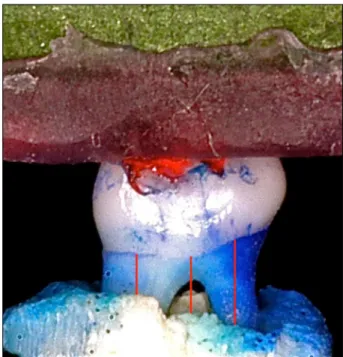
The buccal/palatal morphometric analysis was performed by evaluating standard digital photo-graphs. An examiner, calibrated and blinded to the experimental groups, performed the measurements of the linear distances of the CEJ to the bone crest, using Image Tool 3.0. software (UTHSCPA, San Antonio, USA). Five measurements were performed on each surface (two on the distal root, two on the mesial root and one on the furcation). The measure-ments in pixels were then converted into millimeters using the markings of the endodontic ruler to which the hemimaxillae were attached as reference. Each hemimaxillae generated a mean value for analysis at a later time. Figure 1 shows the specimen and the measurements made on the buccal surface in the site with no ligature.
Proximal site morphometric analysis
After morphometric analysis of buccal/pala-tal surfaces, the hemimaxillae were cut with hand pieces and diamond burs under refrigeration, in mesial and distal aspects of the second molar, pre-serving the bone crest. The CEJ was restained with 1% methylene blue, and alveolar bone loss was evaluated in three regions (buccal root, center of the proximal surface and palatal root) of each proximal surface (mesial and distal). Before cutting, a refer-ence mark was made in order to identify the mesial surface. Each hemimaxillae generated a mean value for later analysis. Figure 2 shows the measurements for the site with ligature on the proximal aspects.
Blinding
Examiner blinding was assured before the mor-phometric analysis. After the photographs were tak-en, they were all codiied by an external examiner, so that the investigators would not know to which group each specimen belonged when analyzed. After measurement was concluded, the code was broken and the photographs were renamed according to the respective group.
Figure 1 - Linear measurements made on digital photo-graphs of the buccal surface of hemimaxillae attached to an endodontic ruler in the site without ligature.
Reproducibility
Reproducibility was checked by testing and re-testing, with a one-week interval between measure-ments. Scatter Graphics and Intraclass Correlation Coeficient (ICC) were used to evaluate reproduc-ibility. The ICC of these measurements was 0.96 and 0.89 for buccal/palatal and proximal surfaces, respectively. These values are considered excellent by the literature.
Data analysis
Parametric tests were used to determine possible correlations and statistical differences between the experimental groups, insofar as the data presented normal distribution. Pearson Correlation Coefi-cient (r) was used to determine the grade of corre-lation between measurements of buccal/palatal and proximal surfaces. Cutoff points were > 0.70: strong correlation; 0.30 to 0.70: moderate correlation; < 0.30: weak correlation. All data analyses were performed using Stata© 10.1 for Macintosh software
(Stata Corporation, College Station, USA).
Results
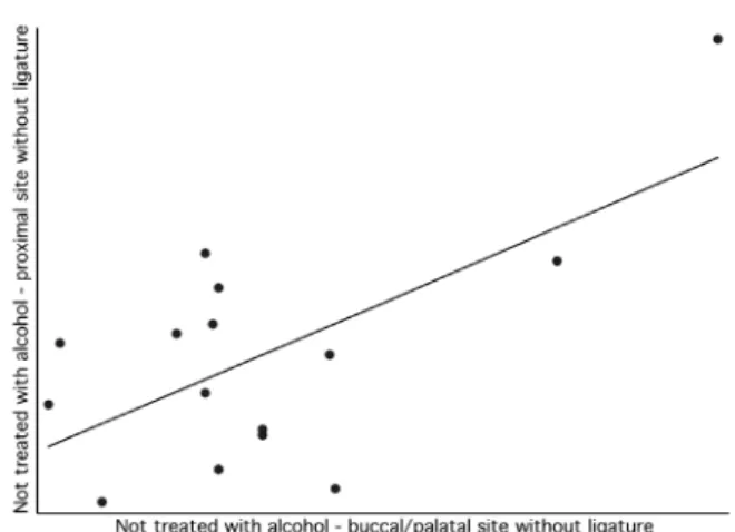
The results of the present study are shown in Figures 3–6. Correlations were assessed separately between buccal/palatal and proximal sites for teeth with and without ligature in the hemimaxillae of rats treated or not treated with alcohol.
The correlation between the variables presented
a positive/ascendant direction (Figures 3–6) for all comparisons. However, there were different values for the Pearson Correlation Coeficient, depending on the comparisons. In general, Figures 3, 5 and 6 presented a moderate correlation, whereas Figure 4 presented a low correlation. The Pearson Correla-tion Coeficient between measurements of buccal/ palatal and proximal surfaces was 0.35 and 0.05 in the groups treated with alcohol, in sites with or without ligature, respectively. When groups that were not treated with alcohol are considered, higher correlations were observed between measurements of buccal/palatal and proximal surfaces, with an r = 0.59 and r = 0.65 in sites with and without liga-ture, respectively.
Discussion
The objective of the present study was to corre-late measurements of ligature-induced alveolar bone loss in Wistar rats, in buccal/palatal and proximal surfaces, using specimens from a controlled experi-mental study. It was observed that the measurements for animals not treated with alcohol presented good correlation. It is important to emphasize that 100% of the specimens were evaluated, with no losses. The CEJ was evident in all specimens, both in the buc-cal/palatal and in the proximal surfaces. This indi-cates that it is possible and reliable to prepare the specimens without losing information. There were no dificulties in analyzing the proximal surfaces.
Figure 3 - Scatter plot correlating buccal/palatal and proxi-mal surfaces in the group treated with alcohol and with liga-ture.
Experimental studies in animals are fundamen-tal in the context of etiopathogenic research of the morbid processes. This situation relies on the idea that it is important to know the different interfer-ences that may occur in the causal chain of diseases. These studies cannot be performed in humans be-cause of ethical reasons, and also bebe-cause it is im-possible to isolate the variables, which is im-possible to do in animal models.15,2 Nonetheless, it is important
to clarify that data obtained in animals represent a step toward gaining the needed knowledge, but must not be directly extrapolated to clinical situations.16
One of the most important aspects to be high-lighted in the present study is that the potential ben-eits of understanding the pathogenesis of periodon-tal destruction in rats can transcend dentistry. In the health sciences, it is very dificult to quantify in-lammation and its effects with objective and repro-ducible measurements. This is done today mostly by analyzing the quantity and intensity of inlamma-tory cells in different manners. In the speciic case of periodontal disease, the inlammatory process leads to tissue destruction, an effect that is quantiiable by objective criteria. Therefore, the experimental mod-els of periodontal disease induction in animals have the potential to be used for studying other chronic inlammatory processes, if what is intended is to un-derstand the chain of events and to isolate variables. These measurements are reproducible and, in the present study, the Intraclass Correlation Coeficients
for buccal/palatal surfaces and proximal surfaces were about 0.9, a igure considered excellent. Ex-perimental models such as that used in the present study are useful, because they allow adequate exam-iner blinding and repeated analyses. This increases the quality of the information generated.
It is also important to highlight that the way al-veolar bone loss was measured in the present study is also at issue. It is possible to measure periodon-tal destruction by histometric, morphometric and also image examinations. Studies published by Fernandes et al.,10 Park et al.8 and Wilensky et al.9
demonstrated that these three outcome-measuring methodologies are possible and capable of detecting differences among the factors studied.
Different studies have addressed the measure-ment issue in distinct ways. For instance, some stud-ies have measured the destruction in the furcation area, others in proximal sites, some in buccal and lingual areas, others with only one measurement, or repeated measurements, etc.14,17-24 To the best of our
knowledge, there are no studies evaluating ligature-induced alveolar bone loss in proximal and buccal/ palatal surfaces in rats. Although ligature-induced alveolar bone loss measurement in proximal sur-faces may be dificult to perform in morphometric analyses, the focus of the present study was to ex-amine differences in the prevalence and extension of periodontal disease in proximal surfaces in humans, which could also occur in animals.25
Figure 6 - Scatter graph correlating buccal/palatal and proximal surfaces in the group not treated with alcohol and without ligature.
The results obtained in the present study showed a positive correlation between the measurements of buccal/palatal and proximal surfaces in groups not treated with alcohol, in sites both with and without ligature. In the group treated with alco-hol, these correlations were less intense. This may be explained by the fact that other factors may in-terfere in the outcomes more easily when there is a modiier. Interestingly, there is a study that has shown differences between the groups only in the site without ligature.6 This has been examined in the
research on etiopathogenesis of periodontal diseases in animal models, and explanations are still scarce. In the present study, an r = 0.59 was obtained for the group not treated with alcohol, in the site with ligature, and a value of r = 0.65 was obtained for the site without ligature. The higher value obtained for this correlation coeficient corroborates that the chances of higher associations between the measure-ments increase when there are no interferences.
The results of the present study are challenging
and show that it is possible to detect destruction of the periodontium in buccal/palatal and also in prox-imal surfaces after an experimental period. Thus, it is optional where the researcher conducts the mea-surements; the best place would be that which is most suitable to posit the hypothesis of his research. In addition, the results show that it is not impor-tant to conduct two distinct methods, i.e., analysis in buccal/palatal and proximal surfaces, in order to evaluate alveolar bone loss; either one or the other may be conducted.
Conclusion
In models of periodontal destruction induced by ligature, there is correlation in alveolar bone loss between buccal/palatal and proximal surfaces. The correlation is stronger in animals not treated with alcohol. In areas with and without ligature-induced periodontal disease, it is possible to detect alveolar bone loss in buccal/palatal and proximal surfaces.
References
1. Page RC, Kornman KS. The pathogenesis of human periodon-titis: an introduction. Periodontol 2000. 1997 Jun;14(1):9-11. 2. Susin C, Rösing CK. O rato como modelo para o estudo das
repercussões do estresse nas doenças periodontais. Rev Perio-dontia. 2002;13(6):5-10.
3. Semenoff TA, Semenoff-Segundo A, Bosco AF, Nagata MJ, Garcia VG, Biasoli ER. Histometric analysis of ligature-induced periodontitis in rats: a comparison of histological section planes. J Appl Oral Sci. 2008 Jul-Aug;16(4):251-6. 4. Cavagni J, Soletti AC, Gaio EJ, Rosing CK. The effect of
dexamethasone in the pathogenesis of ligature-induced peri-odontal disease in Wistar rats. Braz Oral Res. 2005 Oct-Dec;19(4):290-4.
5. Daudt LD, Cavagni J, Gaio EJ, Souza A, Torres IL, Ferreira MB, et al. Effect of inhaled corticosteroid on TNF-alpha pro-duction and alveolar bone loss in Wistar rats. Arch Oral Biol. 2011 Nov;56(11):1398-403.
6. Liberman DN, Pilau RM, Gaio EJ, Orlandini LF, Rosing CK. Low concentration alcohol intake may inhibit spontane-ous alveolar bone loss in Wistar rats. Arch Oral Biol. 2011 Feb;56(2):109-13.
7. Semenoff Segundo A, Semenoff TA, Borges AH, Pedro FL, Sakai VT. Methodological model of chronic stress associated with ligature-induced periodontitis in rats: a radiographic study. Braz Oral Res. 2010 Oct-Dec;24(4):455-9.
8. Park CH, Abramson ZR, Taba Jr M, Jin Q, Chang J, Kreider JM, et al. Three-dimensional micro-computed tomographic imaging of alveolar bone in experimental bone loss or repair. J Periodontol. 2007 Feb;78(2):273-81.
9. Wilensky A, Gabet Y, Yumoto H, Houri-Haddad Y, Shapira L. Three-dimensional quantification of alveolar bone loss in Por-phyromonas gingivalis-infected mice using micro-computed tomography. J Periodontol. 2005 Aug;76(8):1282-6. 10. Fernandes MI, Gaio EJ, Oppermann RV, Rados PV, Rosing
CK. Comparison of histometric and morphometric analyses of bone height in ligature-induced periodontitis in rats. Braz Oral Res. 2007 Jul-Sep;21(3):216-21.
11. Li CH, Amar S. Morphometric, histomorphometric, and microcomputed tomographic analysis of periodontal in-flammatory lesions in a murine model. J Periodontol. 2007 Jun;78(6):1120-8.
12. Kuhr A, Popa-Wagner A, Schmoll H, Schwahn C, Kocher T. Observations on experimental marginal periodontitis in rats. J Periodontal Res. 2004 Apr;39(2):101-6.
13. Liberman DN, Pilau RM, Orlandini LF, Gaio EJ, Rosing CK. Comparison of two methods for alveolar bone loss measure-ment in an experimeasure-mental periodontal disease model in rats. Braz Oral Res. 2011 Jan-Feb;25(1):80-4.
per-centages caused by periodontitis in rats in different locations. J Appl Oral Sci. 2010 Sep-Oct;18(5):493-7.
15. Graves DT, Fine D, Teng YT, Van Dyke TE, Hajishengallis G. The use of rodent models to investigate host-bacteria interac-tions related to periodontal diseases. J Clin Periodontol. 2008 Feb;35(2):89-105.
16. Oz HS, Puleo DA. Animal models for periodontal disease. J Biomed Biotechnol. 2011;2011:754857. Epub 2011 Feb 10. 17. Barros SP, Silva MA, Somerman MJ, Nociti Jr FH. Parathyroid
hormone protects against periodontitis-associated bone loss. J Dent Res. 2003 Oct;82(10):791-5.
18. Bjornsson MJ, Velschow S, Stoltze K, Havemose-Poulsen A, Schou S, Holmstrup P. The influence of diet consistence, drinking water and bedding on periodontal disease in Sprague-Dawley rats. J Periodontal Res. 2003 Dec;38(6):543-50. 19. Cesar-Neto JB, Benatti BB, Haiter Neto F, Sallum AW, Sallum
EA, Nociti Jr FH. Smoking cessation may present a positive impact on mandibular bone quality and periodontitis-related bone loss: a study in rats. J Periodontol. 2005 Apr;76(4):520-5. 20. Duarte PM, Goncalves PF, Sallum AW, Sallum EA, Casati
MZ, Nociti Jr FH. Effect of an estrogen-deficient state and
its therapy on bone loss resulting from an experimental peri-odontitis in rats. J Periodontal Res. 2004 Apr;39(2):107-10. 21. Fine DH, Schreiner H, Nasri-Heir C, Greenberg B, Jiang S,
Markowitz K, et al. An improved cost-effective, reproducible method for evaluation of bone loss in a rodent model. J Clin Periodontol. 2009 Feb;36(2):106-13.
22. Mitsuta T, Horiuchi H, Shinoda H. Effects of topical ad-ministration of clodronate on alveolar bone resorption in rats with experimental periodontitis. J Periodontol. 2002 May;73(5):479-86.
23. Niikura K, Takeshita N, Chida N. A novel inhibitor of vacu-olar ATPase, FR202126, prevents alvevacu-olar bone destruction in experimental periodontitis in rats. J Toxicol Sci. 2005 Dec;30(4):297-304.
24. Vacas MI, Amer M, Chiarenza AP, Luchelli MA, Mandalunis PM, Elverdin JC. Influence of submandibulectomy on alveolar bone loss in rats. J Periodontol. 2008 Jun;79(6):1075-80. 25. Albandar JM, Brunelle JA, Kingman A. Destructive