Comparison of histometric and
morphometric analyses of bone height in
ligature-induced periodontitis in rats
Análise comparativa da altura óssea
histométrica e morfométrica na periodontite
induzida por ligadura em ratos
Abstract: The purpose of this study was to compare histologic and morphometric pro-cedures of bone height measurement. Microscopic measurements are the most frequent methods in periodontal studies with animals, but have limited capacity to identify bone levels associated with both healthy tissues and periodontal disease. Ligatures were placed in the maxillary left second molars of 10 male 60-day-old Wistar rats for 30 days. Left and right maxillary sides of 5 rats were processed for histologic analysis (H), sectioned buccolingually, and stained with HE. The maxillae of the other 5 rats were deleshed and used for morphometric analysis (M). Histometric measurements from the cementoe-namel junction to the bone crest were performed. Standardized photographs were used for morphometric analysis. The t test was used for dependent or independent samples (α= 0.05%). Distances from cementoenamel junction to bone crest were 0.95 ± 0.25 and 1.07 ± 0.30 mm for H and M, respectively. Buccal measurements were 0.92 ± 0.16 and 1.08 ± 0.35 mm for H and M. The values obtained using H and M for areas without liga-tures were 0.44 ± 0.15 and 0.47 ± 0.11 mm for lingual measurements and 0.23 ± 0.08 and 0.41 ± 0.10 mm for buccal measurements. No signiicant differences were found between the two methods in the detection of bone height associated with the placement of ligatures in rats.
Descriptors: Histology; Alveolar bone loss; Periodontium.
Resumo: O objetivo deste estudo foi comparar procedimentos de medida da altura óssea histológica e morfométrica. O método mais freqüente de mensuração da altura óssea em estudos em animais são as medidas microscópicas. No entanto, tem limitada capacidade de identiicar os níveis ósseos associados com os tecidos saudáveis e doença periodontal. Foram colocadas ligaduras no segundo molar superior esquerdo de 10 ratos machos Wis-tar com 60 dias de vida durante um período de 30 dias. Hemimaxilas direitas e esquerdas de 5 ratos foram processadas para a análise histológica (H), cortadas no sentido vestí-bulo-lingual e coradas com HE. Hemimaxilas dos outros 5 ratos tiveram toda a matéria orgânica removida para análise morfométrica (M). Medidas histométricas da junção ce-mento-esmalte (JCE) à crista óssea alveolar foram realizadas. Fotograias padronizadas foram utilizadas na análise morfométrica. O teste t foi utilizado para amostras dependen-tes e independendependen-tes (α = 0,05%). As distâncias entre a JCE e a crista óssea na face palatina foram de 0,95 ± 0,25 e 1,07 ± 0,30 mm para H e M, respectivamente. As medidas vesti-bulares foram 0,92 ± 0,16 e 1,08 ± 0,35 mm para H e M. Os valores obtidos usando H e M para áreas sem ligadura foram 0,44 ± 0,15 e 0,47 ± 0,11 mm por lingual e 0,23 ± 0,08 e 0,41 ± 0,10 mm por vestibular, respectivamente. Não houve diferenças estatisticamente signiicativas entre os dois métodos na detecção da altura óssea associada à colocação de ligaduras em ratos.
Descritores: Histologia; Perda óssea alveolar; Periodonto. Marilene Issa Fernandes(b)
Eduardo José Gaio(a)
Rui Vicente Oppermann(b)
Pantelis Varvaki Rados(b)
Cassiano Kuchenbecker Rosing(b)
(a)Graduate Student; (b)Professors, Graduate
Program in Dentistry – School of Dentistry, Federal University of Rio Grande do Sul.
Corresponding author:
Eduardo José Gaio
Av. Montenegro, 160/701, Bairro Petrópolis Porto Alegre - RS - Brazil
CEP: 90460-160
E-mail: [email protected]
Introduction
Rats have been used in periodontal disease stud-ies4,9,10,12,14 because their periodontal tissue structure
in the molar region is similar to that of human be-ings.20 Moreover, rats can be kept in germfree
con-ditions1,5 and have their immunologic capacity
com-promised,11 which makes them an attractive model
for better understanding the pathogenesis of peri-odontal diseases.
Several methods have been used to evaluate peri-odontal bone loss in rats. Studies have described lesions macroscopically,15,28 histologically3,17,18 or
radiographically.2,10,15 The histologic method is
cur-rently the criterion standard, but it has drawbacks, especially because the pattern of bone resorption in periodontitis is not uniform.14,24 In addition,
prob-lems during histologic processing, dificulties in car-rying it out, its high cost and long time requirements are well-known disadvantages of histologic analy-ses. However, histologic sections can be used not only for the investigation of alveolar bone loss, but also for other studies, such as loss of attachment,6
inlammatory iniltrate,7 tissue characteristics such
as collagen type,23 and immunohistochemical
analy-ses.25
Differences in bone resorption may be important for the evaluation of the methods under investigation in this study. As bone loss is one of the outcomes of periodontal disease, the development of an alterna-tive method to determine the exact anatomy of bone defects is necessary. Therefore, this study compared the measurement of bone height in healthy and dis-eased periodontal tissue using histologic sections and morphologic analysis of dry bone.
Material and Methods
Animal model
Ten male 60-day-old Wistar rats weighing 250 to 350 grams were used. The rats were kept in plas-tic cages and received food (Nuvilab, Curtiba, PR, Brazil) and water ad libitum.The animals were kept in a gnotobiotic environment in the Biomedical Sci-ences Institute, Federal University of Rio Grande do Sul. This study was approved by the Ethics Commit-tee, School of Dentistry, Federal University of Rio Grande do Sul, RS, Brazil.
Experiment
For the placement of ligatures (3-0 cotton su-ture – Ethicon, Johnson & Johnson, São Paulo, SP, Brazil) to induce bone loss, the animals were anesthetized with IM administration of ketamine hydrochloride (Dopalen, Agribrands Brasil Ltda., Paulínea, SP, Brazil) and xylazine hydrochloride (Rompun, Bayer S.A., São Paulo, SP, Brazil). Liga-tures were placed in the maxillary left second molar (test side) and were kept for 30 days. Contralateral teeth, without ligatures, were used as controls. The rats were euthanized by cervical dislocation. The maxillae were removed and sectioned medially.
Specimen preparation and histologic analysis
Ten maxillary sides (test and control sides) were ixed in 10% buffered formalin (Sigma-Aldrich, Saint Louis, MO, USA) for 48 hours. The specimens were kept in Anna Morse solution for 40 days (20% sodium citrate and 50% formic acid) for decalcii-cation. The decalciied specimens were individually neutralized in sodium bicarbonate and prepared for histologic analysis. The specimens were embedded, and three 3-µm buccolingual sections (E. Leitz, Wetzlar, Hesse, Germany) from the maxillary sec-ond molar area8 were obtained from each specimen.
The sections were stained with HE13 (Pró-Cito,
Porto Alegre, RS, Brazil).
Specimen preparation and morphologic analysis
Ten maxillary sides (test and control sides) were deleshed after immersion in 8% sodium hypo-chlorite (Q’boa, Osasco, SP, Brazil) for 4 hours. The specimens were washed in running water and immediately dried with compressed air. To outline the CEJ, 1% methylene blue (Sigma-Aldrich, Saint Louis, MO, USA) was applied to the specimens for 1 minute. Photographs were obtained with a 6.1-megapixel digital camera (Nikon D100, Ayutthaya, Ayutthaya, Thailand) on a tripod to keep the camera parallel to the ground at the minimal focal distance. The specimens were ixed in wax and kept with their occlusal plane parallel to the ground and their long axis perpendicular to the camera. Photographs of the buccal and lingual aspects were made. To vali-date measurement conversions, a millimeter ruler was photographed together with all specimens. The ImageTool software was used by an observer blind-ed to the group to which the slide belongblind-ed to obtain
3 linear measurements from the bone crest to the cementoenamel junction (CEJ). The longest measure was selected for analysis of the results (Figure 2).
Statistical analysis
Mean (± standard deviation) distances from the CEJ to the bone crest were obtained for the test and control sides of the maxillae analyzed by both the histologic and morphometric methods. For in-tragroup morphometric or histologic analysis, the measures of the ligature sides and the sides without ligatures were compared with the t test for paired samples. For the analysis between groups, compar-ing morphometry with histology, the test and con-trol sides were compared with the t test for indepen-dent samples. Signiicance level was established at
α = 0.05.
Results
The results are shown in Table 1 and Graph 1. In all the buccal and lingual areas evaluated by the histometric and morphometric analyses, the liga-tures generated a greater alveolar bone loss, and the differences were statistically signiicant (t test for paired samples; p < 0.05).
Table 1 compares the histometric and morpho-metric methods. The lingual areas of the teeth with or without ligature and the buccal areas of the teeth with ligatures showed no signiicant differences be-tween methods (t test for paired samples; p > 0.05). However, a mean value (± standard deviation) of 0.23 (± 0.08) mm for the buccal area of the teeth without ligature was found in the histometric
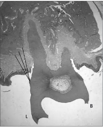
analy-Figure 1 - Histologic analysis: linear measurement from bone crest to cementoenamel junction. B: buccal side; L: lingual side.
Figure 2 - Morphometric analysis: linear measurement from bone crest to cementoenamel junction.
L
sis. This result was statistically different from the mean obtained for corresponding areas when the morphometric method was used (0.41 ± 0.10 mm).
Discussion
This study compared two methods – histometry and morphometry – to evaluate ligature-induced al-veolar bone loss in Wistar rats. As different forms of bone loss evaluation have been reported in litera-ture, the purpose of this study was to develop a reli-able study method.
Histologic evaluation is the standard method used in most studies. Susin, Rösing26 (2003) used Wistar
rats and evaluated bone height histologically in a lin-ear procedure; they used the interproximal space of irst and second molars and measured the distance from the CEJ up to the apical-most point of the bone crest. Nociti et al.18 (2000) and Nogueira-Filho et al.19 (2004) used histology to measure bone loss area
(mm²) in the irst molar furcation of Wistar rats.
Other forms of bone loss evaluation have been used recently. Rivaldo et al.21 (2005) used
de-leshed mandibles of Mus domesticus rats to evalu-ate changes in bone height and measured bone loss as the exposed root surface area (mm²) in irst and second molars. Bone loss was measured as follows: mesially, a line from the CEJ to the bone crest in the mesial root of the irst molar, and distally, a line for the CEJ to the bone crest in the distal root of the second molar; coronally, from the bone crest to the apical-most portion of the CEJ, and apically, from the CEJ to the apical-most point of the bone crest. Kuhr et al.16 (2004) evaluated
ligature-in-duced bone loss in deleshed mandibles of rats using two methods. In one method, the distance from the CEJ to the bone crest was measured linearly in dif-ferent sites; in the other, the exposed root surface area was measured. Their study concluded that the method of measuring the linear distance should be preferred to the method of area evaluation.
The present study used 5 Wistar rats to evaluate induced bone loss and compare two methods: his-tologic evaluation, measuring bone loss linearly,26
and morphometric evaluation, measuring bone loss linearly in deleshed maxillae.16 The observer was
previously trained to conduct the measurements and was blinded to which experimental group the images belonged.27 The study and control groups were used
to compare the capacity of the induction method to generate alveolar bone loss and to compare maxillae in which bone loss was induced using two different methods of analysis.
The main purpose of this study was to compare the histometric and morphometric methods. There-fore, sections were buccolingual and not mesiodistal, differently from most studies.3,22 The morphometric
analysis used digital photographs of the buccal and
Table 1 - Evaluation of induced bone loss: comparison between histologic and morphologic methods for obtaining linear mea-surements (mm) from bone crest to CEJ, for buccal and lingual sides with and without ligatures.
Buccal with ligature Lingual with ligature Buccal without ligature Lingual without ligature
Histometry 0.92±0.16 0.95±0.25 0.23±0.08* 0.44±0.15
Morphometry 1.08±0.35 1.07±0.30 0.41±0.10* 0.47±0.11
*Statistically significant difference between Histometry and Morphometry (t test for independent samples).
Graph 1 - Evaluation of induced bone loss: comparison between histologic and morphologic methods for buccal and lingual sides with and without ligatures. *Statistically significant different mean values between teeth with and without ligatures – paired sample t test.
0.0 0.2 0.4 0.6 0.8 1.0 1.2
Buccal histometry
Buccal morphometry
Lingual histometry
Lingual morphometry Without ligature
With ligature 0.92
0.23
*
*
*
1.08
0.41
*
*
*
0.95
0.44
*
*
*
1.07
0.47
*
*
*
Linear
meas
u
rements
f
ro
m
bo
ne
cre
st
to
C
E
J
lingual aspects of the dry maxillae. The histometric analysis used histologic sections whose images were digitized for evaluation.
The results of this study showed that both meth-ods are capable of detecting vertical periodontal bone loss in test and control teeth. The statistically signiicant differences found between teeth with and without ligatures in both methods demonstrated that the placement of ligatures generated bone loss, which validates the use of ligatures in this study. Moreover, bone loss generated by ligatures was found in both histometric and morphometric analyses (Graph 1).
The comparison between methods, however, re-vealed statistically signiicant differences between the groups without ligature when the buccal sur-face was analyzed. No differences between methods were found in the analysis of the other tooth sur-faces (Table 1).
Future studies should investigate an explana-tion for this inding. However, the low values (mean = 0.23 mm) found for the control group (without ligature) in the histometric analysis of the buccal surfaces were substantially lower than those reported for control groups in studies with rats in which the same distance was measured. Moreover, the mean value for the group without ligature in the morphometric analysis was 0.41 mm, similar to the
values obtained in the histometric and morphomet-ric analyses of the lingual areas. This inding was discrepant with indings for the other groups in this study, and might have been caused by an unwanted inclination of the histologic sections. Despite the care taken, such inclination may have generated an occlusal image of the alveolar bone crest. This is one of the problems of histometry, particularly when maxillae, for which parallel positioning may be dif-icult to obtain, are the object of investigation. The concomitant use of morphometric analysis may mit-igate such effect. However, this objective limitation of histometric measurements of ligature-induced al-veolar bone loss in rats should not preclude its use. Other positive characteristics of histologic analysis should be explored, such as the possibility of identi-fying cell elements and components of soft tissue.
This study contributed to the understanding that the use of morphometric analysis may be an alterna-tive to histometric evaluations because it is faster, easier and less expensive, particularly when the only purpose of the study is to measure bone loss.
Conclusion
Both histometric and morphometric analyses are capable of detecting ligature-induced differences in bone height in rats.
References
1. Amstad-Jossi M, Schroeder HE. Age-related alterations of periodontal structures around the cemento-enamel junction and of the gingival connective tissue composition in germ-free rats. J Periodontal Res. 1978;13(1):76-90.
2. Bjornsson MJ, Velschow S, Stoltze K, Havemose-Poulsen A, Schou S, Holmstrup P. The influence of diet consistence, drinking water and bedding on periodontal disease in Sprague-Dawley rats. J Periodontal Res. 2003;38(6):543-50. 3. Breivik T, Thrane PS, Gjermo P, Fonnum F. Postnatal
gluta-mate-induced central nervous system lesions alter periodontal disease susceptibility in adult Wistar rats. J Clin Periodontol. 2001;28(10):904-9.
4. Chang KM, Ramamurthy NS, McNamara TF, Genco RJ, Gol-ub LM. Infection with a gram-negative organism stimulates gingival collagenase production in non-diabetic and diabetic germfree rats. J Periodontal Res. 1988;23(4):239-44. 5. Crawford JM, Taubman MA, Smith DJ. The natural history
of periodontal bone loss in germfree and gnotobiotic rats
in-fected with periodontopathic microorganisms. J Periodontal Res. 1978;13(4):316-25.
6. Dayan S, Stashenko P, Niederman R, Kupper TS. Oral epithe-lial overexpression of IL-1alpha causes periodontal disease. J Dent Res. 2004;83(10):786-90.
7. Dumitrescu AL, Abd-El-Aleem S, Morales-Aza B, Donaldson LF. A model of periodontitis in the rat: effect of lipopoly-saccharide on bone resorption, osteoclast activity, and local peptidergic innervation. J Clin Periodontol. 2004;31(8):596-603.
8. Fernandes MI, Gaio EJ, Rösing CK, Oppermann RY, Rados PV. Avaliação qualitativa microscópica do tempo de fixação e meio de descalcificação do periodonto em maxila de ratos. Braz Oral Res. 2003;17(Suppl 2):206.
10. Heijl L, Wennström J, Lindhe J, Socransky SS. Periodontal disease in gnotobiotic rats. J Periodontal Res. 1980;15(4):405-19.
11. Holzhausen M, Garcia DF, Pepato MT, Marcantonio E Jr. The influence of short-term diabetes mellitus and insu-lin therapy on alveolar bone loss in rats. J Periodontal Res. 2004;39(3):188-93.
12. Jordan HV, Keyes PH, Bellack S. Periodontal lesions in ham-sters and gnotobiotic rats infected with Actinomyces of human origin. J Periodontal Res. 1972;7(1):21-8.
13. Junqueira LCU, Junqueira LMMS. Técnicas básicas de citologia e histologia. São Paulo: Santos; 1983.
14. Klausen B, Evans RT, Sfintescu C. Two complementary meth-ods of assessing periodontal bone level in rats. Scand J Dent Res. 1989;97(6):494-9.
15. Klausen B, Hougen HP, Eriksen WH, Fiehn NE. Introduction of periodontal bone loss in athymic (nude) rats monoinfected with Streptococcus mutans. J Periodontal Res. 1986;21(1):5-12.
16. Kuhr A, Popa-Wagner A, Schmoll H, Schwahn C, Kocher T. Observations on experimental marginal periodontitis in rats. J Periodontol Res. 2004;39(2):101-6.
17. Nishikawa S, Nagata T, Morisaki I, Oka T, Ishida H. Patho-genesis of drug-induced gingival overgrowth. A review of studies in the rat model. J Periodontol. 1996;67(5):463-71. 18. Nociti FH Jr, Nogueira-Filho GR, Primo MT, Machado MA,
Tramontina VA, Barros SP et al. The influence of nicotine on the bone loss rate in ligature-induced periodontitis. A histo-metric study in rats. J Periodontol. 2000;71(9):1460-4. 19. Nogueira-Filho GR, Froes Neto EB, Casati MZ, Reis SR,
Tunes RS, Tunes UR et al. Nicotine effects on alveolar bone
changes induced by occlusal trauma: A histometric study in rats. J Periodontol. 2004;75(3):348-52.
20. Page RC, Schroeder HE. Periodontitis in man and other ani-mals. A comparative review. Basel: Karger; 1982.
21. Rivaldo EG, Padilha DP, Hugo FN. Alveolar bone loss and aging: a model for the study in mice. J Periodontol 2005;76(11):1966-71.
22. Rovin S, Costich ER, Gordon HA. The influence of bacteria and irritation in the initiation of periodontal disease in germ-free and conventional rats. J Periodontal Res. 1966;1(3):193-204.
23. Sato Y, Kikuchi M, Ohata N, Tamura M, Kuboki Y. Enhanced cementum formation in experimentally induced cementum defects of the root surface with the application of recombi-nant basic fibroblast growth factor in collagen gel in vivo. J Periodontol. 2004;75(2):243-8.
24. Schmidt LJ, Rowe DJ. The effects of diphosphonates on al-veolar bone loss. Quintessence Int. 1987;18(7):497-501. 25. Sculean A, Berakdar M, Windisch P, Remberger K, Donos N,
Brecx M. Immunohistochemical investigation on the pattern of vimentin expression in regenerated and intact monkey and human periodontal ligament. Arch Oral Biol. 2003;48(1):77-86.
26. Susin C, Rösing CK. Effect of variable moderate chronic stress on ligature-induced periodontal disease in Wistar rats. Acta Odontol Scand. 2003;61(5):273-7.
27. Susin C, Rösing CK. Praticando Odontologia Baseada em Evi-dências. Canoas: Ulbra; 1999.