PONTIFÍCIA UNIVERSIDADE CATÓLICA DO RIO GRANDE DO SUL INSTITUTO DE GERIATRIA E GERONTOLOGIA
PROGRAMA DE PÓS-GRADUAÇÃO EM GERONTOLOGIA BIOMÉDICA MESTRADO EM GERONTOLOGIA BIOMÉDICA
MARIA LETÍCIA RASSI GUIMARÃES
EFEITO DA APLICAÇÃO LOCAL DE CÉLULAS MONONUCLEARES ADULTAS AUTÓGENAS EM CIRURGIA DE PERDA ÓSSEA ALVEOLAR
MARIA LETÍCIA RASSI GUIMARÃES
EFEITO DA APLICAÇÃO LOCAL DE CÉLULAS MONONUCLEARES ADULTAS AUTÓGENAS EM CIRURGIA DE PERDA ÓSSEA ALVEOLAR
INDUZIDA EM CAMUNDONGOS.
Dissertação do Curso de Mestrado em Gerontologia Biomédica, do Instituto de
Geriatria e Gerontologia da Pontifícia Universidade Católica do Rio Grande do Sul como parte dos requisitos
necessários à obtenção do grau de Mestre em Gerontologia Biomédica.
Orientadora: Profª. Dra. Dalva Maria Pereira Padilha Co-orientadora: Profª. Dra. Denise Cantarelli Machado
AGRADECIMENTOS
Ao Prof. Dr. Antônio Carlos Araújo de Souza (in memoriam), pelo apoio,
carinho e amizade incondicionais. Agradeço pela dedicação e confiança em mim depositadas e pelos incentivos constantes para meu engrandecimento pessoal e profissional.
À minha orientadora, Profº. Dra. Dalva Maria Pereira Padilha, por ter acreditado em meu potencial, pelas contribuições, colaboração e incentivo profissional. Pelo conhecimento e defesa dos ensinamentos da Odontogeriatria e Gerontologia.
À minha co-orientadora, Profº. Dra. Denise Cantarelli Machado, pela confiança, incentivo e apoio durante a realização desta pesquisa.
Às minhas grandes amigas, Profº. Dra. Maria Luisa Tagliaro e Raquel Mattos, pelo apoio, carinho, companheirismo e convivência diária nestes 2 ½ anos em Porto Alegre. Amigas que apoiaram, auxiliaram e contribuíram para a conclusão deste Mestrado e que, antes de mais nada, me ensinaram que mesmo morando em cidades tão distantes, nossa amizade continua inabalável.
Ao Profº. Dr. Fernando Neves Hugo, pelo apoio no desenvolvimento desta pesquisa, ampliando meus conhecimentos e me auxiliando nos momentos necessários. Agradeço também pela sua amizade nesta minha temporada em POA.
Aos meus colegas de pesquisa e laboratório, Christian Viezzer, Fernanda da Cruz, pelo apoio e auxílio nesta pesquisa. Sem o auxílio, dedicação e principalmente a amizade de vocês, este trabalho não teria se concretizado.
Ao Programa de Pós-Graduação em Gerontologia Biomédica, pela oportunidade de estudo e desenvolvimento da presente pesquisa.
RESUMO
A associação entre envelhecimento e perda óssea alveolar em humanos é de difícil avaliação e a reparação dos tecidos periodontais e constitui um desafio a Odontologia. O objetivo este estudo foi analisar a resposta do tecido ósseo alveolar frente à aplicação local de células mononucleares adultas autógenas, com análises morfométricas e análise da expressão da osteocalcina em RT-PCR. Foram utilizados camundongos machos BALB/c de 3 (n=5) e 9 (n=7) meses de idade. A perda óssea alveolar foi induzida por cirurgias de descolamento mucoperiósteo vestibular em mandíbulas nas regiões de molares nos lados direito e esquerdo. O lado direito - sítio experimental, com a aplicação de células mononucleares, e o lado esquerdo - sítio controle, com a aplicação de DPBS. Os resultados da perda óssea foram expressos em mm2 e comparados com teste t de Student pareado. Nas análises morfométricas aos 3 e 9 meses, a área da perda óssea na face vestibular não foi diferente entre os grupos. Entretanto, a perda óssea das amostras controle aos 3 meses, face lingual foi significativamente maior que as amostras experimentais (p<0.03). A expressão da proteína osteocalcina no grupo dos camundongos de 3 meses foi praticamente inexistente. Enquanto na análise dos resultados aos 9 meses a proteína apresenta diferenças significativas de expressão, demonstrando que de 7 sítios experimentais, 5 deles expressaram mRNA de osteocalcina. Nas condições deste experimento os resultados evidenciaram a prevenção da perda óssea alveolar na face lingual dos camundongos mais jovens e uma significante expressão da proteína osteocalcina aos 9 meses.
ABSTRACT
The association in humans between aging and alveolar bone loss is difficulty to evaluation and periodontal tissues repair and is a challenge for dentistry. This study analyzed the behavior of mandibular bone tissue after application of adult autogenous mononuclear cells using morphometric examination and measurements of osteocalcin values with RT-PCR. Male BALB/c mice aged 3 (n=5) and 9 months (n=7) were used. Alveolar bone loss was induced by mucoperiosteal flap surgery of the right and left molar regions of the mandible. The right side - experimental side and received applications of adult autogenous mononuclear cells, and the left side - control side and received applications of DPBS. Results of alveolar bone loss were expressed in mm2 and compared by paired Student t test. Morphometric analyses revealed in 3 and 9-months-old, area of bone loss in the buccal side were not statistically different between groups. However, the mean in lingual side of control samples was significantly larger compared with experimental samples (p<0.03). Osteocalcin expression revealed that the values of the protein were practically negligible in samples of 3-month-old mice. The analysis of the 9-month-old mice, however, showed that the protein values were significantly different: 5 of the 7 experimental samples showed osteocalcin expression in mRNA. Under the experimental conditions the results showed that alveolar bone loss was prevented in the lingual side of younger mice, and that values of osteocalcin expression were significantly different in the samples of 9-month-old mice.
SUMÁRIO
1. REFERENCIAL TEÓRICO...11
2. OBJETIVOS...18
2.1 Objetivo Geral...18
2.2Objetivos Específicos...18
ARTIGO EM INGLÊS...19
3. CONSIDERAÇÕES FINAIS...35
4. REFERÊNCIAS BIBLIOGRÁFICAS...37
1. REFERENCIAL TEÓRICO
A estrutura interna do osso está adaptada às forças mecânicas, mudando continuamente durante o crescimento e alterações funcionais. Nos ossos maxilares, as alterações estruturais estão correlacionadas ao crescimento, erupção dentária, movimentos dentários, desgaste e perda dos dentes, e todos estes processos tornam-se possíveis quando coordenados pelas atividades de reabsorção e aposição ósseas. Enquanto o aumento das forças funcionais conduz à formação de novo osso, a diminuição da função leva a uma diminuição do volume do osso 1.
Alguns fatores, sistêmicos e locais, controlam esta atividade de remodelação óssea. Os fatores sistêmicos são hormônios (como hormônio da paratireóide, calcitonina, insulina, hormônio de crescimento, vitamina D, glicocorticóides, hormônios sexuais, hormônios da tireóide) que atuam em diferentes fases da remodelação com funções definidas para cada um 2. Os fatores locais são secretados pelas células do osso, sendo algumas citocinas secreções de células do sistema imune-hematopoiético presentes no micro ambiente das trabéculas do osso como: fatores de crescimento insulínicos (IGFs), fator transformante de crescimento beta (TGF- ), proteínas morfogenéticas do osso (BMP), fatores de crescimento de fibroblastos (FGF), fatores de crescimento derivados de plaquetas, e citocinas 2.
alveolar. Na mandíbula, esta diminuição progressiva de substância óssea reduz a possibilidade de reabilitação efetiva da função bucal 3.
No entanto, esta atrofia óssea observada durante o envelhecimento não se faz de forma homogênea, pois, antes dos 50 anos, perde-se, sobretudo osso trabecular (principalmente trabéculas de menor importância estrutural) e, após essa idade, principalmente osso cortical (lamelas de menor importância estrutural, localizadas na superfície endosteal) 4.
Para o estudo da reabsorção óssea alveolar e suas conseqüências, existe um modelo de reabsorção óssea em resposta a separação do retalho mucoperiósteo. Este modelo cirúrgico no osso alveolar revela uma reabsorção do aspecto interno do periodonto deste osso opondo-se as superfícies radiculares, podendo progredir em direção a perda total ou parcial das superfícies óssea vestibular ou lingual do osso 5.
Yaffe et al (1994) realizaram um experimento com 60 ratos Wistar com o
Na cirurgia de descolamento do retalho mucoperiósteo, durante o procedimento de dissecação, os tecidos moles da papila gengival, incluindo os feixes de fibras colágenas, são incisadas e o periósteo é separado do osso alveolar, particularmente na área de união gengival, estendendo-se à área da junção mucogengival 7,8. O rompimento das fibras colágenas do periósteo marginal desencadeia a atuação dos fibroblastos, que residem nas fibras, a iniciarem a cascata de eventos e interações celulares, promovendo a reabsorção óssea alveolar. Uma considerável quantidade de atividade osteoclástica é evidenciada junto a toda extensão de osso alveolar e também, por toda a superfície do ligamento periodontal 6,7 e uma significante perda óssea é iniciada no ligamento periodontal quando a gengiva marginal é cortada e separada do mucoperiósteo pelo acesso coronal 7.
Esta atividade de reabsorção ocorre no osso adjacente aos dentes da mandíbula e, uma maior atividade de reabsorção ocorre durante a primeira semana de cirurgia, com um decréscimo de 22% de retenção de cálcio, e continua nos 21 dias seguintes a cirurgia de descolamento do retalho mucoperiósteo 6,9.
Esta cirurgia periodontal estimula a atividade osteoclástica com vários valores de perda da crista óssea alveolar. Fatores locais como infecção, trauma oclusal e geometria óssea podem afetar quantitativamente o estágio de perda óssea bem como o estágio de regeneração. O aumento da mobilidade imediatamente após a cirurgia periodontal e a deiscência óssea, quando o osso da área é muito fino, pode ser resultado do processo de indução desta reabsorção óssea alveolar, criando porosidades no osso cortical devido ao rompimento da atividade de reabsorção em vários sítios 8.
Rivaldo et al (2004) realizaram um estudo com 72 camundongos CF1 Mus domesticus e estes animais foram divididos em três grupos: machos, fêmeas virgens e
objetivo foi desenvolver um modelo de estudo para a cirurgia de perda óssea alveolar em camundongos durante o envelhecimento. Os resultados demonstraram que ocorre uma reabsorção óssea similar, com exposição de área radicular, nos camundongos de 3 e 12 meses. Os autores concluíram então que o camundongo de 3 meses de idade, independente do sexo e status reprodutivo, pode ser um modelo útil para o estudo da perda óssea alveolar induzida no processo de envelhecimento 10.
Diversas substâncias têm sido freqüentemente estudadas e utilizadas em tratamentos tópicos, sistêmicos, ou a combinação de ambos, para prevenir ou minimizar a perda óssea alveolar 5,8,9,10,11,12,13,14,15. Atualmente, estudos têm sido realizados com o objetivo de avaliar e identificar a possibilidade de atuação de células tronco na regeneração do tecido periodontal e tecido ósseo alveolar 16,17,18,19.
O periodonto é um tecido complexo compreendido de dois tecidos duros (cemento e osso) e dois moles (gengiva e ligamento periodontal). É constituído por populações heterogêneas de células, como os osteoblastos, fibroblastos e cementoblastos, e células tronco mesenquimais, que permanecem dentro do ligamento periodontal e são responsáveis pela hosmeostase tecidual, servindo como fonte renovável de células progenitoras gerando tipos celulares locais ao longo da vida adulta. Em um evento de injúria ao periodonto estas células tronco mesenquimais podem ser ativadas para uma diferenciação terminal e reparação ou regeneração tecidual para as áreas de osso alveolar e cemento adjacente ao defeito. Mudanças inflamatórias no periodonto podem influenciar a distribuição das células tronco nestes tecidos 20, 21.
molares foram criados defeitos de furca grau III. O grupo experimental recebeu atelocolágeno com células tronco mesenquimais e o grupo controle gel de colágeno sem células tronco mesenquimais. Os resultados demonstraram que no grupo experimental houve uma significativa formação de novo osso e de ligamento periodontal. A superfície radicular desnuda foi totalmente coberta por novo cemento. A área de furca também apresentou formação de cemento por toda a superfície, além de fibras de Sharpey’s inseridas no cemento. Entretanto, não houve completa reconstrução do osso alveolar. Os autores concluíram que o auto-transplante de células tronco mesenquimais da medula óssea é uma possibilidade para a regeneração do tecido periodontal 16.
As células tronco são células multipotentes e usualmente, entre uma célula tronco e sua progene terminalmente diferenciada existe uma população intermediária de progenitores com limitada capacidade proliferativa e restrito potencial de diferenciação 22,23 que estão presentes, especialmente em organismos complexos 23. As células tronco e suas células progenitoras são definidas pela habilidade de auto-renovação podem constituir diferentes classes de células sob diferentes regulações moleculares em diversos tipos teciduais 23.
Uma possível definição para a plasticidade das células tronco é que as células tronco adultas de um tecido específico através da incumbência de originar uma linhagem celular pode, sob certas condições do micro-ambiente, adquirir a habilidade para se diferenciar em células de um tecido diferente 24.
Em organismos de animais mamíferos, existem evidências de que os fatores de crescimento e as interações célula-célula podem influenciar no resultado do destino das decisões destas células pela multipotencialidade de seus progenitores 25.
perde sua habilidade de proliferação e perde a capacidade de manter um organismo por toda sua vida 26. O estado de senescência destas células tronco mesenquimais pode também ser evidenciado pela diferenciação em adipócitos, pois com o envelhecimento, a medula óssea é parcialmente substituída por tecido adiposo 27.
A densidade celular das células tronco mesenquimais é um importante fator para a regeneração tecidual. Fluidos teciduais suficientes e suprimento sanguíneo podem ser necessários para a sobrevivência das células tronco mesenquimais após o transplante e subseqüente diferenciação em células periodontais 16.
2. OBJETIVOS
OBJETIVO GERAL
Verificar a influência da injeção local de células mononucleares adultas autógenas no defeito ósseo alveolar induzido em camundongos de 3 e 9 meses de idade.
OBJETIVOS ESPECÍFICOS:
- Mensurar a área de perda óssea alveolar (mm2);
Effect of local application of adult autogenous mononuclear cells on
alveolar bone loss induced by mucoperiosteal flap surgery in mice.
Guimarães MLR 1, Machado DC 2,3, Viezzer C 3, Oliveira RM 4, Hugo FN 5, Padilha DMP 2.
1 Biomedical Gerontology Post-graduate Program, Pontifical Catholic University of Rio Grande do Sul (PUCRS), Porto Alegre, Brazil.
2 Institute of Geriatrics and Gerontology, PUCRS. 3 Institute of Biomedical Research, PUCRS. 4 Faculty of Biosciences, PUCRS.
5 Faculty of Dentistry, Piracicaba, University of Campinas, UNICAMP, São Paulo, Brazil.
Introduction
Periodontal regeneration is a challenge in dentistry because periodontal tissue has a limited regeneration capacity 1.
Many substances have been tested for the regeneration of periodontal tissue, such as chitosan and collagen sponge (Park et al. 2003) 2, vitamin C (Bsoul and Terezhalmy 2004) 3, bisphosphonates (Yaffe et al. 1995; Tenenbaum et al. 2002) 4,5, bisacylphosphonates (Yaffe et al. 2000) 6, sodium alendronate applied locally (Meraw et al. 1999 ; Yaffe et al. 1999; Binderman et al. 2000) 7,8,9, and calcium phosphate cement (Hayashi et al. 2006) 10 .
Many types of cells are found in the periodontal ligament, such as cementoblasts, osteoblasts, fibroblasts, myofibroblasts, endothelial cells, neuron cells and epithelial cells. Populations of mesenchymal stem cells in periodontal tissues raised interest in its use in periodontal therapy.1
Adult stem cells are functionally multipotent cells capable of unlimited self-renovation 14,15,16,17, also, they produce at least one type of highly differentiated progeny 15,16 or many differentiated cellular types of in vivo tissue 14,17.
The transplantation of mesenchymal stem cells from bone marrow may express a differentiation potentiality in periodontal tissues, and alveolar bone regeneration may, therefore, be stimulated by transplantation of mesenchymal stem cells without pre-induction or treatment for differentiation 18.
The objective of this study was to evaluate the effect of local application of adult autogenous mononuclear cells on the prevention of mandible alveolar bone loss induced in mice by mucoperiosteal flap surgery.
Materials and Methods
Study design
mice were kept in individual cages and received distilled water and food ad libitum. Standard conditions of light (12/12 light/dark cycles) and temperature (20°C) were kept during the experiment. Animals were monitored daily. Mice were weighed before surgery for induction of alveolar bone loss, before injection with mononuclear cells or DPBS, and before euthanasia.
Procedure to obtain mononuclear cells
Induction of alveolar bone loss and treatment with stem cells
At 3 or 9 months of age, recipient mice were weighed and intramuscularly anesthetized. A mucoperiosteal flap was raised on the buccal aspect of mandibular right and left molars to induce alveolar bone loss. The mucosa was separated from the underlying bone after an incision in the marginal gingiva with the aid of a small elevator, and was immediately placed back without any suture. Seven days after the surgery, during the period of accelerated bone resorption (Yaffe et al., 1994 11), the mice were anesthetized as described above and a solution containing mononuclear cells was injected in the right side of the mandible, whereas the left side received an injection of the same amount of DPBS.
Samples processing
Twenty-one days after the surgical procedure, during the period of maximum alveolar bone loss (Yaffe et al., 1994 11), animals were killed under anesthesia.
Bone Loss Analysis
One independent and previously trained examiner performed two measurements of each sample at an interval of one week. In the buccal aspect, the area of alveolar bone loss was measured in the first molar region, whereas in the lingual aspect, the area of alveolar bone loss was measured in the region of root exposure of first and second molars using the landmarks suggested by Tatakis and Guglielmoni 2000 20 and modified by Rivaldo et al. 2005 12 (Figure 1 - A and B).
A B
Figure 1. Illustration of the landmarks used to define the area of alveolar bone loss. A) Bone loss, lingual surface; B) Bone loss, buccal surface.
Statistical analysis
Procedure for mRNA expression osteocalcin analysis
Ribonucleic acid extraction (RNA) and purification of messenger RNA (mRNA)
The region of first and second molars of the alveolar bone tissue was extracted and macerate for local extraction of bone cells. Total RNA of these cells was isolated, and a Trizol LS (Invitrogen, USA) reagent solution was added to them. The samples were washed once with 1 ml DPBS. After that, 200 µl DPBS and 800 µl Trizol LS were added, and the samples were incubated for 5 minutes at room temperature for nuclear and cellular membrane destruction and nucleic acid liberation. After incubation, was added 160 µl/tube cold chloroform. The samples were homogenized for 15 seconds, incubated for 10 minutes at room temperature, and centrifuged (Eppendorf 5417R, USA) at 14.000 rpm for 15 minutes at 4ºC. When centrifugation was complete, the supernatant were transfer to a new tube, to which 400µl cold isopropyl alcohol was added, and incubated for at least two hours at -20°C. After that, the samples were centrifuged for 10 minutes under the same conditions. The precipitates were washed with 500 µl 75% cold ethanol and centrifuged at 14.000 rpm for 15 minutes at 4°C.
The supernatant was aspirated and the tubes were kept for about 30 minutes at room temperature for the precipitate to evaporate. Once evaporated, RNAs were soluble in 20 µl of H2O diethylpyrocarbonate (DEPC 0.1%, Invitrogen, USA) and treated with 1µl RNase inhibitor (40 U/µl; Invitrogen, USA). The integrity of total RNA samples extract was analyzed in 1.5% agar gel stained with bromide ethidium (EtBr), after electrophoresis in TAE buffer.
with 10 U of the enzyme in 22 µl final volume. The samples were incubated at room temperature for 15 minutes. After that, the enzyme was inactivated by addition of 1µl of 25 mM EDTA and incubated for 10 minutes at 65ºC.
Reverse transcription to obtain cDNAs
After treated with DNase I, a reverse transcription reaction was performed for the first band syntheses of complementary DNA (cDNA) using a Superscript II enzyme and originator oligonucleotides oligos-dT12-18 (Invitrogen, USA). For each reaction, 22 µl RNA total extraction, 0.5 µl Superscript II enzyme (200 U/µl; Invitrogen, USA), 0.2µl oligos- dT12-18, 4.5 µl DTT (0.1 M; Invitrogen, USA), 4.5 µl dNTPs (100 mM; Invitrogen, USA), 4.5 µl MgCl2 (50 mM), and 9 µl enzyme buffer (5x; Invitrogen, USA) were added. Volume of reaction solution was brought to 45 µl by adding MQ water. The samples were incubated in a termocycler (Peltier Thermocycler, MJ Research, USA) for 10 minutes at 25°C, 45 minutes at 42°C, and 3 minutes at 99°C. After that, 5 U of ribonuclease H inhibitor (2 U/µl; Invitrogen, USA) was added to each tube, and the samples were incubated at 37°C for 20 minutes. cDNAs were stored at -20°C until use for PCR reaction.
Polymerase chain reaction (PCR) for Osteocalcin
PCR was performed in a termocycler under the following conditions: 95°C for 3 minutes, 30 consecutive cycles at 95°C for 30 seconds, 55°C for 45 seconds and 72°C for 1 minute. A final cycle at 72°C for 5 minutes was used to ensure amplicons full extension.
PCR products were separated by horizontal electrophoresis in 2% agar gel in TAE buffer, stained with bromide ethidium, and visualized under UV light.
Results
Animals weight results
The mean weight of the 3 and 9 months-old mice on analysis of variance, before surgery, before injections and before euthanasia weren’t statistically significant.
Morphometric analysis of alveolar bone
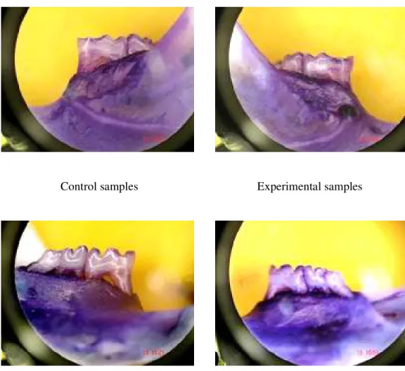
Control samples Experimental samples
Control samples Experimental samples
Figures 3. Pictures of buccal surfaces (superior pictures) and lingual surfaces (inferior pictures) in control groups (left side) and experimental groups (right side) in 9-month-old mice.
Table 1. Mean (± standard deviation) root exposure area in molar region of mice (in mm2).
AGE
DPBS MONONUCLEAR
CELLS
P
3 Months ( n=5) buccal area 0.22 (±0.05) 0.24 (±0.05) 0.57
lingual area 0.35 (±0.05) 0.27 (±0.04) 0.03* 9 Months ( n=7) buccal area 0.33 (±0.07) 0.34 (±0.08) 0.9
lingual area 0.43 (±0.05) 0.34 (±0.1) 0.09 * p < 0.05
In animals that received mononuclear cells, root exposure areas did not differ between buccal and lingual surfaces of 3- and 9-month-old mice (Tables 2 and 3). In the control group, larger root exposure areas were found in the lingual surfaces of both 3- and 9-month-old animals.
Table 2. Mean (± standard deviation) root exposure area in buccal and lingual surfaces of molar region of 3-month-old mice (in mm2).
3 MONTHS ( n=5)
buccal area lingual area P
DPBS 0.22 (±0.05) 0.35 (±0.05) 0.001* MONONUCLEAR
CELLS
Table 3. Mean (± standard deviation) root exposure area in buccal and lingual surfaces of molar region of 9-month-old mice (in mm2).
9 MONTHS ( n=7)
buccal area lingual area P
DPBS 0.33 (±0.08) 0.43 (±0.05) 0.001* MONONUCLEAR
CELLS
0.34 (±0.08) 0.34 (±0.1) 0.9
* p < 0.05
Analysis of osteocalcin expression
The analysis results for osteocalcin expression after application of adult autogenous mononuclear cells in area of alveolar bone loss caused by mucoperiosteal flap surgery showed that mRNA osteocalcin expression quantity was different in the groups under study.
Table 4. mRNA osteocalcin expression in BALB/c mice submitted to bone alveolar loss and treaded with adult autogenous mononuclear cells or DPBS.
3 MONTHS (n=5)
9 MONTHS (n=7)
MONONUCLEAR CELLS
- - - - + + - - + + + +
DPBS
- - + - + - + - + + - -
Discussion
In this study, we evaluated the effect of local application of adult autogenous mononuclear cells on areas of induced alveolar bone loss in the molar region of the mandible of 3- and 9-month-old mice.
Morphometric analysis revealed that alveolar bone loss exposed molar roots of 3- and 9-month-old animals, which confirmed the results that Yaffe et al. (1994) 11 found in rats and Rivaldo et al. (2005) 12 found in mice.
In the control sides of 3- and 9-month-old animals, alveolar bone loss was larger on the lingual aspect, which was also found in previous studies 11, 12, 13. Bone loss did not differ between buccal and lingual aspects in the animals that received local application of mononuclear cells.
The expression of osteocalcin (OC), a non-collagenous protein of the bone matrix synthesized by osteoblasts, was also analyzed in this study. OC is a marker of bone turnover when resorption and formation are coupled, and it is elevated in cases of rapid turnover, such as in fracture repair 21.
Our findings confirmed a difference in bone behavior between younger and older animals 12,22. In 9-month-old mice, a later beginning of repair could explain the high osteocalcin expression 3 weeks after mucoperiostal flap, which was not observed in 3-month-old animals. Reinhardt et al. (2004) found a high negative correlation between OC/ALB ratios found in bone wash and subsequent bone density in healing. They suggest that sites with high bone turnover may heal slowly 23.
A study by Srouji and Livne (2005) reported that an osteogenic subpopulation of bone marrow mesenquimal stem cells selected in vitro produced a bone specific matrix that was positive for osteocalcin 24. Their finding may confirm our detection of an initial healing stage in 9-month-old mice, which is marked by a larger root exposure, not different from the one found in control samples, and a higher expression of osteocalcin. Our findings suggest that repair may be delayed in older animals when compared with younger recipient mice.
It is unclear whether the application of adult autogenous mononuclear cells prevented the resorption induced by a mucoperiosteal flap or accelerated the repair in younger animals. Further studies to analyze the behavior of alveolar bone repair after application of adult autogenous mononuclear cells in stages before 3 weeks may help to clarify this question.
of 9-month-old animals. Another limitation was the fact that we were not able to perform specific tests to determine the fate or differentiation of implanted cells.
Another difficulty was the size of the experimental animals used in this study. Although they are easy to handle, the final area of surgical lesion was less than 0.5 mm, which decreased the number of cells injected and increased the possibility of application errors.
In summary, the results of this study showed that BALB/c mice that underwent mucoperiosteal flap surgery suffered alveolar bone loss. Measurements of root exposure showed that the side of 3-month-old animals that received a local application of adult autogenous mononuclear cells from bone marrow had less alveolar bone loss than the control side. The technique used in this study is based on the extraction of mononuclear cells with no stem cell separation or differentiation, and it is easy, fast and less expensive than traditional techniques to study cell implantation in the process of alveolar bone repair.
References
1 - Bartold PM, Shi S, Gronthos S. Stem cells and periodontal regeneration. Periodontology 2000, 2006, 40: 164-172.
2 - Park JS, Choi SH, Moon IS, Cho KS, Chai JK, Kim CK. Eight-week histological analysis on the effect of chitosan on surgically created one-wall intrabony defects in beagle dogs. J Clin Periodontol, May 2003, 30(5): 443-53.
3 - Bsoul SA, Terezhalmy GT. Vitamin C in health and disease. J Contemp Dent Pract, May 2004, 5(2): 1-13.
5 - Yaffe A, Fine N, Alt I, Binderman I. The Effect of Bisphosphonate on Alveolar Bone Resorption Following Mucoperiosteal Flap Surgery in the Mandible of Rats. J Periodontol, November 1995, 66(11): 999-1003.
6 - Yaffe A, Golomb G, Breuer E, Binderman I. The Effect of Topical Delivery of Novel Bisacylphosphonates in Reducing Alveolar Bone Loss in the Rat Model. J Periodontol, October 2000, 71(10): 1607-1612.
7 - Meraw SJ, Reeve CM, Wollan PCJ. Use of alendronate in peri-implant defect regeneration. Periodontol, Feb 1999, 70(2): 151-8.
8 - Yaffe A, Binderman I, Breuer E, Pinto T, Golomb G. Disposition of alendronate following local delivery in a rat jaw. J Periodontol 1999, 70: 893-895.
9 - Binderman I, Adut M, Yaffe A. Effectiveness of Local Delivery of Alendronate in Reducing Alveolar Bone Loss Following Periodontal Surgery in Rats. J Periodontol, August 2000, 71(8): 1236-1240.
10 - Hayashi C, Kinoshita A, Oda S, Mizutani K, Shirakata Y, Ishikawa I. Injectable calcium phosphate bone cement provides favorable space and a scaffold for periodontal regeneration in dogs. J Periodontol, June 2006, 77(6): 940-6.
11 - Yaffe A, Fine N, Binderman I. Regional Accelerated Phenomenon in the Mandible Following Mucoperiosteal Flap Surgery. J Periodontol, January 1994, 65(1): 79-83. 12 - Rivaldo EG, Padilha DMP, Hugo FN. Alveolar Bone Loss and Aging: A Model for the Study in Mice. J Periodontol, November 2005; 1-6.
13 - Binderman I, Adut M, Zohar R, Bahar H, Faibish D, Yaffe A. Alveolar Bone Resorption Following Coronal Versus Apical Approach in a Mucoperiosteal Flap Surgery Procedure in the Rat Mandible. J Periodontol, October 2001, 72(10): 1348-1353.
14 - Morrison SJ, Shah NM, Anderson DJ. Regulatory Mechanisms in Stem Cell Biology. Cell, February 1997, 88(7): 287-298.
15 - Park IK, Morrison SJ, Clarke MF. Bmi1,stem cells, and senescence regulation. The Journal of Clinical Investigation, January 2004, 113(2): 175-179.
16 - Watt FM, Hogan BLM. Out of Eden: Stem Cells and Their Niches. Science, February 2000, 287(25): 1427-1430.
17 - Kooy DVD, Weiss S. Why Stem Cells? Science, February 2000, 287(25): 1439-1441.
19 - Rivaldo EG, Padilha DMP, Hugo FN, Hilgert JB, Rybu BR. Reproducibility of a hemi mandible positioning device and a method for measuring alveolar bone loss area in mice . J Oral Sci, 2007, 49: 13-17.
20 - Tatakis DN, Guglielmoni P. HLA-B27 transgenic rats are susceptible to accelerated alveolar bone loss. J Periodontol 2000, 71: 1395-1400.
21 - Slovik DM, Gundberg CM, Neer RM, Lian JB. Clinical evaluation of bone turnover by serum osteocalcin measurements in a hospital setting. Journal of Clinical Endocrinology and Metabolism, 1984, 59: 228-230.
22- Lu C, Miclau T, Hu D, Hansen E, Tsui K, Puttlitz C, Marcucio RS. Cellular basis for age-related changes in fracture repair. Journal of Orthopaedic Research , 2005, 23: 1300-1307.
23 - Reinhardt RA, Sanderfer VJ, Meinberg TA, Nummikoski P, Lee H-M, Marx DB. Local biochemical markers of bone turnover: relationship to subsequent density of healing alveolar bone defects. J Clin Periodontol, 2004, 31: 223-228.
24 - Srouji S, Livne E. Bone marrow stem cells and biological scaffold for bone
repair in aging and disease. Mechanisms of Ageing and Development , 2005, 126: 281-287.
25 - Polimeni G, Koo K-T, Qahash M, Xiropaidis AV, Albandar JM, Wikesjö UME. Prognostic factors for alveolar regeneration: effect of tissue occlusion on alveolar bone regeneration with guided tissue regeneration. J Clin Periodontol, 2004, 31: 730-735. 26 - Acneil RL, Somerman M. Development and regeneration of the periodontium: parallels and contrasts. Periodontology 2000, 1999, 19: 8-20.
3. CONSIDERAÇÕES FINAIS
O objetivo da terapia periodontal é a regeneração dos tecidos periodontais perdidos e a reconstrução dos tecidos envolvidos. A formação de tecido conectivo anexo, cemento com inserção de fibras colágenas, em superfícies radiculares desnudas e o crescimento de novo osso alveolar podem ser alcançados com terapias regenerativas através da utilização de substitutos ósseos diversos, em procedimentos cirúrgicos periodontais para possíveis cicatrização e regeneração de feridas cirúrgicas 28.
A engenharia tecidual óssea é uma terapia alternativa para promover a cicatrização óssea, utilizando métodos diversos para a regeneração e reparação do tecido ósseo com uma variada combinação de polímeros, células e fatores de indução para estimular ativamente a formação do tecido 29.
Eventos associados com a regeneração periodontal envolvem o recrutamento de derivados locais de células progenitoras para o sítio ao qual ocorrerá a diferenciação subseqüente em células do ligamento periodontal, cementoblastos ou osteoblastos 29.
O objetivo da terapia periodontal não é apenas impedir o avanço da doença periodontal, mas também na regeneração do tecido periodontal destruído e habilitar para sua função normal 30.
Estudos futuros com análise de diferentes proteínas ósseas, verificando os processos de reabsorção ou formação óssea, e análises histológicas, deverão ser realizados.
4. REFERÊNCIAS BIBLIOGRÁFICAS
1 - Histologia e embriologia oral de Orban, S. N. Bhaskar, 8° edição, editora Artes Médicas, São Paulo, pp.484.
2 - Lindomar Guimarães Oliveira. “Osteoporose: Guia para Diagnóstico, Prevenção e Tratamento” Editora Revinter, 2002, Rio de Janeiro, p. 431 (capítulo 09 p.52-58). 3 - Torres SVS. Saúde Bucal: Alterações Fisiológicas e Patológicas do Envelhecimento. In: Freitas, E. V. de, Py L., Néri, A. L., Cançado, F. A. X., Gorzoni, M. L., Rocha, S. M. da e colaboradores “Tratado de Geriatria e Gerontologia” editora Guanabara Koogan, Rio de Janeiro, 2002, pps.1187.
4 - Rossi E, Sader CS. Envelhecimento do Sistema Osteoarticular. In: Freitas, E. V. de, Py L., Néri, A. L., Cançado, F. A. X., Gorzoni, M. L., Rocha, S. M. da e colaboradores “Tratado de Geriatria e Gerontologia” editora Guanabara Koogan, Rio de Janeiro, 2002, pps.1187.
5 - Yaffe A, Golomb G, Breuer E, Binderman I. The Effect of Topical Delivery of Novel Bisacylphosphonates in Reducing Alveolar Bone Loss in the Rat Model. J Periodontol, October 2000, 71(10):1607-1612.
6 - Yaffe A, Fine N, Binderman I. Regional Accelerated Phenomenon in the Mandible Following Mucoperiosteal Flap Surgery. J Periodontol, January 1994, 65(1): 79-83. 7 - Binderman I, Adut M, Zohar R, Bahar H, Faibish D, Yaffe A. Alveolar Bone Resorption Following Coronal Versus Apical Approach in a Mucoperiosteal Flap Surgery Procedure in the Rat Mandible. J Periodontol, October 2001, 72(10): 1348-1353.
8 - Yaffe A, Fine N, Alt I, Binderman I. The Effect of Bisphosphonate on Alveolar Bone Resorption Following Mucoperiosteal Flap Surgery in the Mandible of Rats. J Periodontol, November 1995, 66(11): 999-1003.
9 - Binderman I, Adut M, Yaffe A. Effectiveness of Local Delivery of Alendronate in Reducing Alveolar Bone Loss Following Periodontal Surgery in Rats. J Periodontol, August 2000, 71(8):1236-1240.
10 - Rivaldo EG, Padilha DMP, Hugo FN. Alveolar Bone Loss and Aging: A Model for the Study in Mice. J Periodontol, November 2005: 1-6.
11 - Park JS, Choi SH, Moon IS, Cho KS, Chai JK, Kim CK. Eight-week histological analysis on the effect of chitosan on surgically created one-wall intrabony defects in beagle dogs. J Clin Periodontol, May 2003, 30(5):443-53.
13 - Tenenbaum HC, Shelemay A, Girard B, Zohar R, Fritz PCJ. Bisphosphonates and periodontics: potential applications for regulation of bone mass in the periodontium and other therapeutic/diagnostic uses. Periodontol, July 2002, 73(7):813-22.
14 - Meraw SJ, Reeve CM, Wollan PCJ. Use of alendronate in peri-implant defect regeneration. Periodontol, February 1999, 70(2):151-8.
15 - Hayashi C, Kinoshita A, Oda S, Mizutani K, Shirakata Y, Ishikawa I. Injectable calcium phosphate bone cement provides favorable space and a scaffold for periodontal regeneration in dogs. J Periodontol, June 2006, 77(6):940-6.
16 - Kawaguchi H, Hirachi A, Hasegawa N, Iwata T, Hamaguchi H, Shiba H, Takata T, Kato Y, Kurihara H. Enhancement of periodontal tissue regeneration by transplantation of bone marrow mesenchymal stem cells. J Periodontol, September 2004, 75(9):1281-1287.
17 - Christgau M, Moder D, Hiller K-A, Dada A, Schmitz G, Schmalz G. Growth factors and cytokines in autologous platelet concentrate and their correlation to periodontal regeneration outcomes. J Clin Periodontol, 2006, 33:837-845.
18 - Chen SC, Marino V, Gronthos S, Bartold PM. Location of putative stem cells in human periodontal ligament. J Periodont Res, 2006, 547-553.
19 - Matsubara T, Suardita K, Ishii M, Sugiyama M, Igarashi A, Oda R, Nishimura M, Saito M, Nakagawa K, Yamanaka K, Miyazaki K, Shimizu M, Bhawal UK, Tsuji K, Nakamura K, Kato Y. Alveolar Bone Marrow as a Cell Source for Regenerative Medicine: Differences Between Alveolar and Iliac Bone Marrow Stromal Cells. J Bone Miner Res, 2005, 20:399-409.
20 - Bartold PM, Shi S, Gronthos S. Stem cells and periodontal regeneration. Periodontology 2000, 2006, 40:164–172.
21 - Chen SC, Marino V, Gronthos S, Bartold PM. Location of putative stem cells in human periodontal ligament. J Periodont Res, 2006:547–553.
22 - Watt FM, Hogan BLM. Out of Eden: Stem Cells and Their Niches. Science, February 2000, 287(25):1427-1430.
23 - Kooy DVD, Weiss S. Why Stem Cells? Science, February 2000, 287(25):1439-1441.
24 - Verfaillie CM, Pera MF, Lansdorp PM. Stem Cells: Hype and Reality. Hematology 2002:369-391.
25 - Morrison SJ, Shah NM, Anderson DJ. Regulatory Mechanisms in Stem Cell Biology. Cell, February 1997, 88(7):287-298.
27 - Prockop DJ. Marrow Stromal Cells as Stem Cells for Nonhematopoietic Tissues. Science, April 1997, 276(4).
28 - Devecio lu D, Tözüm TF, engün D, Nohutcu RM. Biomaterials in Periodontal Regenerative Surgery: Effects of Cryopreserved Bone, Commercially Available Coral, Demineralized Freeze-dried Dentin, and Cementum on Periodontal Ligament Fibroblasts and Osteoblasts. Journal of Biomaterials Applications, October 2004, 19: 107-120.
29 - Hstong SX, Mooney DJ. Regeneration of vascularized bone. Periodontology 2000, 2006, 41: 109-122.
30 - Yun JH, Hwang SJ, Kim CS, Cho KS, Chai JK, Kim CK, Choi SH. The