GLUCOSE UTILISATION DURING STATUS EPILEPTICUS
IN AN EPILEPSY MODEL INDUCED BY PILOCARPINE
A qualitative study
Fulvio Alexandre Scorza
1, Ricardo Mario Arida
2, Margareth Rose Priel
3,
Lineu Calderazzo
4, Esper Abrão Cavalheiro
5ABSTRACT - Status epilepticus (SE) is a medical emergency and it is associated to brain damage. 2-deoxy-[14C]
glucose (2-DG) procedure has been used to measure the alterations in the functional activity of the brain induced by various pharmacological and toxicological agents. The aim of this study was to determine which changes occur in the seizure anatomic substrates during the SE induced by pilocarpine (PILO) using [14C]-2
deoxyglucose functional mapping technique. Wistar male adult rats were submitted to SE PILO-induced for 6h and received [14C] 2-deoxyglucose injection via jugular vein 45 min before the 6th hour of SE. The control
animals were submitted to all procedures but received saline and not pilocarpine. Brain sections were prepared and exposed X-ray film about seven days. The optical density of each region was obtained using a solid state digital analyser. The analysis revealed that 14C-2DG utilisation was pronounced in the SE rats on the areas
corresponding to the hippocampal formation (+50.6%), caudate-putamen (+30.6%), frontoparietal cortex (+32.2%), amygdala (+31.7%), entorrinal cortex (+28.2%), thalamic nucleus (+93.5%), pre-tectal area (+50.1%) and substantia nigra (+50.3%) when compared to control. Our results suggest that the different activation levels of the distinct structures may be particularly important for understanding triggering and spreading mechanisms underlying epileptic activity during status epilepticus.
KEY WORDS: deoxyglucose, pilocarpine, seizure, status epilepticus.
Utilização de glicose durante o estado de mal epiléptico no modelo de epilepsia induzido pela pilocarpina: um estudo qualitativo
RESUMO - O estado de mal epiléptico (SE) é uma emergência médica e está associado a lesão cerebral. O procedimento da [14C] desoxiglicose tem sido utilizado para avaliar as alterações da atividade funcional cerebral
induzidas por agentes farmacológicos e toxicológicos. O objetivo deste estudo foi verificar as alterações metabólicas do cérebro de ratos durante o SE induzido pela pilocarpina, para tanto, utilizamos a técnica de mapeamento funcional da [14C] desoxiglicose. Ratos machos da raça Wistar foram submetidos ao SE induzido
pela pilocarpina durante período de 6 horas; 45 minutos antes de se completar 6 horas de SE, tais animais receberam uma injeção de [14C] desoxiglicose por via venosa (veia jugular). Os animais pertencentes ao grupo
controle foram submetidos aos mesmos procedimentos, no entanto, receberam solução salina e não pilocarpina. As fatias cerebrais foram preparadas e expostas em filme de raioX por um período de sete dias. A análise da densidade óptica de cada região foi obtida por analisador digital de estado sólido. Tal análise revelou aumento no consumo de glicose durante o SE nas seguintes regiões: formação hipocampal (+50,6%), núcleo caudado-putamen (+30,6%), córtex frontoparietal (+32,2%), amigdala (+31,7%), córtex entorrinal (+28,2%), complexo talâmico (+93,5%), área pré-tectal (+50,1%) e substância negra (+50,3%), quando comparadas com os animais pertencentes ao grupo controle. Nossos resultados sugerem que a ativação dessas estruturas deve ser particularmente importante nos mecanismos de desencadeamento e alastramento da atividade epiléptica durante o estado de mal epiléptico.
PALAVRAS-CHAVE: desoxiglicose, pilocarpina, crises epilépticas, estado de mal epiléptico.
Laboratório de Neurologia Experimental, Universidade Federal de São Paulo/Escola Paulista de Medicina (UNIFESP/EPM), São Paulo SP, Brasil: 1Professor Adjunto de Neurofisiologia da Universidade de Mogi das Cruzes; 2Pesquisador Associado UNIFESP/EPM; 3Pesquisadora
Associada UNIFESP/EPM; 4Professor Adjunto da Disciplina de Neurologia Experimental UNIFESP/EPM; 5Professor Titular da Disciplina de
Neurologia Experimental UNIFESP/EPM. Agências Financiadoras: CNPq, FAPESP, PRONEX. Received 2 August 2001, received in final form 24 October 2001. Accepted 31 October 2001.
Status epilepticus (SE) has been defined as recur-rent epileptic seizures without full recovery of sciousness before the next seizure beginning, or con-tinuous clinical and/or electrical seizure activity last-ing for more than 30 minutes whether or not con-sciousness is impaired1. The fundamental pathophysi-ology of SE involves a failure of mechanisms that normally abort an isolated seizure. This failure can arise from abnormally persistent, excessive excita-tion or ineffective recruitment of inhibiexcita-tion. The rela-tive contribution of these factors is poorly under-stood. The temporal and spatial determinants of SE are also relatively unknown; experimental studies suggest that there is induction of reverberating sei-zure activity between, for example, hippocampal and parahippocampal structures and that seizures pro-gress through a sequence of a distinct electrogra-phic changes2,3.
It is likely that numerous mechanisms are invol-ved, depending on the underlying cause. Our best insights come from cases in which SE was caused by an exogenous toxin. The most notable example in-volved the ingestion in 1987 of mussels contami-nated with domoic acid, an analogue of glutamate4. Some patients had prolonged and profound SE5. This occurrence suggests that excessive activation of ex-citatory amino acid receptors can cause prolonged seizures and suggests that excitatory amino acids have a causative role in SE6. SE can also be caused by penicillin and related compounds that antagonise the effects of g-aminobutyric acid (GABA)2. Engel (1995) suggests that the failure of inhibition may be due in some cases to a shift in the functional prop-erties of the GABAa receptor that occurs, as seizures become prolonged7.
SE lasting approximately 30 to 45 minutes can
cause cerebral injury, specially in limbic structures such as the hippocampus, and seizure activity itself is sufficient to damage the central nervous system (CNS)8,9. This damage is partially a consequence of glu-tamate-mediated excitotoxity and does not appear to be due primarily to an excessive metabolic demand imposed by repetitive neuronal firing. The superim-position of systemic stresses such as hyperthermia, hypoxia or hypotension exacerbates the degree of neuronal injury in animal models of SE, a finding consistent with empirical observations in humans10,11. Alterations in the functional activity of the brain induced by various pharmacological and toxicologi-cal agents can be measured by the 2-deoxy-[14C] glu-cose (2-DG) procedure developed by Sokoloff et al. in 1977. The autoradiographic technique has been used to trace regional glucose utilisation by the brain tissue in normal conscious state under physiological and pathological experimental conditions12.
The measurement of brain metabolism during seizures and interictal periods has been used to iden-tify the CNS structures responsible for the genera-tion, propagation and control of the epileptic activ-ity12. Local cerebral glucose utilisation has been shown to change dramatically during SE13,14, and marked regional hypermetabolism has been shown to cor-relate with the development of neuronal damage in various models of seizures in adult rodents and pri-mates13-15. In the amygdaloid kindling, a normal deo-xyglucose uptake without any behavioural seizure activity (e.g. in the initial stage of the kindling pro-cess) was described16. However, rats with generalised motor seizures (final stage of kindling) exhibited an increase in deoxyglucose uptake by the substantia nigra, rostral globus pallidus and neocortex16. Elec-trical stimuli to amygdala induces both an enhanced
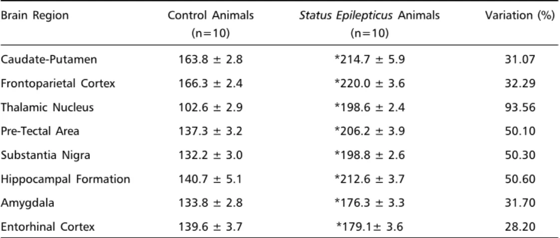
Table 1. Basal levels of cerebral energy metabolism of the status epilepticus of adult wistar rats.
Brain Region Control Animals Status Epilepticus Animals Variation (%)
(n=10) (n=10)
Caudate-Putamen 163.8 ± 2.8 *214.7 ± 5.9 31.07
Frontoparietal Cortex 166.3 ± 2.4 *220.0 ± 3.6 32.29
Thalamic Nucleus 102.6 ± 2.9 *198.6 ± 2.4 93.56
Pre-Tectal Area 137.3 ± 3.2 *206.2 ± 3.9 50.10
Substantia Nigra 132.2 ± 3.0 *198.8 ± 2.6 50.30
Hippocampal Formation 140.7 ± 5.1 *212.6 ± 3.7 50.60
Amygdala 133.8 ± 2.8 *176.3 ± 3.3 31.70
Entorhinal Cortex 139.6 ± 3.7 *179.1± 3.6 28.20
deoxyglucose uptake and co-localised seizure activ-ity that initially spreads from the stimulated site to a restricted circuitry and later involves the whole sys-tem17. Several alterations in the local deoxyglucose uptake induced by kainic acid, bicuculine and metrazol-induced seizures were described in rats18. On the other hand, in patients with temporal lobe epilepsy, the EEG, neuropathological and positron emission tomography (PET) studies have all empha-sised the primary involvement of the anterior hip-pocampal formation and the amygdaloid complex in the pathogenesis of most complex partial seizures19. The systemic administration of a potent muscar-inic agonist pilocarpine (PILO) in rats promotes a sequential behavioural and electrographic changes that can be divided into three distinct periods: (a) an acute period that built up progressively into lim-bic SE and that lasts 24h, (b) a silent period with a progressive normalisation of EEG and behaviour which varies from 4 to 44 days, and (c) a chronic period with spontaneous recurrent seizures (SRSs). These spontaneous seizures recur 3-5 times per week per animal and its main features resemble those of human complex partial seizures. Therefore, the pilo-carpine model of epilepsy provides unique experi-mental conditions for studying the human disorder20. The purpose of the present study was to determine with the 14C-2 deoxyglucose functional mapping technique which changes occurs in the seizure anato-mic substrates during the SE induced by pilocarpine.
METHOD
Adult male Wistar rats (n=40), weighting 280-300g were housed under standard controlled conditions (7:00 A.M./7:00 P.M. light/dark cycle; 20-22°°C; 45-55% humid-ity) with food and water ad libitum. The animals were separated in two groups: a) 6h SE (n=10) and b) Control group (n=10). All the animals were anaesthetised with a pentobarbital/chloral mixture. A polyethylene catheter (PE-10, Clay Adams) was permanently implanted into one ex-ternal jugular vein. To minimise the stress and facilitate the injection of 14C-2DG, the catheter was fixed over the
skull and often flushed with heparinized saline after the surgical procedure. After 3h recovery from anesthesia, SE was induced by pilocarpine injection20.
14C-2DG (50-55 µCi/mol; Sigma) was prepared in
etha-nol, evaporated under N2 stream and ressupended in normal saline. 14C-2DG (200 µg/kg) was administrated by
rapid intravenous infusion. The animals were killed 45 minutes after the 14C-2DG infusion by decapitation and
the brains were rapidly removed, frozen in cooled methyl-butane (-40°C), and stored at -70°C.
For autoradiographical analysis, 20 µm thick coronal brain sections were prepared in cryostat (Jung CM 1800-Leica) at -20°C. Brain sections were mounted on cover slips
and dried immediately at 55°C on a hot plate. Autoradio-grams were then prepared and exposed to a Kodak SB-5 X-ray film (Sigma Chemical Company) for about seven days. Then, the film was processed in a developer solution (Kodak, GB). Cerebral glucose utilisation rate by different structures was evaluated through the analysis of radioac-tive labelling on the autoradiograms. The optical density of each region were obtained using a solid state digital analyser consisting of a charge-coupled device scanner and a computer (NIH Image version 1.57, Scanner-Epson ES 1000C).
Analysis of glucose uptake were determined in the mainly regions involved in the pathophysiology of this epilepsy model, hippocampal formation, caudate-puta-men, frontoparietal cortex, amygdala, hypothalamus, entorhinal cortex, thalamic nucleus, endopiriform nucleus, primary olfactory cortex, pre-tectal area, substantia nigra. Differences in deoxyglucose uptake between pilo-carpine-treated and control animals were statistically evalu-ated with the Student’s T-test for p < 0.05.
RESULTS
Pilocarpine administration induced both ictal and interictal epileptic activity in hippocampal and corti-cal electrographic recordings which was correlated with the sequence of behavioural alterations, as pre-viously described21. In summary, 2-10 minutes after pilocarpine injection, a predominant theta rhythm could be observed in the hippocampal recording accompanied by slow-voltage fast activity in the cor-tex. Subsequent high-voltage fast activity and iso-lated spikes were seen in the hippocampus, which spread rapidly to the cortex leading to the synchro-nisation of both recordings. This kind of activity evol-ved to a pattern of isolated electrographic seizures, which culminated in SE.
At the same time, behavioural changes could be observed. Immediately after pilocarpine administra-tion, animals started to show akinesia, ataxic lurch-ing and facial automatism. This behaviour progressed to motor limbic seizures, which culminated in SE 20-50 minutes pilocarpine injection.
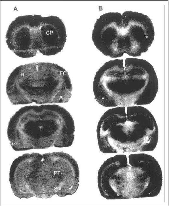
Fig 1. Representative 14C-2DG autoradiographs. Sections have been selected at the coronal level of the
caudate-putamen (CP), frontoparietal cortex (FC), hippocampal formation (H), thalamic nucleus (T), substan-tia nigra (SN), pre-tectal area (PT), amygdala (A) and entorhinal cortex (EC). A:14C-2DG autoradiographs
prepared from a control animal. B:14C-2DG autoradiographs prepared from a status epilepticus animal.
Note the activation of all the structures.
DISCUSSION
The present study evaluates cerebral metabolic rate in rats during the acute period of the pilocarpine model of epilepsy by 14C-2DG autoradiography. The observation of increased glucose utilisation by the hippocampal formation, caudate-putamen, fronto-parietal cortex, amygdala, entorhinal cortex, thalamic nucleus, pre-tectal area and substantia nigra sug-gests that these areas, contiguously and intensely
activated, have a strong tendency to act together as a single functional entity.
In animals, investigations with 14C-2-deoxyglucose autoradiographic technique12 have implicated the involvement of the anterior hippocampal formation and the amygdaloid complex in the pathogenesis of most complex partial seizures in a variety of experi-mental models of epilepsy12,15,18.
focal epilepsy and generalised epilepsy have all indi-cated marked differences in the involvement of dif-ferent brain regions22. A study of SE induced by PTZ (pentylenetetrazol) demonstrated that after 1, 5, 15, 20, 60, and 90 min of seizure activity (120-150 mg/ kg, intrarterially) marked increases occur in certain areas. These included, hippocampus, most areas of the cerebral cortex, the striatum and the reticular formation of the brainstem. Furthermore, the pat-tern of activation, once established, tended to per-sist while the seizure lasted. The striking exception was the substantia nigra. This nucleus showed ini-tially a marked increase in glucose use after 1, 5 and 15 min of seizure activity and a marked lack of activ-ity 90-min after seizure induction22.
The anatomic substrates activated during seizure activity differ from those activated during interictal intervals23. We have recently showed that increased metabolic rate in the lateral posterior thalamic nu-clei during the interictal period may be a result of the activation of cerebral circuits controlling SRSs initiation and/or generalisation.
The patterns of 14C-2-deoxyglucose uptake asso-ciated with the less severe forms of status activity primarily involve parts of the amygdaloid complex and the ventral hippocampal formation, while the more severe, convulsive levels also involve wide-spread structures throughout the ventral fore-brain17,24,25. White and Prince26 have characterised the neural substrates of both subconvulsive and convul-sive forms of SE. The major finding is that persistent focal seizure activity induced by the electrical stimu-lation of the amygdala or the olfactory cortex leads to the development of one of four discrete levels of self-sustaining SE. Each of these is consistently asso-ciated with the activation of distinct anatomical structures26. They showed that the amygdalo-hippoc-ampal area is the focus of the most restrictive form of limbic status, type I. The other, regarding more expansive forms of types II and III status, also in-volve the amygdalo-hippocampal area, but engage more widely distributed structures throughout the ventral forebrain. The anatomical relationships among these structures suggest that the basal nu-cleus of amygdala and the ventral hippocampal for-mation should be important for the generation and expression of these more widespread seizure states. Their subsequent experiments were designed to iden-tify the source or sources of the epileptiform activity in type II and III status, with special emphasis on the roles of the basal nucleus and the ventral hippoc-ampal formation. The major findings are that the
basal nucleus of the amygdala is the primary epilep-togenic focus of both seizure states, and that the ventral hippocampus is additionally involved in the development of sustained ictal discharges with fa-cial and forelimb clonus26.
On the other hand, the increased metabolic rate in the caudate putamen (striatum) may be a result of neuronal circuits involved in seizure control. The cau-date putamen represents the largest receiving area of the basal ganglia. This region transforms motor information coming from the cortex and conveys it to output nuclei, e.g., the substantia nigra, the entopeduncular nucleus and the globus pallidus27.
The increase in glucose metabolism within the substantia nigra suggests that take part in an im-portant circuit for the initiation and propagation of seizure activity within the limbic system. Alternatively, pathways interconnecting substantia nigra with the limbic forebrain are responsible for its modulator effect on the limbic seizure threshold. They may dif-fusely innervate almost all parts of the limbic sys-tem and thus render it capable of controlling the neuronal activity and regulating the neuronal excit-ability throughout the brain. Surprisingly, 14C-2DG autoradiographic monitoring of limbic seizures in rats produced by focal application of picrotoxin or penicillin into the entorhinal cortex shows that the substantia nigra becomes activated relatively late in the course of seizures28, making the first proposal rather unlikely. In fact, autoradiographic studies on the functional anatomy of limbic seizures disclosed that amygdala acts as an exit for propagation of lim-bic seizures to extrapiramidal pathways, where the paroxysmal activity can be relayed forward result-ing in emergence of motor phenomena29.
The pre-tectal area (PT) is interconnected with superior colliculus and zona incerta30. These structu-res receive projections from the cortex and have widespread projections that might influence seizure activity. In the other hand, since the activation of this structure (PT) also correlated with maximal con-vulsive activity, it is possible that PT play a role in seizure motor expression, in view of their numerous descending projections to cerebellum15.
In summary, our results suggest that these dif-ferences may be particularly important for under-standing triggering and spreading mechanisms un-derlying epileptic activity during status epilepticus and recurrent seizures.
REFERENCES
1. Treiman DM. Generalized convulsive status epilepticus. In Engel J Jr, Pedley T.A (eds). Epilepsy: a comprehensive textbook. Philadelphia: Lippincott - Raven Press: 1997.
2. Lothman EW, Bertram EH, Bekenstein JW, Perlin JB. Self-sustaining limbic status epilepticus induced by ‘continuous’ hippocampal stimu-lation : electrographic and behavioral characteristics. Epilepsy Res 1991;3:107-119.
3. Treiman DM, Walton NY, Wickboldt C, DeGiorgio C, Predictable se-quence of EEG changes during generalized convulsive status epilepticus in man and three experimental models of status epilepticus in the rat. Neurology 1987;37(Suppl 1):244-245.
4. Perl TM, Bedard L, Kosatsky T, Hockin JC, Todd ECD, Remis RS. An outbreak of toxic encephalopathy caused by eating mussels contami-nated with domoic acid. N Engl J Med 1990;322:1775-1780.
5. Teitelbaum JS, Zatorre RJ, Carpenter S. Neurological sequelae of domoic acid intoxication due to the ingestion of contaminated mussels. N Engl J Med 1990;322:1781-1787.
6. Lothman EW. The biochemical basis and pathophysiology of status epilepticus. Neurology 1990;40(Suppl 2):13-23.
7. Engel J Jr. Inhibitory mechanisms of epileptic seizure generation. In Fahn s, Hallett M, Lüders HO, Marsden CD. Advances in neurology. Vol. 67. Philadelphia Lippincott-Raven Publishers: 1995.
8. Corsellis JAN, Bruton, CJ. Neuropathology of status epilepticus in humans. In Delgado-Escueta AV, Wasterlain CG, Treiman DM, Porter RJ (eds). Advances in Neurology Vol 34: Status epilepticus: mechanisms of brain damage and treatment. New York: Raven Press, 1983:129-139.
9. Sloviter R. Epileptic brain damage in rats induced by sustained electri-cal stimulation of the perforant path. I. Acute electrophysiologielectri-cal and light microscopic studies. Brain Res Bull 1983;10:675-697.
10. Meldrum BS, Vigouroux RA, Brierley, JB. Systemic factors and epilep-tic brain damage: prolonged seizures in paralyzed, artificially venti-lated baboons. Arch Neurol Psychol 1973;29:82-87.
11. Meldrum BS, Brierley JB. Prolonged epileptic seizures in primates: is-chemic cell change and its relation to ictal physiological events. Arch Neurol 1973;28:10-17.
12. Sokoloff L, Reivich M, Kennedy C, et al. The [14C] deoxyglucose method
for the measurement of cerebral glucose utilization: theory, procedure, and normal values in the concious and anesthetized albino rat. J Neurochem 1977;28:897-916.
13. Ingvar M. Cerebral blood flow and metabolic rate during seizures: Re-lationship to epileptic brain damage. Ann NY Acad Sci 1986;462:194-206. 14. Meldrum B. Metabolic factors during prolonged seizures and their
re-lation to nerve cell death. In Delgado-Escueta AV, Wasterlain CG, Treiman DM, Porter RJ (eds). Advances in Neurology, Vol. 34: Status epilepticus. New York: Raven Press, 1983:261-275.
15. Handforth A, Treiman DM. Functional mapping of the late stages of
status epilepticus in the lithium - pilocarpine model in rat: a [14C] 2
-deoxyglucose study. Neuroscience 1995;64:1075-89.
16. Engel J Jr, Kuhl DE, Phelps ME, Mazziotta JC. Interictal cerebral glu-cose metabolism in partial epilepsy and its relation to EEG changes. Ann Neurol 1982;12:510-517.
17. Handforth A, Ackerman RF. Functional [[14C]] 2 deoxyglucose
map-ping of progressive states of status epilepticus induced by amygdala stimulation in rat. Brain Res 1988;460: 94-102.
18. Ben-Ari Y, Tremblay E, Riche D, Ghilini G, Naquet R. Electrographic, clinical and pathological alterations following systemic administration of kainic acid, bicuculine or pentrazole: metabolic mapping using the deoxyglucose method with special reference to the pathology of epi-lepsy. Neuroscience 1981;6:1361-1391.
19. Goldring S, Edwards I, Harding GW, Bernardo KL. Results of anterior temporal lobectomy that spares the amygdala in the patients with com-plex partial seizures. J. Neurosurg 1992;77:185-93.
20. Cavalheiro EA. The pilocarpine model of epilepsy. Ital J Neurol Sci 1995;16:33-37.
21. Turski W, Cavalheiro EA, Schwarz M, Czucwar SJ, Kleinrok Z, Turski L. Limbic seizures produced by pilocarpine in rats: behavioral, eletroencephalographic and neuropathological study. Behav Brain Res 1983;9:315-335.
22. Howse DC. Status espilepticus: cerebral energy metabolism during experimental status epilepticus. In Delgado-Escueta AV, Wasterlain CG, Treiman DM, Porter RJ (eds). Advances in Neurology Vol. 34: New York: Raven Press, 1983:209-215.
23. Scorza FA, Sanabria ERG, Calderazzo L, Cavalheiro EA. Glucose utili-zation during interictal intervals in an epilepsy model induced by pilo-carpine: a qualitative study. Epilepsia 1998,39:1041-1045.
24. McIntyre DC, Don JC, Edson N. Distribution of [14C]2-deoxyglucose
after various forms and durations of status epilepticus induced by stimulation of a kindled amygdala focus in rats. Epilepsy Res 1991,10:119-133.
25. Van Landinghan KE, Lothman EW. Self-sustaining limbic status epi-lepticus, acute and chronic cerebral metabolic studies: limbic hyper-metabolism and neocortical hypohyper-metabolism. Neurology 1991,41:1942-1949.
26. White LE, Prince JL. The functional anatomy of limbic status epilepticus in the rat: II. The effects of focal deactivation. J Neurosci 1993;13:4810-4830.
27. De Long MR, Georgopoulos AP. Motor function of the basal ganglia. In Brookhart JM, Mountcatle VB, Brooks VB (eds). Handbook of physi-ology. Bethesda The Nervous System. American Physiological Soci-ety, 1981:1017-1061.
28. Watson RE, Troiano R, Poulakos J, Weiner S, Block CH, Siegel A. A 2-[[14C]] deoxyglucose analysis of the functional neural pathways of the
limbic forebrain in the rat I. The amygdala. Brain Res. Rer 1983;5:1-44. 29. Collins RC, Tearse RG, Lothman EW. Functional anatomy of limbic seizures: focal discharges from medial enthorinal cortex in rat. Brain Res 1983;280:25-40.
30. Watanabe K, Kawana E. The cells of origin of the incertofungal projec-tions to the tectum, thalamus, tegmentum and spinal cord in the rat: a study using the autoradiographic and horseradish peroxidase meth-ods. Neuroscience 1982;7:2389-2406.
31. Andy OJ, Jurko MF. Diencephalic seizures: case report. Appl Neuro-physiol 1983;46:62-67.