© 2015 Associação Brasileira de Otorrinolaringologia e Cirurgia Cérvico-Facial. Published by Elsevier Editora Ltda. All rights reserved. www.bjorl.org
Brazilian Journal of
OTORHINOLARYNGOLOGY
Coordination
Wilma T. Anselmo-Lima e Eulalia Sakano
Participants
André Alencar, Atílio Fernandes, Edwin Tamashiro,
Elizabeth Araújo, Érica Ortiz, Fabiana Cardoso Pereira Valera, Fábio Pinna, Fabrizio Romano, Francini Padua, João Mello Jr., João Teles Jr., José E. L. Dolci, Leonardo Balsalobre, Macoto Kosugi, Marcelo H. Sampaio, Márcio Nakanishi, Marco César, Nilvano Andrade, Olavo Mion, Otávio Piltcher, Reginaldo Fujita, Renato Roithmann, Richard Voegels, Roberto E. Guimarães, Roberto Meireles, Shirley Pignatari, Victor Nakajima
For the purpose of citation
Wilma Terezinha Anselmo Lima, Eulalia Sakano, Edwin Tamashiro, Elizabeth Araújo, Érica Ortiz, Fábio Pinna, Fabrizio Romano, Francini Padua, João Mello Jr., João Teles Jr., José E. L. Dolci, Leonardo Balsalobre, Macoto Kosugi, Marcelo H. Sampaio, Márcio Nakanishi, Marco César, Nilvano Andrade, Olavo Mion, Otávio Piltcher, Reginaldo Fujita, Renato Roithmann, Richard Voegels, Roberto E. Guimarães, Roberto Meireles, Victor Nakajima, Fabiana Cardoso Pereira Valera, Shirley Pignatari
Introduction
Rhinosinusitis (RS) is an inlammatory process of the nasal mucosa, and according to the evolution of signs and symp-toms, it is classiied as acute (ARS; < 12 weeks) or chronic (CRS; ≥ 12 weeks). According to the severity of the condition, it is classiied as mild, moderate, or severe. Disease severity is graded using a visual analog scale (VAS) (Fig. 1), from 0 to 10 cm. Patients are asked to quantify, from 0-10 at the VAS, the degree of discomfort caused by their symptoms, with 0 meaning no discomfort and 10 the highest discomfort. Sever-ity is then classiied as: mild; 0-3 cm; moderate; > 3-7 cm; and severe; > 7-10 cm.1
Although VAS has only been validated for CRS in adults, the European Position Paper on Rhinosinusitis and Nasal Polyps (EPOS) 20121 also recommends its use in ARS. There are sev-eral speciic questionnaires for rhinosinusitis, but in practice, most have limited application, particularly in acute cases.2-4
Acute rhinosinusitis
Deinition
ARS is an inlammatory process of the nasal mucosa of sud-den onset, lasting up to 12 weeks. It may occur one or more times in a given period of time, but always with complete remission of signs and symptoms between episodes.
Classiication
There are several classiications for RS. One of the most often used is the etiological classiication, which is based mainly on symptom duration:1
• Common cold or viral ARS: a condition that is usually self-limited, in which symptoms last less than ten days; • Post-viral ARS: deined when there is symptom
wors-ening after ive days of disease, or when symptoms persist for more than ten days;
• Acute bacterial RS (ABRS): a small percentage of pa-tients with post-viral ARS can develop ABRS.
Viral ARS or common cold symptoms traditionally last less than ten days. Symptom worsening around the ifth day, or persistence beyond ten days (and less than 12 weeks), can represent a case of post-viral RS. It is estimated that a small percentage of post-acute viral RS (around 0.5% to 2% of cases) develop into a bacterial infection.
Regardless of duration, the presence of at least three of the signs/symptoms below may suggest ABRS:
• Nasal discharge (with unilateral predominance) and purulent secretion in the nasopharynx;
• Local intense pain (with unilateral predominance); • Fever > 38ºC;
• Elevated erythrocyte sedimentation rate or C-reactive protein levels;
Rhinosinusitis: evidence and experience
October 18 and 19, 2013 - São Paulo
CONSENSUS
• “Double worsening”: acute relapse or deterioration after the initial stage of mild symptoms.
Associated factors
Environmental exposure
Exposure to increasing levels of humidity, but not fungi, has been associated with ARS.5 Seasonal variations have also
been reported in the literature, with increased incidence of ARS during the winter months.5-9 Exposure to air
pollu-tion,10-12 irritants used in the production of
pharmaceu-ticals,13 in photocopiers,14 and smoke from forest ires,15
have all been associated with increased prevalence of ARS symptoms.
Anatomical factors
Anatomical variations including Haller cells, concha bullosa, nasal septal deviation, choanal atresia, pharyngeal tonsil hypertrophy, nasal polyps, hypoplastic sinuses, and odonto-genic origin of infections may be associated with ARS.10,16-18
Allergy
The role of allergy in ARS is controversial. There have been studies that assessed the association between allergic rhi-nitis and ARS,19-35 while others dismissed such an
associa-tion.35-37
Ciliary injury
Ciliary injury has been considered a characteristic of viral and bacterial RS.38 It includes the loss of cilia and
cili-ated cells, as well as alteration of the normal mucocili-ary transport. However, smoking and allergies have also been implicated in the alteration of the mucociliary trans-port,39,40 and the alteration in the mucociliary clearance in patients with allergic rhinitis has been shown to predis-pose to ARS.22
Primary ciliary dyskinesia (PCD)
This is a rare autosomal recessive disease, in which the cil-ia are either immotile or beat with a pattern incompatible with mucus transport in the airway. PCD is associated with chronic upper airway symptoms such as rhinorrhea, episodic facial pain, anosmia, and bronchiectasis.41 Newborns may present rhinorrhea from the irst day of life.42,43 There are no data on the frequency of ARS episodes in this group of pa-tients. According to the European Respiratory Society Task Force on Primary Ciliary Dyskinesia, recurring ARS is rare in patients with PCD, although the episodes should be treated with appropriate antibiotics and for a prolonged period of time.44,45
Smoking
Children living in environments with adult smokers are more prone to episodes of ARS than those who are not exposed to this environment.46 Active smokers with ongoing allergic in-lammation have increased susceptibility to ARS when com-pared to non-smokers during the course of allergic
inlam-mation, suggesting that exposure to cigarette smoke and allergic inlammation are mediated by different pathways and possible synergistic mechanisms.47
Smoking (active and passive) has been shown to alter the normal bacterial lora present in the nasopharynx, resulting in greater potential for colonization of pathogens than in non-smokers.48 Once smoking is discontinued, the microbial popu-lation begins to show the same pattern found in nonsmokers.49
Gastroesophageal relux
Little is known about the association between ARS and gas-troesophageal relux. Although studies conducted between 1997 and 2006 have observed a signiicant association be-tween the two diseases,50 a recent systematic review found
a weak association between acid relux, nasal symptoms, and ARS.51
Anxiety and depression
States of impaired mental health, anxiety, or depres-sion are often associated with increased susceptibility to ARS.52 However, the involved mechanisms remain
un-clear.
Antimicrobial resistance
The main pathogens of ABRS include S. pneumoniae, H.
inluenzae, S. pyogenes, M. catarrhalis, and S. aureus.38
Despite the problems related to bacterial resistance, it is estimated that approximately 80% of cases of mild ARS respond to amoxicillin at a dose of 70 to 90 mg/kg/day. A study by Principi and Esposito53 demonstrated that most
cases of ARS caused by H. inluenzae and M. catarrhalis
and approximately 15% of those caused by S. pneumoni-ae resolve spontaneously. Lin et al. observed that 70% of
S. pneumoniae and 71.4% of H. inluenzae cases isolated
from 69 children were resistant to amoxicillin and clavu-lanate.19
Concomitant chronic disease
Concomitant chronic disease (bronchitis, asthma, cardio-vascular disease, diabetes mellitus, or malignant tumor) in children has been associated with an increased incidence of ARS after inluenza.54
Clinical diagnosis
Signs and symptoms
At primary health care levels and for epidemiological purpos-es, ARS can be diagnosed based on symptoms alone, without detailed otorhinolaryngological assessment and/or with- out imaging studies.
recurrence of symptoms just before evaluation is frequent. Health care professional should realize that, in most cases, this may represent the evolution of the same disease, from a viral to a post-viral ARS, rather than two distinct infec-tions. Subjective evaluation of patients with ARS and their diagnosis is based on the presence of two or more of the following cardinal symptoms:1
• Nasal obstruction/congestion;
• Anterior or posterior nasal discharge/rhinorrhea (most often, but not necessarily, purulent);
• Facial pain/pressure/headache; • Disorder of olfaction.
In addition to the symptoms described above, odyno-phagia, dysphonia, cough, and ear fullness and pressure, as well as systemic symptoms such as asthenia, malaise, and fever, may also occur. The few studies on the frequen-cy of these symptoms in ARS in the community have shown great variability.55-57 The possibility of ABRS is greater in the presence of three or more of the following signs and symptoms:1
• Nasal secretion/presence of pus in the nasal cavity with unilateral predominance;
• Local pain with unilateral predominance; • Fever > 38°C;
• Deterioration/worsening of symptoms after the initial period of the disease;
• Elevated erythrocyte sedimentation rate (ESR) and C-reactive protein (CRP) levels.
ARS symptoms have a characteristically abrupt onset, without a recent history of RS symptoms. In the acute ex-acerbation of CRS, the diagnostic criteria and treatments similar to those used for ARS should be used.1 “Cough”,
al-though considered an important symptom according to most international guidelines, is not one of the cardinal symp-toms in this document. Nonetheless, in the pediatric pop-ulation, cough is considered one of the four cardinal symp-toms, replacing olfaction disorders.1,58 Gwaltney et al.,59
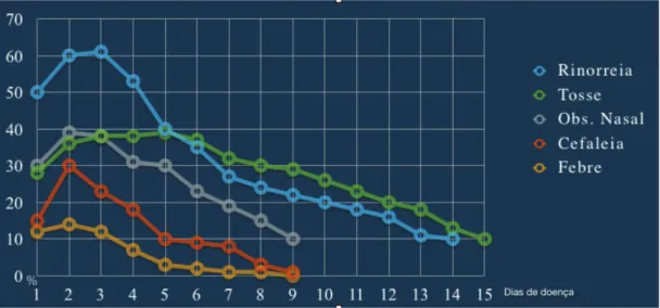
when studying the symptoms of spontaneous rhinosinusal infections by rhinovirus in relation to the time of onset and duration, observed that the peak of typical symptoms such as nasal obstruction, rhinorrhea, and cough occurs between
the second and third days of infection (Fig. 2), with a ten-dency to decrease thereafter. Symptoms can, however, last for 14 days or more.
Nasal obstruction is one of the important symptoms of ARS and should be assessed together with other patient complaints. In spite of the scarcity with which methods of objective evaluation of nasal obstruction (such as rhino-manometry, nasal peak inspiratory low, and acoustic rhi-nometry) are applied in daily practice in patients with ARS, studies have shown a good correlation between the symp-toms reported by patients and the objective measurements obtained by these methods.1
Purulent rhinorrhea is often interpreted in clinical practice as indicative of bacterial infection and need for antibiotic use.60,61 However, evidence of this association is limited. Although it is a symptom that appears to increase the chances of positive bacterial culture, in isolation it does not characterize ABRS.62 Purulent rhinorrhea with unilateral predominance and pus in the nasal cavity have a positive predictive value of only 50% and 17%, respec-tively, for positive bacterial culture obtained by maxillary sinus aspirate.63 Thus, the presence of purulent rhinorrhea does not necessarily indicate the existence of a bacterial infection and should not be used as an isolated criterion for the prescription of an antibiotic.62-64 Decreased olfaction is one of the most dificult symptoms to quantify in clinical practice and usually only is evaluated subjectively. Com-plaints of hyposmia and anosmia are commonly associated with ARS, and can be assessed with good correlation by em-ploying validated objective tests with subjective scales.65,66 It is important that these tests of olfactory function are translated and culturally and socioeconomically adapted for their use in different populations.67
Facial pain and pressure commonly occur in ARS. When unilateral, facial or dental pain has been considered a pre-dictor of acute maxillary sinusitis.55,68 The complaint of dental pain in the upper teeth abutting on the maxillary sinus showed a statistically signiicant association with the presence of positive bacterial culture obtained from sinus aspirates, with a predominance of S. pneumoniae and H.
inluenza.69 However, in another study, the predictive
itive value of unilateral facial pain for bacterial infection was only 41%.68
Several studies and guidelines have sought to deine the combination of symptoms that best determine the highest probability of bacterial infection and antibiotic response.1
In the study by Carenfelt and Berg,68 the presence of two or more indings (purulent rhinorrhea and unilaterally predom-inant local pain, pus in the nasal cavity, and bilateral puru-lent rhinorrhea) showed 95% sensitivity and 77% speciicity for the diagnosis of ABRS.
The clinical examination of a patient with ARS should initially comprise assessment of vital signs and physical examination of the head and neck, with special attention aimed at the presence of localized or diffuse facial edema. At oroscopy, posterior purulent oropharyngeal secretions58
are important. Anterior rhinoscopy is a part of the physical examination that should be performed in the primary as-sessment of patients with rhinosinusal symptoms; although it provides limited information, it may reveal important as-pects of the nasal mucosa and secretions.1
Fever may be present in some patients with ARS in the irst days of infection59 and, when higher than 38°C, it is
regarded as indicative of more severe disease and may in-dicate the need for more aggressive treatment, especially when associated with other severe symptoms. Fever is also signiicantly associated with positive bacterial culture ob-tained from nasal aspirate, especially S. pneumoniae and H. inluenzae.
In patients with ARS, the presence of edema and pain on palpation of the maxillofacial region may be indicative of more severe disease requiring antibiotics, despite the limit-ed data available in literature.60
At the primary health care level, nasal endoscopy is usu-ally not routinely available and is not considered a man-datory examination for ARS diagnosis. When available, it allows the specialist to better visualize the nasal anatomy and to obtain a topographic diagnosis and material for mi-crobiological analysis.1
At the assessment and clinical examination of patients, possible variations between geographical regions and differ-ent populations should be considered. Among other factors, climatic, social, economic, and cultural differences, as well as opportunity of access to health care, can change the sub-jective perception of the disease and potentially generate peculiar clinical features. The importance of this variability is unknown; more studies are needed to establish this.
Complementary examinations
Nasal endoscopy
As previously mentioned, it is not a mandatory examination for the diagnosis of ARS, but it may be useful for the assess-ment of the nasal anatomy, biopsy, and culture. Several mi-crobiological studies have shown a reasonable correlation between the indings collected by puncture from the middle meatus, allowing for a microbiological conirmation of the agent and its therapeutic response. Some authors recom-mend diagnostic conirmation through nasal endoscopy and culture, as many patients with clinical or radiological evi-dence of ABRS do not have a positive culture.1,70
C-reactive protein (CRP)
Low or normal levels of this protein can identify patients with low likelihood of bacterial infection, preventing un-necessary antibiotic use. Treatment guided by polymerase chain reaction (PCR) has been associated with a reduction in antibiotic use, without affecting the outcome. Although more studies are still required to include this routine di-agnostic examination for ABRS, some studies have shown that CRP levels are strongly associated with the presence of changes in computed tomography (CT), and that high CRP levels can be considered predictive of positive bacterial cul-ture from punccul-ture or sinus lavage.69,71,72
Erythrocyte sedimentation rate (ESR)
Inflammatory markers such as ESR and plasma viscosity are elevated in ARS, and may reflect disease severity and the need for more aggressive treatment. Their lev-els are associated with the presence of CT alterations in ARS and values greater than 10 are considered predic-tive of fluid level or opacity at CT. High values are also predictive of positive bacterial culture by puncture or lavage.1,73,74
CT
It should not be used in the initial diagnosis of ARS, although it is indicated in special situations, such as unilateral signs and symptoms, suspected complications, and treatment failure. It must be considered in severe disease and immu-nosuppressed patients. Recent studies suggest that routine use of CT in patients with ARS adds little information to their management.1,75,76
Simple X-ray
It has low sensitivity and speciicity, being of little use in the diagnosis of ABRS due to the high number of false-positive and false-negative results.1
Ultrasonography (USG)
USG of the paranasal sinuses has low sensitivity and very limited usefulness in the diagnosis of ARS, due to the high number of false-positive and false-negative results.1
Treatment
There is a worldwide concern regarding the indiscriminate use of antibiotics and bacterial resistance. It is estimated that approximately 50 million unnecessary antibiotic pre-scriptions for RS are given in the US and used to treat viral infections. When a more selective algorithm for antibiotic therapy is followed, the beneit is greater and only three patients need to be treated for one to achieve the expected result.77 Thus, there is a worldwide trend to treat ARS ac-cording to disease severity and duration.
Antibiotic therapy
the disease;78 they are never indicated for symptomatic treatment and their indiscriminate use should be avoid-ed, since that can increase the risk for the development of bacterial resistance.79
Clinical studies have demonstrated that approximate-ly 65% of patients diagnosed with ABRS show spontaneous clinical resolution80 sometimes within the irst few days;78 therefore, the initial adjuvant treatment without antibiot-ics is a viable option in cases of mild and/or post-viral si-nusitis. The introduction of antibiotics should be considered when there is no improvement after adjuvant therapy or if symptoms exacerbate. Antibiotics are indicated in cases of moderate to severe ABRS; in patients with severe symptoms (fever > 37.8°C and in the presence of severe facial pain); in immunocompromised patients, regardless of disease du-ration; and in cases of mild or uncomplicated ABRS that do not improve with initial treatment with topical nasal corti-costeroids.81,82
There are no studies that deine the optimal duration of antibiotic treatment. In general, treatment duration varies from seven to ten days for most antimicrobial agents and 14 days for clarithromycin. Amoxicillin is considered the antibiotic agent of irst choice in primary health centers, due to its effectiveness and low cost. Macrolides have comparable eficacy to amoxicillin and are indicated for patients allergic to β-lactams.79,82,83 In cases of suspected penicillin-resistant S. pneumoniae, severe cases, and/or
associated comorbidities, broader-spectrum antimicrobi-als are indicated.
Intranasal topical corticosteroids
Patients older than 12 years with post-viral RS, or with un-complicated ABRS with mild or moderate symptoms81
with-out fever or intense facial pain,82 beneit from topical nasal
corticosteroids as monotherapy. In addition to relieving the symptoms of rhinorrhea, nasal congestion, sinus pain, and facial pain/pressure,81 topical corticosteroids minimize the
indiscriminate use of antibiotics, thus reducing the risk of bacterial resistance.82
Studies suggest that topical nasal corticosteroids in combination with appropriate antibiotic therapy results in faster relief of general and speciic symptoms of RS, especially congestion and facial pain,84-89 and acceler-ates patient recovery, even when there is no signiicant improvement in the radiological image.87,88,90 However, the optimal dose and treatment duration still need to be established.85-88 Although there are no studies comparing
the effectiveness of several types of nasal corticosteroids in ARS, many of them (such as budesonide, mometasone furoate, and luticasone propionate) have shown bene-its.90 Their use is recommended for at least 14 days to
effect improvement in symptoms.
Oral corticosteroids
The use of oral corticosteroids for adults with ABRS and intense facial pain is recommended, as long as there are no contraindications to their use.91,92 Oral corticosteroids
should be used for three to ive days, in the irst few days of the acute event only, and always associated with anti-biotic therapy, in order to shorten the duration of facial
pain91 and decrease the need for analgesics.92 Evaluation
after ten to 14 days of treatment shows no signiicant differences in symptom resolution or treatment failure when comparing antibiotic therapy alone and antibiotics with oral corticosteroids.92 The few studies in the
liter-ature using oral corticosteroids in the treatment of ABRS showed favorable results with methylprednisolone and prednisone.
Nasal lavage
Despite the frequent use of isotonic or hypertonic saline solution in nasal lavage of patients with rhinitis and RS, lit-tle is known about their real beneits in ARS.
Randomized trials93 comparing nasal saline and
hy-pertonic solutions showed greater intolerance to hyper-tonic solution. A meta-analysis of placebo-controlled, randomized, double-blinded trials showed evidence of limited beneit of nasal saline irrigation in adults, with no difference observed between case and control groups. A single study showed a mean difference of improved time to symptom resolution of 0.3 days, without statistical sig-niicance.94
In another meta-analysis of patients younger than 18 years with ARS, there was no clear evidence that antihis-tamines, decongestants, and nasal lavage were effective in children with ARS.95
Despite little evidence of clinical beneit, the use of na-sal na-saline lavage is generally recommended in patients with ARS. It promotes improvement of ciliary function, reduces mucosal edema and inlammatory mediators, and helps to cleanse the nasal cavity, by removing the infectious secre-tions, and saline lavage has no reported side effects.96
Oral and topical decongestants
The use of oral decongestants alone or associated with an-tihistamines in patients with ABRS does not signiicantly change the clinical or radiological evolution, either in chil-dren97 or in adults.98
Topical nasal decongestants (topical vasoconstrictors), such as 0.1% xylometazoline, are not indicated alone for the treatment of ABRS,99 but they do provide subjective and
objective improvement of nasal obstruction in patients with viral ARS. In cases of patients with ABRS as a complication of persistent rhinitis, the use of topical nasal vasoconstrictors may relieve nasal obstruction100 and increase inspiratory
na-sal low.101 Even in this restricted population, it is important
to consider the complications caused by interactions with other drugs, as well as the possibility of adverse effects on hypertension, glaucoma, diabetes mellitus, thyroid disease, urinary retention, and benign prostatic hyperplasia (BPH).99
Due to the rebound effect, the use of topical nasal vaso-constrictors should be restricted to a maximum of ive days. They should not be used by children younger than 2 years.
Nonsteroidal anti-inlammatory drugs (NSAIDs)
colds. Nonetheless, their analgesic effect is beneicial, with improvement of headache, ear pain, and muscle and joint pain, and without evidence of increased adverse effects in this population. Therefore, they can be used for the sympto-matic improvement in patients with common cold.102
In spite of their analgesic effect in acute inlammato-ry processes of the ear, orophainlammato-rynx, and paranasal sinus-es,103 NSAIDs are not recommended as the only treatment
of ABRS, and should be used with caution even when asso-ciated with antibiotics, due to the increase in possible side effects.104,105
Mucolytics
The association of mucolytics in the treatment of ARS is still controversial. It is believed that they reduce nasal secre-tion viscosity due to their mucoregulatory activity, resulting in fragmentation of acid mucopolysaccharide (AMPS) ibers and, therefore, facilitating mucociliary transport and their elimination through the nose and paranasal sinuses.106 When combined with antibiotics, they may facilitate penetration into the paranasal sinus mucosa and improvement of the inlammatory process.107 There have been some studies us-ing oral bromhexine combined with oral antibiotics and ace-tylcysteine combined with topical nasal antibiotics.106-108 However, those studies did not clearly state the time and severity of RS; therefore, their results should be analyzed with caution. Studies with oral erdosteine showed no signif-icant beneit in children.109
Phytotherapics
There are few placebo-controlled, randomized, and dou-ble-blinded studies of herbal medicines in the treatment of ARS. In spite of the beneits demonstrated by some of them, their use in clinical practice should be approached cautious-ly because of the scarcity of published evidence regarding the pharmacokinetics and pharmacodynamics of these com-ponents and their mechanisms.
• Pelargonium sidoides:110 A study with Cochrane
collabora-tion for the treatment of acute respiratory infeccollabora-tions con-cluded that it can be effective in alleviating the symptoms of the common cold and post-viral ARS in adults.
• Myrtle Essential Oil: which is extracted from Pinus
spp. (pine), Citrus aurantifolia (lime) and Eucalyptus globulus. A controlled, randomized, multicenter trial
reported a statistical difference in symptom improve-ment score of post-viral ARS (from 10.5 to 9.2) when compared to placebo, reducing the need for antibiot-ics (20% in patients who used the medication vs. 40% in those who used a placebo). In Germany, it is recom-mended for the treatment of ARS.111
Probiotics
A Cochrane review112 with ten studies demonstrated that
probiotics are superior to placebo in reducing the number of patients with upper respiratory tract infection episodes, number of episodes per participant, and antibiotic use. Therefore, they may be indicated for the prevention of the common cold.
Immunomodulators
A systematic review113 of eight randomized controlled trials
(RCTs) in children with more than three episodes of upper airway infections per fall/winter (six months) who used OM-85 BV extract demonstrated that these children had fewer episodes of upper airway viral infections when compared to the placebo group (38% vs. 52%; p < 0.001), and that the beneits are greater for patients with risk factors for recur-rent infections.
Acute rhinosinusitis complications
RS complications are caused by acute or chronic infections; although they are more common in children, they may also occur in adults and can be orbital-palpebral, bone, and in-tracranial.
Epidemiology
Most RS complications originate from ethmoid sinus infec-tions. It is estimated that prior to the advent of antibiot-ics, the rate of blindness arising from complications was up to 20%, and is currently around 11% of cases. Mortality from meningitis of sinus origin in the past was approximately 17%; it currently ranges from 1% to 2.5%.1,114-116 The mortality rate from intracranial complications is around 20% to 40%,114,117 and from neurological deicits, 25%.117,118 The incidence var-ies by geographic region. In the Netherlands, for instance, the complication rate is estimated at 1:12,000 ARS in chil-dren and 1:36,000 ARS in adults,119 whereas in the United
States it ranges from 2.7 to 4.3:1,000.000;120 and in France,
2.5:1,000,000/year, excluding pediatric patients.121 It is
more frequent in males. In children, complications usually occur from the acute processes, whereas in adults, they are more often seen with CRS with or without polyposis.119,120,122
There are no exact prevalence data for the several types of complications. Orbital complications comprise from 60% to 75%; intracranial, from 15% to 20%; and osseus, 5% to 10%.123 Childhood sinusal disease is the presumed cause of
10% of intracranial suppuration, 10% of preseptal cellulitis, and 90% of orbital cellulitis, subperiosteal and intraorbital abscesses.124 Antibiotic prescription does not appear to re-duce the incidence of complications.5,119
Physiopathogenesis
Dissemination occurs by direct extension, bone erosion, through diploic veins and hematogenously through venous involvement.125 Certain anatomical characteristics are
im-portant in the genesis of these complications:1,114
• the thin boney lamina papyracea that separates the orbital contents from ethmoid cells;
• in children, a number of larger neurovascular forami-na and several boney sutures that remain open in the medial orbital wall and facilitate the dissemination of infection; and
ophthal-mic veins, which communicate with intraorbital ves-sels and directly with the cavernous sinus.
Classiication of orbital complications
Existing classiications are based on anatomical-clinical cri-teria, but none is universally accepted. It is important to remember that the orbital septum consists of a delection or extension with change in direction, laterally forming the lateral palpebral ligament, and medially, the medial pal-pebral ligament, behind the lacrimal sac. It functions as a protective barrier against infections for the internal orbital area.116,118,123 The earliest classiication was that of Hu-bert, which dates from 1937.118 In 1970, Chandler et al.123
proposed a classiication that is still the most cited in the world literature, which takes into account the orbital sep-tum:
• Group 1 – periorbital cellulitis: eyelid inlammation with edema, without dissemination into the orbit; • Group 2 – Orbital cellulitis: the infection crosses the
orbital septum and penetrates the orbital cavity; • Group 3 – subperiosteal abscess: post-septal abscess
between the lamina papyracea and the periosteum, contained by the latter;
• Group 4 – orbital abscess: true orbital abscess, puru-lent secretion inside the orbit, within the extrinsic eye musculature, near the optic nerve;
• Group 5 – thrombosis of the cavernous sinus. Due to failures observed in this classiication revealed by imaging studies (CT and magnetic resonance imaging [MRI]), Mortimore and Wormald126 suggested removing the cavern-ous sinus thrombosis group from orbital complications and placing it into the cranial complications group.
Group 1 – preseptal infection;
Group 2 – subperiosteal post-septal infection; Group 3 – intraconal post-septal infection.
In Brazil, Velasco et al.127 proposed a simpler classiica-tion, with only three groups, considering preseptal cellulitis as a palpebral rather than orbital infection:
• Orbital cellulitis; • Subperiosteal abscess; • Orbital abscess.
Among all classiications, most authors still use that pro-posed by Chandler.116,128-131
Bacteriology
Regarding the bacteriology in orbital complications, the most common microorganisms are the same that are iden-tiied in RS.128 The widespread use of the heptavalent
pneumococcal conjugate vaccine (PCV7) has reduced the frequency of S. pneumoniae in RS complications, with a subsequent increase in infections by S. aureus, as well as in the prevalence of methicillin-resistant S. aureus (MRSA)
associated with orbital infections.132
Orbital-palpebral cellulitis
The presence of palpebral edema, erythema, localized pain, nasal obstruction, rhinorrhea, dificulty opening the eyes, and possibly fever, can be observed in cases of
or-bital-palpebral cellulitis. It is caused by venous obstruction created by the pressure on the ethmoid vessels,116,118 and can progress into palpebral abscess and rarely, to cutaneous istula. Visual acuity and ocular motility are preserved and this assessment is dificult in some children.133 Inlammation
of the eyelid and conjunctiva is observed on CT as edema-tous tissue.134 It occurs as a complication of viral upper res-piratory tract infection, acute dacryocystitis, skin infection and, less commonly, RS.135-138 It has a favorable prognosis
with antibiotics and often requires no imaging tests, being treated as simple acute ethmoiditis.121
Orbital cellulitis
It is characterized by edema extending into the post-septal region. It appears most often as a complication of acute RS.137,138 It presents exophthalmia, chemosis, and conjunc-tival hyperemia,130 and affects the orbital adipose tissue
without forming an abscess. Visual acuity and ocular motili-ty are usually preserved, but a slight decrease of the latter may occur, and some children initially may lose the ability to distinguish green and/or red colors.126,139,140 Ophthalmo-logic evaluation and emergency CT are necessary, and treat-ment should be aggressive and immediate.
Subperiosteal abscess
The clinical picture presents with high fever (39.5°C or higher), chills, changes in general status, exophthalmos with exophoria, decreased ocular motility, severe pain, pre-served visual acuity (although decreased in some cases),141 and leukocytosis with a shift to the left.141 The CT discloses the presence of purulent collection in the medial orbital wall, between the periorbital and the orbital bone, with an extraconal location and, thus, outside the ocular mus-cles.116 The most common microorganisms are Streptococci in children and anaerobic bacteria in adults. Total vision loss can occur, especially in diabetic adults. Abscesses located more superiorly can result in intracranial complications by extending into the frontal lobe.
Orbital abscess
It is an intraconal lesion, commonly the consequence of late diagnosis or immunosuppression.142 The clinical picture is more severe with irreducible, painful exophthalmos with se-vere chemosis, complete ophthalmoplegia, and marked de-crease in visual acuity.130 The CT image shows purulent
col-lection in the soft tissues around the eyeball. It may remain localized or extend through the orbital septum, emerging as a loating mass in the eyelid. It is a severe condition that can lead to amaurosis. The visual impairment depends on the orbital pressure and optic neuritis. Thromboembolism may occur in the vascular supply of the nerve, choroid, and retina. With increasing pressure, there is retinal artery oc-clusion, which, if lasting over 90 minutes, leads to irreversi-ble degeneration of the optic nerve and retina.116,118
and optic foramen.116,118 Clinically, the eyeball is ixed and pupils are dilated and nonreactive to light; ptosis, and palpebral, corneal, and conjunctival hypoesthesia are also observed. When there is a concomitant lesion in the optic foramen, ophthalmoplegia, amaurosis, severe ocular pain, and sensory deicits from anesthesia to neuralgia are seen in the distribution of the ophthalmic nerve. Since the pos-terior orbital bone is thicker than the anpos-terior bone, these indings are rare and, when present, are more common in sphenoethmoiditis.
Cavernous sinus thrombosis
It consists in the dissemination of an infection along the op-tic canal or intravenously to the cavernous sinus. It causes blindness, abolition of the pupillary relex to light, corneal anesthesia, and paralysis of nerves III and VI. The following are also observed: high fever, altered sensorium, prostra-tion, severe deep retro-orbital pain, bilateral involvement, and central nervous system signs. The accompanying photo-phobia and neck stiffness may be mistaken for meningitis. The mortality rate is approximately 30%.114
Diagnosis
The diagnosis of complications should involve otorhinolaryn-gologic, ophthalmic, and neurological evaluations, as well as neurosurgical assessment, when necessary. Imaging stud-ies, particularly CT with contrast and MRI, play an important role. High-resolution CT is the technique of choice when orbital complications are suspected. MRI better character-izes the local extent of disease or its dissemination beyond the nasal and paranasal cavities. A combination of CT and MRI is useful in cases of dificult diagnosis.143 It usually dis-closes swelling of the medial rectus muscle, periorbital lat-eralization, and downward and lateral displacement of the eyeball. When obliteration of the extraocular muscle detail is evident and the optic nerve appears as conluent mass, an orbital abscess is present. Imaging studies may also detect air bubbles produced by anaerobic bacteria. The predictive accuracy of the clinical diagnosis is 82% and of the CT is 91%.144-146
Laboratory analysis usually shows leukocytosis with a left shift; an elevated CRP level is associated with more severe outcomes and may suggest or indicate the need for more aggressive treatment in the early phase.71
Differential diagnosis
Patients with RS and proptosis may have a subperiosteal orbital hematoma; 13 cases have been reported in the lit-erature.147 Orbital lymphatic malformations can lead to proptosis, compressive optic neuropathy, vision loss, and cellulitis. The MRI shows a well-outlined intraorbital mass with a heterogeneous signal.148
General treatment standards
Treatment is medical for orbital-palpebral or periorbital cellulitis. It requires hospitalization, careful observation, and intravenous antibiotic therapy. Clindamycin or
amoxi-cillin + clavulanate potassium with metronidazole and/or, particularly in children, oxacillin + ceftriaxone can be used in the treatment. Most patients respond well to the con-servative treatment, and surgical intervention is not neces-sary.115,116,118 It is always recommended to discuss with the local Hospital Infection Committee which antibiotic is the most appropriate.
The identiication of abscesses on the CT, orbital or pro-gressive visual indings, or lack of response to intravenous antibiotics, are all indications for surgical exploration. In-tensive ophthalmological control is crucial.149
Children with small and medium-sized subperiosteal ab-scesses, without signiicant ocular signs, may be success-fully treated medically. Surgical drainage is indicated for medium-sized to large abscesses with severe visual loss, and in cases with inadequate response to medical treatment.150
Usually, a medium-sized subperiosteal abscess that does not improve with medical therapy can be drained endoscopical-ly, while a lateral or intraconal abscess may require an open procedure.151
There are controversies regarding the surgical indica-tion in subperiosteal abscesses. For the initial treatment,141 many studies have documented an improvement in young children with medical therapy alone.133,142,152 If medical treatment is chosen, it is essential that clinical improve-ment occurs within 24 to 48 hours; that there is no visual impairment; that the abscess volume is less than 0.5 to 1.0 mL; the abscess is located medially; that there are no sys-temic symptoms and that the child is less than four years of age.153 Surgical drainage should be strongly considered
when an older child has a subperiosteal abscesses with sig-niicant ocular indings, when improvement is not observed after 48 hours of medical treatment, when the abscess vol-ume > 0.5 mL, the length > 17 mm, and the width > 4.5 mm.154 In general, immediate surgical drainage is indicated in the following situations: the abscess is not in a medial location, or there is visual loss, clinical deterioration or an absence of clinical improvement in 24 to 48 hours.114,116,141
Based on the diagnosis of a subperiosteal abscess, in which there is no purulent secretion after opening the lam-ina papyracea, an orbital abscess should be suspected, and incisions should be performed along the periosteum to re-lease the purulent material from the orbit.155 Some authors
always recommend surgical treatment for subperiosteal ab-scess, with drainage of the abscess and sinuses involved.141 The endoscopic approach is always safer and more effec-tive, but associated external approaches may be necessary. Acute sphenoid RS may cause thrombosis of the ipsilat-eral or contralatipsilat-eral cavernous sinus. Early surgical sphe-noidotomy and aggressive medical treatment are the bases of the successful management of this life-threatening com-plication.156
Intracranial complications
occur, a capsule develops, forming the abscess. There is a high incidence of anaerobic bacteria and mixed lora. Mi-croorganisms most frequently mentioned in the literature include Streptococcus millieri and S. anginosus, Fusobac-terium sp. and S. aureus.114,128,158,159S. anginosus causes more severe infections, higher rates of neurological com-plications, more neurosurgical interventions, and more cen-tral nervous system sequelae.160 Polymicrobial cultures are obtained in 50% of patients.161
Meningitis
In decreasing order of frequency, the paranasal sinuses related to the origin of meningitis, are the sphenoid, fol-lowed by the ethmoid, frontal, and maxillary sinus. Clin-ical manifestations include fever, severe headache, neck stiffness, irritability, and behavioral disorders. CT deines and delimits the disease and can identify the presence of additional complications. Lumbar puncture125 reveals
in-creased proteins and cells, and a culture and sensitivity test should be performed. Lumbar puncture is contraindi-cated in the presence of intracranial hypertension (ICH) or abscess.125 The treatment is medical, and sinus
interven-tion is reserved for refractory cases. The mortality rate is around 5%.116,118
Extradural abscess
This consists of a purulent collection between the dura mater and the cranium. Occasionally, it is associated with frontal osteomyelitis. The clinical manifestations are vague, with few or no neurological signs, which, when present, include persistent headache, fever, and rarely, behavioral changes. The diagnosis is usually delayed because of a fail-ure to recognize the signiicance of the clinical indings. By the time of diagnosis there is usually ICH with worsening headache, vomiting, and behavioral changes.116,118
Subdural abscess
Subdural abscess is characterized by the presence of puru-lent collection between the dura mater and the pia-arach-noid. Patients present severe headache, fever, and de-creased level of consciousness. CT shows a decreasing image, not extending beyond the midline, thus differentiat-ing from the extradural abscess. Surgery is performed at the neurosurgeon’s discretion.116,118
Brain abscess
The incidence of cases of sinus origin varies greatly, ranging from 3% to 11% up to 66%. The most common location is in the frontal lobe. Focal symptoms and increased intrac-ranial pressure appear late with poor general condition, coma, and cranial nerve palsy. The frontal lobe is an area of clinical silence that yields inconstant symptoms. Fever, ICH, seizures, waking period disorders, coma, motor deicit, sensory disturbances, and altered vision may occur.
Imaging studies show a rounded lesion with a hypodense center and peripheral enhancement that initially is irregu-lar but becomes more well deined as the necrotic portion
progresses. It can be multilocular. Lumbar puncture is con-traindicated due to the risk of brain stem decompression and herniation. The initial treatment during the phase of cerebritis is based on antibiotic therapy, although empir-ical. Once an abscess is formed, surgical drainage is indi-cated by puncture or craniotomy159,162,163 combioned with concomitant paranasal sinus drainage.162 The latter, alone, does not substitute for intracranial drainage.157 Antibiotics should be maintained for four to eight weeks.128
Third-gen-eration cephalosporins can be used in combination with with metronidazole, mannitol, hyperventilation, and dexa-methasone, with or without anticonvulsants.128
Bone complications
They occur with the extension of the infectious process to the bone, possibly involving the brain and nervous sys-tem. The most common associated sinus infections are in the frontal and maxillary sinuses.1 In the frontal
re-gion, there is a diploic spongy boney layer, with a rich vascular network including diploic veins that course be-tween the external and internal bone laminae. These veins do not have valves and allow unimpeded passage of blood between the spaces of the sinus mucosa and the skull.1 Series of patients with complications of sinusitis
demonstrated that osteomyelitis occurs in 9%146 to 32% of cases.164 A peculiar clinical form of localized frontal osteomyelitis may be focal or circumscribed, often with progression to a cutaneous istula. The diffuse or dissem-inated frontal form is characterized by thrombophlebitis of the diploic veins, which progresses to the frontal bone and the cranial cavity, leading to avascular necrosis, bone sequestration, and expansion to subperiosteal infection. It is more common in young individuals with extensive, pneumatized, and vascular diploe, increasing the risk of infection.165 A softened, loating tumor may be observed, without signs of inlammation, called Pott’s tumor.118
It corresponds to a subperiosteal abscess of the frontal bone associated with underlying osteomyelitis.117,166 Ra-diographically, it has three phases: 1) condensation with bone sequestration; 2) rarefaction, when there is bone necrosis; and 3) decalciication or absence of bone tissue in irregular areas, interspersed with islands of calciica-tion and areas of bone sequestracalciica-tion. A CT scan conirms the diagnosis. Scintigraphy with technetium 99 for diag-nosis and with gallium 67 for the follow-up is useful, but not essential.165 Treatment consists in the administration of clindamycin and abscess drainage through coronal ac-cess, with reconstruction.
Osteomyelitis of the maxillary sinus is often a compli-cation of odontogenic infection, more common in infants.
Atypical rhinosinusitis complications
most likely hypotheses were: the patient’s position during surgery, duration of surgery, or minor trauma to the lamina papyracea during perforation of the mastoid.177
Chronic rhinosinusitis
Deinition and epidemiology
CRS is an inlammatory disease of the sinonasal mucosa that persists for at least 12 weeks. In speciic cases, exclusive sinus involvement can be observed, as in odontogenic sinus-itis or fungal ball.
CRS can phenotypically be divided further into two main en-tities: CRS without nasal polyps (CRSsNP) and CRS with nasal polyposis (CRSwNP). Currently, there is evidence to suggest that these two entities have distinct physiopathogenic mechanisms.
CRS is a common disease in the population, and studies about its epidemiological data are important to evaluate its distribution, analyze its risk factors, and promote public health policies. However, such data are scarce in the lit-erature. Additionally, data comparison is hindered by the different deinitions and methodologies used in the studies.
This disease has a high direct cost for public health, which includes medical visits, supplementary and radiological exams, hospitalization, surgery, and drug treatment, as well as indi-rect costs, such as decreased work productivity and absentee-ism.178-181 In the United States, the estimated expenditure on these patients is US$ 8.6 billion per year,182 of which US$ 150
million are related to antibiotic use.183 Additionally, overall
quality of life and disease-speciic questionnaires show great impact of CRS on patients’ quality of life.184-187
In 2007, the European Position Paper on Rhinosinusitis and Nasal Polyps (EPOS)188 was published, and a CRS
deini-tion was introduced for epidemiological purposes, charac-terized by the presence of two or more of these symptoms for more than 12 weeks, a) nasal obstruction/congestion; b) rhinorrhea (anterior or posterior); c) facial pain/pressure; d) reduction or loss of olfaction. One of the two symptoms had to be either a) or b) above. Supplementary examina-tions, such as nasal endoscopy or imaging studies were not required for diagnosis.
The annual study by the National Center for Health Statistics (NCHS) of the United States population by means of household surveys observed a prevalence of self-reported medical diag-nosis of sinusitis of 13% of the adult population in 2010 and a response rate of 60.8%. However, there was no distinction be-tween ARS and CRS in this study, as the criteria that deined CRS in this questionnaire was an afirmative answer to the question: “In the last 12 months, have you had sinusitis diagnosed by a physician or healthcare professional?”189 However, this
preva-lence is used in most published studies that refer to CRS. In Canada, an epidemiological study of complex sam-pling design, with a national response rate of 82%, was performed through telephone interviews with individu-als aged 12 years and older, with symptoms of chronic diseases for more than six months.190 Individuals were
considered as having CRS when the following question was answered affirmatively: “Do you have sinusitis diag-nosed by a healthcare professional?” In that study, the prevalence of self-reported RS was 5%.190
In South Korea, a nationwide study was performed through complex cluster and multistage sampling. A medical team that included an otorhinolaryngologist visited house-holds and performed interviews with participants aged 12 years or older. The diagnosis of CRS was deined by a positive response to symptoms of nasal obstruction and rhinorrhea for more than three months and an endoscopic examination with indings of polyps or secretion in the middle meatus. The estimated CRS prevalence in South Korea was 6.95%.191
Hastan et al.192 published part of the results of the
Glob-al Allergy and Asthma Network of Excellence (GA2LEN) Euro-pean multicenter study related to the investigation of CRS epidemiology. A questionnaire was mailed to a randomized sample of adults between 15 and 75 years in 19 centers in Europe, covering 12 countries, using as a diagnostic cri-terion the epidemiological deinition published in EP3OS 2007 (The European Position Paper on Rhinosinusitis and Nasal Polyps)188 The estimated prevalence of CRS in Europe
was 10.9% (6.9% to 27.1%), but the overall response rate was 48%, with wide variation between centers (23.2% to 80.3%).213 Tomassen et al.193 reported the consistency and
validity of the epidemiological criterion of CRS deined by EP3OS 2007m188 using data from the GA2LEN study.
Pilan et al.,194 in a recent study in the city of São Paulo, Brazil, with complex sampling design incorporating stratiication and multiple selection stages to obtain a representative sample of the population, used the ep-idemiological deinition of CRS recommended by EP3OS 2007.188 The questionnaire involving this deinition was
applied in 2006, through household interviews, to indi-viduals aged 12 years or older, and a prevalence of CRS of 5.51% and high response rate of 87.8% was observed.194 No statistically signiicant difference was found in alence by gender. In that study, there was a higher prev-alence of CRS in patients who had asthma and rhinitis. However, there was no signiicant association with smok-ing. For the purpose of comparison with the methodology used in the study by Pleis et al.,189 the same question was
inserted: “In the last 12 months, have you had sinusitis diagnosed by a physician?” The self-reported prevalence of RS diagnosed by a physician (with no distinction be-tween acute and chronic) was 16.55%.
Physiopathogenesis
Microbiology
In contrast to the etiopathogenesis of ABRS, which involves a continuum of changes promoted by a viral infection fol-lowed by bacterial superinfection, the role of microbial agents in the pathogenesis of CRS is not yet fully elucidated.
Viral participation
Currently, there is little research regarding viral involve-ment in the physiopathogenesis of CRS. Despite the high frequency of acute infections of the upper airways, it is not very clear yet whether viruses act as a source of chronic stimulation or trigger the initial inlammatory process. As they have the capacity to incorporate into the host DNA as the episomal form, the virus may persist chronically in the respiratory mucosa. Recent studies of viral genomic detec-tion have demonstrated from none195 to signiicant rates of
the main respiratory viruses, especially rhinovirus196 and metapneumovirus.197 However, there is still no evidence whether these viruses are involved in a latent infection without cytopathic effects for the host, or whether they are active, producing antigens and replicating.
Fungal participation
Among the different classiications of sinonasal chronic in-lammatory processes involving a fungal etiology, it is indis-putable that, in some conditions, such as fungal ball and the invasive chronic forms, the role of fungi is essential.198,199
However, the participation of fungi in the forms of idiopath-ic CRS, those without apparent cause, is still a subject of much controversy.
The theory of a fungal etiology for CRS200 was
height-ened by the correlation of the high incidence of the detec-tion of fungi in CRS patients, associated with a high number of eosinophils in tissue and secretions. Several in vitro stud-ies have demonstrated that stimulation of lymphocytes by fungal antigens could produce increased amounts of 5, IL-13, and IFN-gamma201 and stimulate eosinophil
degranula-tion.202,203 However, other investigators failed to reproduce
such indings or even found divergent results.204-206 The attempt to prove the fungal theory through clini-cal trials with topiclini-cal and systemic antifungals did not pro-duce encouraging results. Controlled studies have failed to demonstrate the eficacy of oral207 and topical antifungals for the treatment of CRS.208-213
The fact that the ubiquity of fungal elements could act as a constant stimulator of innate immunity receptors and, in turn, could lead to stimulation of speciic inlammatory responses cannot be ignored.214,215 In light of the present evidence, fungi appear to have universal participation in CRS, and play a modulatory role in some patients.216
Bacterial participation
Studies involving conventional bacterial growth and identi-ication techniques have been widely performed in patients with CRS. Most Brazilian217,218 and international studies219-222
observed a higher prevalence of S. aureus, Gram-negative,
and anaerobic bacteria in patients compared to controls, or even those with ARS. However, the identiication of bac-teria by the traditional method, through in vitro culture, has some sensitivity and speciicity limitations. In general, the conventional method only shows positivity for dominant microorganisms or those with favorable growth on that me-dium, representing only the collection site (middle meatus, nasal cavity, and paranasal sinus) with a risk of contamina-tion from other regions (such as the nasopharynx and the
nasal vestibule), or does not allow the differentiation of colonizing microbes from pathogenic microorganisms (for instance, S. epidermidis).
In order to overcome such limitations of lora interpreta-tion in individuals with CRS, more sensitive and speciic tech-niques have been used for the characterization of nasal lora in healthy individuals and those with CRS. Recent studies using molecular techniques have shown high prevalence of bacteria, with a predominance of S. aureus, P. aeruginosa, and anaer-obic bacteria, characteristically polymicrobial.223-225 These
studies have demonstrated that individuals with CRS have the same bacterial load as their healthy peers, but with lower lora diversity, indicating a possible microbiota disor-der.226 Broader studies including the analysis of the human microbiome are still necessary to assess the importance of the quantity and biodiversity of these bacteria in patients and healthy individuals, considering that the genetic, ge-ographic, and environmental characteristics may inluence the microbiota in different health scenarios.
Based on conventional microbiology and some molecular studies, it was observed that S. aureus is the main bacterial
agent found in Western patients with CRS, both in pre- and postoperative conditions,227 with a lower prevalence in the Chinese population;228 it is more frequently identiied in
patients with extensive sinonasal polyps than in controls or even individuals with CRSsNP.229
A peculiar characteristic of S. aureus is its capaci-ty to produce exotoxins with superantigen properties. There is evidence that staphylococcal superantigens may play a role in the physiopathogenesis of CRS, especially in CRSwNP, with induction of speciic polyclonal IgE and mast cell stimulation;230 increases in IL-4, IL-5,
eosino-phils, and eosinophil cationic protein;231-234 and associ-ation with severe asthma.230,235-238 However, the mere
presence of enterotoxin-producing S. aureus in the nasal cavity is not suficient to produce a chronic inlammatory reaction and polyp formation.239 It is believed that the
primary action of superantigens is to modulate inlam-mation in the upper airways, depending on the distinct reactions of each individual.240
Another bacterial form, which has been demonstrated in CRS, is bacterial bioilms. Despite the great variability in the prevalence of bioilms in different studies, probably due to the different techniques used, it is estimated that at least 25% of cases are associated with their presence.241,242
In general, patients with CRS have signiicantly higher rates of bioilm when compared to healthy individuals. How-ever, similar to planktonic bacteria, it is unclear what the real role of bioilms is in the physiopathogenesis of CRS and it is not possible to determine whether the colonization of bioilms would be the cause or the consequence of chronic inlammation.243
In addition to the possible involvement of multiple spe-cies of bacteria in bioilms, simultaneous fungal and bacte-rial colonization has also been observed.244
In terms of pathogenic mechanisms, two independent studies, using different detection methods for different populations, showed opposite results on polarization of the inlammatory response, whether to Th1 (neutrophils, IFN-gamma, interferon-gamma, macrophage inlammato-ry protein-1 [MIP-1], granulocyte colony stimulator factor [G-CSF])250 or Th2 (IL-4, IL-5, eosinophil cationic protein
[ECP])251 in patients with bioilm. Recent studies have
shown that the presence of bioilm is associated with in-creased positivity of tumor necrosis factor (TNF) receptor expression types I and II and increased plasma cells and eo-sinophilic markers, both in CRSwNP and CRSsNP.252
In CRSwNP, the presence of bioilms modiies the pattern of antigen-presenting cells in the subepithelial layer, with a possible change in the stimulatory mode of adaptive re-sponses and consequent production of speciic inlammatory mediators.253 Finally, the presence of bacterial bioilms in
CRS is associated with increased expression of toll- like re-ceptor-4 (2) and nuclear factor kB (NF-kB), but not TLR-4, possibly with activation of innate immunity in different ways than in CRS without bioilm.254
In addition to the bacterial forms that colonize the sur-face of the sinonasal mucosa, viable intracellular bacteria have also been identiied in the respiratory mucosa of pa-tients with CRS, especially S. aureus.255,256
The presence of viable intracellular bacteria could jus-tify another form of bacterial persistence in the respira-tory mucosa, especially in chronic and recurrent disease. Although the mechanisms that lead to the internalization and intracellular survival of S. aureus are not known, curi-ously the intracellular niche of microcolonization is asso-ciated with lower adjacent inlammatory triggering, with reduced recruitment of surrounding T lymphocytes and eosinophils.257 Also, Tan et al.258 demonstrated a
signii-cant correlation between the intracellular presence of S. aureus with bacterial bioilms on the mucosal surface of
individuals with CRS, reporting that both the intracellu-lar persistence and the adhesion of bacterial forms on the surface can contribute to the maintenance of the chron-ic inlammatory process. Another relevant fact is that the type of strain of S. aureus can also determine the impact on the host. Thus, the capacity to form bioilms on the sur-face, internalization in speciic cells, and the production of certain pro- and anti-inlammatory cytokines also de-pend on the morphological and functional characteristics of the bacteria.259
The great diversity of sinonasal microbiota, either as planktonic bacteria, bioilm, or intracellular forms, as well as the numerous possibilities of interaction with mecha-nisms of innate and adaptive immunity of the host, probably acts as an important factor of tissue inlammation in CRS, either as a triggering or modulating factor or even as a fac-tor that maintains chronic sinonasal inlammation.
Inlammatory mechanisms
Although similar in their symptoms, CRSsNP and CRSwNP are different at molecular and cellular level. There is grow-ing scientiic evidence that the phenotypic differentiation of CRS is insuficient, making it necessary to differentiate between the different types of CRS based on the disease endotype, i.e., the cellular and molecular markers.260 That
would be useful not only to better predict patient prognosis but also to develop new therapies, prescribed according to the CRS endotype.
Histologically, CRSsNP is characterized by neutrophil in-iltration, increased ibrosis, and collagen deposition in the stroma. The basement membrane is slightly thickened and there are no pseudocyst deposits.261 CRSwNP is character-ized by extensive leukocyte iniltration (eosinophilic in 80% of cases) with the overt presence of pseudocysts with albu-min accumulation and edema, associated with decreased collagen in the stroma; the basement membrane is thick-ened and there are signiicant histological alterations in the epithelium.261
The most recent theories suggest that there is a disorder in the interaction between innate and adaptive immunity in both cases. Adaptive immunity is phylogenetically more recent, coordinated mainly by lymphocytes. This system de-pends on the individual’s prior exposure to this antigen.1
Innate immunity is phylogenetically older and immedi-ately recognizes (without prior exposure) that which does not belong to the body. For example, after one exposure to a single-stranded DNA virus that is not characteristically present in human beings, innate immunity is immediately activated. This system was formerly believed to be extreme-ly rudimentary, but it is now known that it is extremeextreme-ly com-plex and dynamically interacts with adaptive immunity.
Thus, in simplistic terms, it has been suggested that CRS follows irreparable damage to the epithelium and activation of innate immunity. The latter is ultimately responsible for the activation of the individual’s adaptive immunity.262 In this sense, the main cell that initiates this process is the epithelial cell.
The nasal epithelium is important, not only as a mechan-ical barrier against different pathogens and stimuli, but also as an active participant in the innate and adaptive immune processes.263,264 In ideal conditions, the epithelium is able to destroy these particles without activating the adaptive system.265 Therefore, an epithelial lesion is essential for the chronic inlammatory process.
In such an epithelial lesion, the pathogen-associated molecular patterns (PAMPs) bind to pattern recognition receptors (PRRs) present in the cell membrane and cyto-plasm of epithelial cells. These PRRs are activated by the presence of pathogens, antigens, and necrotic cells, among other inciters. The best-known PRRs are currently the toll-like receptor (TLR) and NOD-LR-nucleotide-binding and oligomerization domain-like receptors (NLR). TLRs are the most often studied in the nasal epithelium. There are over ten known TLRs, and each is speciic for a pathogen. As an example, while TLR-2 binds to Gram-positive bacteria and some fungi, TLR-3 predominantly binds to viruses, and TLR4 to Gram-negative bacteria.
Once bound to PAMPs, TLRs induce the secretion of pro-teins in nasal mucus (such as lysozymes and lactoferrins),260 cytokines, and chemokines.266 Cytokines are molecules that promote the inlammatory pattern; chemokines are respon-sible for the recruitment of inlammatory cells toward the injured tissue.