Risk factors for agitation in critically ill patients
INTRODUCTION
Agitation in critically ill patients is a phenomenon that can compromise patient safety and assistance during intensive care unit (ICU) hospitalizations. It is characterized by increased motor and mental activity that manifests as inappropriate behavior, disorganized thoughts and a loss of self-control over actions. Agitation often masks diagnostics, delays treatment onset, which may
have an impact on the morbidity and mortality of this population.(1-3)
he genesis of agitation is multifactorial.(4-6) Some medical conditions
can coexist with or precede agitation episodes. hese factors interact with the underlying disease and may increase the occurrence of hyperactivity
episodes in the population.(1,3,7) Metabolic demand is increased during periods
of agitation, which could compromise energy balance and precipitate organ dysfunction that, in turn, contributes to the loss of homeostasis among Thiago Miranda Lopes de Almeida1, Luciano
Cesar Pontes de Azevedo1, Paulo Maurício Garcia Nosé1, Flavio Geraldo Resende de Freitas1, Flávia Ribeiro Machado1
1. Anesthesiology, Pain and Intensive Care Department, Escola Paulista de Medicina, Universidade Federal de São Paulo -
Sao Paulo (SP), Brazil. of agitation in the irst 7 days after Objective: To evaluate the incidence
intensive care unit admission, its risk factors and its associations with clinical outcomes.
Methods: his single-center
prospective cohort study included all patients older than 18 years with a predicted stay > 48 hours within the irst 24 hours of intensive care unit admission. Agitation was deined as a Richmond Agitation Sedation Scale score ≥ +2, an episode of agitation or the use of a speciic medication recorded in patient charts.
Results: Agitation occurred in
31.8% of the 113 patients. Multivariate analysis showed that delirium [OR = 24.14; CI95% 5.15 - 113.14; p < 0.001], moderate or severe pain [OR = 5.74; CI95% 1.73 - 19.10; p = 0.004],
Conflicts of interest: None.
Submitted on July 21, 2016 Accepted on September 21, 2016
Corresponding author:
Thiago Miranda Lopes de Almeida Anesthesiology, Pain and Intensive Care Department
Rua Napoleão de Barros, 737, 6º andar Zip code: 04024-900 - São Paulo (SP), Brazil E-mail: [email protected]
Responsible editor: Felipe Dal Pizzol
Fatores de risco para desenvolvimento de agitação em pacientes
críticos
ABSTRACT
Keywords: Psychomotor agitation; Risk factors; Delirium; Pain; Respiration, artiicial; Intensive care
mechanical ventilation [OR = 10.14; CI95% 2.93 - 35.10; p < 0.001], and smoking habits [OR = 4.49; CI95% 1.33 - 15.17; p = 0.015] were independent factors for agitation, while hyperlactatemia was associated with a lower risk [OR = 0.169; CI95% 0.04 - 0.77; p = 0.021]. Agitated patients had fewer mechanical ventilation-free days at day 7 (p = 0.003).
Conclusion: he incidence of
agitation in the irst 7 days after admission to the intensive care unit was high. Delirium, moderate/severe pain, mechanical ventilation, and smoking habits were independent risk factors. Agitated patients had fewer ventilator-free days in the irst 7 days.
critically ill patients.(1) here is also an increased chance of self-extubation, removal of devices, falls and injuries
in the presence of agitation.(8-11) Agitation is associated
with a longer duration of mechanical ventilation (MV), an increased length of hospital and ICU stay, higher mortality rates and higher costs.(3,5,8,12-16)
he assessment of risk factors for agitation among critically ill patients may help understand its genesis and clinical context. his knowledge can provide a foundation for further clinical studies to test therapeutic and preventive strategies for agitation in the context of intensive care. hus, the objectives of this study were to evaluate the incidence of agitation in the irst seven days of intensive care unit admission, to identify the risk factors for its development and to assess its associations with poor clinical evolution.
METHODS
his was a single-center, prospective cohort study conducted among patients admitted to a 17-bed general ICU at a university hospital. We included patients who were at least 18 years old within the irst 24 hours of ICU admission and who had a predicted stay of more than 48 hours. Pregnant women, patients with previous psychiatric conditions, patients transferred from another ICU and those who had used haloperidol, dexmedetomidine, risperidone, or quetiapine prior to the study were excluded. he study was approved by the institution’s Research and Ethics Committee (655.838) without the need for informed consent due to its observational nature.
All patients were visited twice daily in the irst 7 days of admission. In this prospective assessment, we considered agitated patients to be those with a Richmond Agitation
Sedation Scale (RASS) score equal to or greater than +2.(17)
We also retrospectively included those who had an episode of agitation recorded in their charts at any time during the day and those who received speciic medications for agitation, such as quetiapine, risperidone, haloperidol or dexmedetomidine, which were exclusively used for agitation according to unit standards. All remaining subjects were considered non-agitated.
During the initial visit, we obtained baseline and demographic data as well as information on previous hospital stays, the type and reason for admission, patient origin, Charlson comorbidity index, presence of other comorbidities, Simpliied Acute Physiology Score 3 (SAPS 3), and the Sequential Organ Failure Assessment
(SOFA).(18) We also recorded the presence of multiple
trauma, deined as trauma in more than two organs or systems, and severe brain injury, deined by a Glasgow Coma Score < 8 on arrival at the hospital.
During subsequent visits in the irst 7 days, data were recorded on the clinical outcomes and potential risk factors for agitation. We administered the Confusion Assessment
Method for Intensive Care Units (CAM-ICU)(19) and an
analog pain scale twice a day. Patients with a RASS > 1 and a positive CAM-ICU were considered to have agitation secondary to hyperactive delirium. We considered pain to be moderate/severe when the score was greater than or equal to 3, within a range from 0 to 10. We documented all times when the scales could not be applied because of unresponsiveness. We also collected data on the SOFA score,
use of anticholinergic medications,(20) sedatives, opioids or
vasopressors, presence of pressure ulcers, sepsis,(21) acute
respiratory distress syndrome,(22) hyperlactatemia (lactate
> 14mg/dL), fever (axillary temperature > 37.8 °C) and the use of invasive devices. he need for MV and renal replacement therapy were also collected. We also recorded information about the presence of a clock in the room and the frequency of family visits.
Among patients without agitation, the total observation period was 7 days. Among agitated patients, only the variables present before the irst episode of agitation were computed. We followed all patients until hospital discharge to assess the pre-deined outcomes. We analyzed ICU-free days and hospital-free days in 28 days, MV-free days and vasopressor-free days in the irst 7 days, and ICU and hospital mortality.
Statistics
he sample size was estimated considering a frequency of agitation of 25% among those exposed to risk factors and 10% among those not exposed. he required size was estimated to be 99 individuals based on a power of 80%
and a 5% signiicance level in a 2-sided hypothesis test.(23)
Comparisons of categorical variables were made using chi-square tests. Continuous variables were presented as the mean ± standard deviation or the median (interquartile range) according to the Kolmogorov-Smirnov normality
test. We used Student’s t-test or Mann-Whitney U test
selection procedure. he results of the multivariate analysis were expressed as odds ratios with 95% conidence
intervals. We used Statistical Package for Social Science
(SPSS) v. 22.0 for Windows (Chicago, IL, USA) for the statistical analysis. In all analyses, we considered p < 0.05 statistically signiicant.
RESULTS
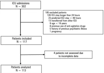
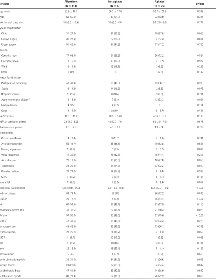
Between April and August 2014, 302 patients were hospitalized at the ICU. Of these, 185 were excluded; the main reasons for exclusion are depicted in igure 1. We included 117 patients, and 4 were not analyzed due to incomplete data collection. hus, our sample consisted of 113 patients. heir main baseline characteristics are described in table 1.
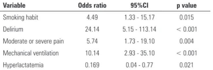
he multivariate analysis included variables with a p ≤ 0.05 in the univariate analysis and those that were considered clinically relevant, namely, smoking, alcoholism, delirium, moderate or severe pain, MV and hyperlactatemia. As observed in table 2, the factors independently associated with a higher incidence of agitation were the presence of delirium, moderate or severe pain, MV, and smoking. he presence of hyperlactatemia remained a protective factor for agitation.
Agitated patients had fewer MV free-days and lower hospital mortality than non-agitated patients (Table 3). However, after adjusting for age and SAPS 3 score, MV free-days remained signiicantly associated with the presence of agitation only, and hospital mortality was no longer signiicant [odds ratio 3.01; CI95% 0.89 - 10.26; p = 0.770].
DISCUSSION
In this study, we found a high incidence of agitation in the irst 7 days of ICU admission. In most cases, patients experienced agitation in the irst 3 days after admission, and the factors associated with its occurrence were the presence of delirium, moderate or severe pain, MV, and a smoking habit. Patients with hyperlactatemia had a lower incidence of agitation. Agitated patients had fewer MV free-days.
he incidence of agitation in our study was lower than those previously reported in similar populations.
Jaber et al. reported an agitation incidence of 52%,(5)
while an even higher incidence (70%) was found by
Fraser et al.(6) A higher incidence has also been reported
in studies including patients under prolonged MV(5) and
in critically ill clinical patients.(9) his variation may be
due to diferences in the criteria used to deine agitation and the use of diferent diagnostic tools, as well as a longer observation period after ICU admission.
As expected, delirium was an independent risk factor for agitation in the irst 7 days of ICU admission. In this time window, delirium occurred in 17.7% of the patients. his incidence was lower than that of other studies in critically ill patients because of our shorter duration of observation. Delirium is a highly prevalent condition in
critically ill patients (20 - 80%).(22-30) Peterson et al.(30)
reported a 71.5% prevalence of delirium, of which 54.9% were mixed type, showing that patients in ICUs frequently
have moments of hyperactivity.(8,29-31)
However, we were able to identify other risk factors for agitation that were not related to the presence of delirium.
Figure 1 - Enrollment flowchart. ICU - intensive care unit.
Table 1 - Characteristics of the study population in the entire group according to agitation status
Variables All patients
(N = 113)
Not agitated (N = 77)
Agitated
(N = 36) p value
Age (years) 55.2 ± 18.7 56.3 ± 17.0 52.7 ± 21.8 0.342
Male 63 (55.8) 40 (51.9) 23 (63.9) 0.234
Prior hospital stays (days) 3.0 (2.0 - 10.5) 3.0 (2.0 - 8.0) 2.0 (2.0 - 6.0) 0.777 Type of hospitalization
Clinic 31 (27.4) 21 (27.3) 10 (27.8) 0.955
Elective surgery 31 (27.4) 22 (28.6) 9 (25.0) 0.691
Urgent surgery 51 (45.1) 34 (44.2) 17 (47.2) 0.760
Location
Operating room 77 (68.1) 51 (66.2) 26 (72.2) 0.524
Emergency room 19 (16.8) 13 (16.9) 6 (16.7) 0.977
Ward 16 (14.2) 13 (16.9) 3 (8.3) 0.225
Other 1 (0.9) 0 1 (2.8) 0.142
Reason for admission
Postoperative monitoring 49 (43.4) 36 (46.8) 13 (36.1) 0.288
Sepsis 16 (14.2) 14 (18.2) 2 (5.6) 0.073
Respiratory failure 11 (9.7) 8 (10.4) 3 (8.3) 0.731
Acute neurological disease* 19 (16.8) 7 (9.1) 12 (33.3) 0.001
Multiple trauma 4 (3.5) 4 (5.2) 0 0.164
Other 14 (12.5) 8 (10.4) 6 (16.7) 0.451
SAPS 3 (points) 44.8 ± 15.2 46.2 ± 14.6 41.6 ± 16.3 0.139 SOFA at admission (points) 2.5 (1.0 - 5.2) 4.0 (2.0 - 7.0) 4.0 (2.0 - 7.0) 0.675 Charlson score (points) 4.0 ± 2.9 4.1 ± 2.8 3.9 ± 3.1 0.719 Comorbidities
Chronic renal failure 14 (12.4) 9 (11.7) 5 (13.9) 0.741 Arterial hypertension 55 (48.7) 36 (46.8) 19 (52.8) 0.551
Hearing impairment 11 (9.7) 5 (6.5) 6 (16.7) 0.089
Visual impairment 41 (36.3) 25 (32.5) 16 (44.4) 0.217
Alcohol abuse 20 (17.7) 10 (13.0) 10 (27.8) 0.055
Tobacco use 23 (20.3) 11 (14.3) 12 (33.3) 0.019
Diabetes mellitus 26 (23.0) 19 (24.7) 7 (19.4) 0.538
COPD 11 (9.7) 7 (9.1) 4 (11.1) 0.736
Severe TBI 11 (9.7) 4 (5.2) 7 (19.4) 0.017
Glasgow at ICU admission 13.5 (10.0 - 14.0) 15.0 (14.0 - 15.0) 13.5 (10.0 - 14.0) < 0.001
Bed clock absent 83 (73.4) 57 (74) 26 (72.2) 0.840
Delirium 20 (17.7) 4 (5.2) 16 (44.4) < 0.001
Pain 60 (53.1) 37 (48.1) 23 (63.9) 0.116
Moderate to severe pain 48 (42.5) 27 (35.1) 21 (58.3) 0.020
MV use† 57 (50.4) 30 (39.0) 27 (75.0) < 0.001
Sepsis 47 (41.6) 35 (45.5) 12 (33.3) 0.223
Vasopressor use† 48 (42.5) 35 (45.5) 13 (36.1) 0.349
Hyperlactatemia 29 (25.7) 24 (31.2) 5 (13.9) 0.050
ARDS 11 (9.7) 10 (13.0) 1 (2.8) 0.088
RRT 11 (9.7) 8 (10.4) 3 (8.3) 0.731
Fever 22 (19.5) 18 (23.4) 4 (11.1) 0.125
Pressure ulcers 5 (4.4) 4 (5.2) 1 (3.2) 0.660
Family absent during visits 35 (31.0) 24 (31.2) 11 (30.6) 0.948 Invasive devices 106 (93.8) 72 (93.5) 34 (94.4) 0.847 Anticholinergic drugs 47 (41.6) 33 (42.9) 14 (38.9) 0.690 Sedatives and opioids 83 (73.4) 57 (76.0) 26 (72.2) 0.668
Table 2 - Risk factors for agitation in intensive care unit patients - multivariate analysis
Variable Odds ratio 95%CI p value
Smoking habit 4.49 1.33 - 15.17 0.015
Delirium 24.14 5.15 - 113.14 < 0.001
Moderate or severe pain 5.74 1.73 - 19.10 0.004
Mechanical ventilation 10.14 2.93 - 35.10 < 0.001
Hyperlactatemia 0.169 0.04 - 0.77 0.021
95%CI - 95% confidence interval. Backward stepwise selection procedure was used for the logistic regression - likelihood ratio. Hosmer and Lemeshow test: p = 0.102.
Table 3 - Hospital outcomes according to agitation status
Variables Not agitated
(N = 77)
Agitated
(N = 36) p value
ICU-free days in 28 days 22.0 (11.5 - 24.5) 20.0 (12.0 - 23.0) 0.226 Hospital-free days in 28 days 9.0 (0 - 19.0) 11.0 (0 - 18.7) 0.228 MV-free days in 7 days 7.0 (3.5 - 7.0) 5.0 (1.2 - 6.7) 0.003 Vasopressor-free days in 7 days 7.0 (5.0 - 7.0) 7.0 (5.0 - 7.0) 0.495
ICU mortality 13 (17.1) 3 (8.3) 0.215
Hospital mortality 21 (28.4) 4 (11.1) 0.043
ICU - intensive care unit; MV - mechanical ventilation. Results are expressed as the number
(%), mean ± standard deviation or median (25% - 75%). Chi-square or Student’s t-tests
were used as appropriate.
his is a relevant inding, as a misdiagnosis of delirium can lead to inadequate treatment for both the underlying cause and for delirium itself. A previous habit of smoking is recognized as a risk factor for agitation, given the risk
of withdrawal syndrome.(32,33) Lucidarme et al.,(32) in a
study that included predominantly critically ill medical patients, showed that smokers had a higher incidence of agitation than non-smokers. Moderate or severe pain was more common among agitated patients. he majority of our studied patients were surgical (72.5%), which means that they had high exposures to pain in the irst 7 days of observation. Previous studies that showed an association between pain and agitation did not assess whether the
patient’s pain occurred before agitation.(13,34-38) In our
study, we clearly showed that pain is a risk factor of agitation, as only episodes occurring before agitation were considered. MV was also associated with a higher risk of
agitation, as previously reported by Woods et al.(9) Potential
reasons for this association include the presence of the endotracheal tube, respiratory secretions and asynchrony with the ventilator. Patients under MV might not be able to communicate their needs to the healthcare team. he inability to communicate has previously been described
as a risk factor for agitation.(11) In our unit, sedation was
maintained as minimal as possible. Our inding suggests that the current no sedation or minimal sedation protocols
need to also include a frequent assessment of pain and discomfort among patients using endotracheal tubes
and MV.(39)
An unexpected inding was the lower incidence of agitation among patients with hyperlactatemia. Although we did not assess the potential mechanisms associated with this relationship, we can hypothesize that patients who develop tissue dysoxia may be more severely ill than
those without signs of abnormal cellular metabolism.(18)
More severe patients might require continuous long-term sedation, which can contribute to a lower incidence of
agitation.(11) Another potential reason is the presence of
neurological impairment or renal or hepatic dysfunction that could lead to a reduction in the level of consciousness, limiting the occurrence of agitation. he presence of neuromuscular weakness might also limit the clinical manifestation of agitation.
We were unable to show an association between age and agitation. Although age has been considered a risk factor for agitation, recent prospective studies have shown that age is a protective factor.(5,6,9) As delirium is frequent among agitated patients and among the elderly, it is possible that the prevalence of the hypoactive subtype among patients older than 65 years inluences the potential association
between age and agitation.(30) We were also not able to
show an association between alcohol abuse and agitation. his relationship was expected, as abstinence is a well-known risk factor for agitation. he lack of association might be a consequence of the low prevalence of alcohol abuse among our patients.
Similar to other studies,(5,9) agitated patients had a
longer duration of MV in the irst 7 days, although no
diference was found in hospital mortality.(5,9) We were
unable to show an association between agitation and increased use of sedatives or higher severity of illness, which could possibly explain this inding. However, we can hypothesize that being agitated might have precluded
attempts to discontinue MV, as suggested by others.(1)
excluded because they had been admitted for more than 24 hours, mostly on the weekends when the study team was not always available. his also led to a high incidence of missing data among the included patients. hird, the high frequency of MV also compromised the pain and delirium assessments. Fourth, we did not collect data on the presence of agitation during the patients’ entire ICU stay, which may have reduced our incidence of agitation. We also prospectively evaluated the presence of agitation only twice per day. he assessment of the entire day was conducted in a retrospective manner, and cases might have been missed. Additionally, we used the administration of antipsychotic drugs to deine the presence of agitation. Although the use of these drugs is well controlled in our unit, misuse for other indications might have occurred. Finally, we did not collect data on agitation treatment,
which might have inluenced the outcome. However, this was not one of our objectives.
he results reinforce the fact that in addition to delirium, there are other independent risk factors for agitation among ICU patients. Good care practices, sedation, analgesia, and management of MV could reduce the incidence of agitation and provide beneits to patients admitted to the ICU.(11,20,40-45)
CONCLUSION
Agitation in the irst 7 days of intensive care unit admission was common. he incidence of delirium, moderate or severe pain, mechanical ventilation, and smoking were independent risk factors for the development of agitation. he presence of agitation was associated with fewer mechanical ventilation-free days.
Objetivo: Avaliar a incidência de agitação nos primeiros 7 dias após admissão à unidade de terapia intensiva, seus fatores de risco e associação com desfechos clínicos.
Métodos: Estudo de coorte unicêntrico prospectivo
que incluiu maiores 18 anos, admitidos à unidade de terapia intensiva há menos de 24 horas e com previsão de permanência superior a 48 horas. Agitação psicomotora foi deinida como pontuação igual ou superior a +2 na Escala de Agitação e Sedação de Richmond ou episódio de agitação, ou registro de uso de medicação especíica na icha clínica.
Resultados: Ocorreu agitação em 31,8% dos 113 pacientes incluídos. Na análise multivariada, delirium (OR = 24,14; IC95% 5,15 - 113,14; p < 0,001), dor moderada ou intensa (OR = 5,74; IC95% 1,73 - 19,10; p = 0,004), ventilação
mecânica (OR = 10,14; IC95% 2,93 - 35,10; p < 0,001) e tabagismo (OR = 4,49; IC95% 1,33 - 15,17; p = 0,015) foram independentemente associados a maior risco de desenvolver de agitação. Por outro lado, hiperlactatemia associou-se a um menor risco de ocorrência de agitação (OR = 0,169; IC95% 0,04 - 0,77; p = 0,021). Pacientes agitados tiveram menor tempo livre de ventilação mecânica em 7 dias (p = 0,003).
Conclusão: A incidência de agitação nos 7 primeiros dias de internação em unidade de terapia intensiva foi elevada. Delirium, dor moderada ou intensa, ventilação mecânica e tabagismo foram fatores de risco independentes para o desenvolvimento de agitação. Pacientes agitados tiveram menor tempo livre de ventilação mecânica nos 7 primeiros dias.
RESUMO
Descritores: Agitação psicomotora; Fatores de risco;
Delirium; Dor; Respiração artiicial; Cuidados intensivos
REFERENCES
1. Chevrolet JC, Jolliet P. Clinical review: agitation and delirium in the critically ill--significance and management. Crit Care. 2007;11(3):214.
2. Crippen D. Agitation in the ICU: part one. Anatomical and physiologic basis for the agitated state. Crit Care. 1999;3(3):R35-46.
3. Zeller SL, Rhoades RW. Systematic reviews of assessment measures and pharmacologic treatments for agitation. Clin Ther. 2010;32(3):403-25. 4. Lindenmayer JP. The pathophysiology of agitation. J Clin Psychiatry.
2000;61 Suppl 14:5-10.
5. Jaber S, Chanques G, Altairac C, Sebbane M, Vergne C, Perrigault P, et al. A prospective study of agitation in a medical-surgical ICU: incidence, risk factors, and outcomes. Chest. 2005;128(4):2749-57.
6. Fraser GL, Prato BS, Riker RR, Berthiaume D, Wilkins ML. Frequency, severity, and treatment of agitation in young versus elderly patients in the ICU. Pharmacotherapy 2000;20(1):75-82.
7. Tramm R, Hodgson C, Ilic D, Sheldrake J, Pellegrino V. Identification and prevalence of PTSD risk factors in ECMO patients: A single center study. Aust Crit Care. 2015;28(1):31-6
8. Ely EW, Shintani A, Truman B, Speroff T, Gordon SM, Harrell FE Jr, et al. Delirium as a predictor of mortality in mechanically ventilated patients in the intensive care unit. JAMA. 2004;291(14):1753-62.
10. Reade MC, O’Sullivan K, Bates S, Goldsmith D, Ainslie WR, Bellomo R. Dexmedetomidine vs. haloperidol in delirious, agitated, intubated patients: a randomised open-label trial. Crit Care. 2009;13(3):R75.
11. Barr J, Fraser GL, Puntillo K, Ely EW, Gélinas C, Dasta JF, Davidson JE, Devlin JW, Kress JP, Joffe AM, Coursin DB, Herr DL, Tung A, Robinson BR, Fontaine DK, Ramsay MA, Riker RR, Sessler CN, Pun B, Skrobik Y, Jaeschke R; American College of Critical Care Medicine. Clinical practice guidelines for the management of pain, agitation, and delirium in adult patients in the intensive care unit. Crit Care Med. 2013;41(1):263-306. 12. Capone Neto A, Dalfior Jr L. Fatores de risco. In: Flores DG, Capone Neto
A. Delirium no paciente grave. São Paulo: Atheneu; 2013. p. 53-9. 13. O’Connor H, Al-Quadheeb NS, White AC, Thaker V, Devlin, JW. Agitation
during prolonged mechanical ventilation at a long-term acute care hospital: risk factors, treatments and outcomes. J Intensive Care Med. 2014;29(4):218-24.
14. Ruokonen E, Parviainen I, Jakob SM, Nunes S, Kaukonen M, Shepherd ST, Sarapohja T, Bratty JR, Takala J; “Dexmedetomidine for Continuous Sedation” Investigators. Dexmedetomidine versus propofol/midazolam for long-term sedation during mechanical ventilation. Intensive Care Med. 2009;35(2):282-90.
15. Xia ZQ, Chen SQ, Yao X, Xie CB, Wen SH, Liu KX. Clinical benefits of dexmedetomidine versus propofol in adult intensive care unit patients: a meta-analysis of randomized clinical trials. J Surg Res. 2013;185(2):833-43.
16. Wan RY, Kasliwal M, McKenzie CA, Nicholas NA. Quetiapine in refractory hyperactive and mixed intensive care delirium: a case series. Crit Care. 2011;15(3):R159.
17. Sessler CN, Gosnell MS, Grap MJ, Brophy GM, O’Neal PV, Keane KA, et al. The Richmond Agitation-Sedation Scale: validity and reliability in adult intensive care unit patients. Am J Respir Crit Care Med. 2002;166(10):1338-44.
18. Vincent JL, Moreno R, Takala J, Willats S, De Mendonça A, Bruining H, et al. The SOFA (Sepsis-related Organ Failure Assessment) score to describe organ dysfunction/failure. On behalf of the Working Group on Sepsis - Related Problems of the European Society of Intensive Care Medicine. Intensive Care Med. 1996;22(7):707-10.
19. Pessoa RF, Nácul FE. [Delirium in the critically ill patient]. Rev Bras Ter Intensiva. 2006;18(2):190-5. Portuguese. [Delirium in the critically ill patient].
20. Campbell N, Perkins A, Hui S, Khan B, Boustani M. Association between prescribing of anticholinergic medications and incident delirium: a cohort study. J Am Geriatr Soc. 2011;59 Suppl 2:S277-81.
21. Bone RC, Balk RA, Cerra FB, Dellinger RP, Fein AM, Knaus WA, Schein RM, Sibbald WJ. Definitions for sepsis and organ failure and guidelines for the use of innovative therapies in sepsis. The ACCP/SCCM Consensus Conference Committee. American College of Chest Physicians/Society of Critical Care Medicine. Chest. 1992;101(6):1644-55. Review.
22. Ferguson ND, Fan E, Camporota L, Antonelli M, Anzueto A, Beale R, et al. The Berlin definition of ARDS: an expanded rationale, justification, and supplementary material. Intensive Care Med. 2012;38(10):1573-82. 23. Lwanga SK, Lemeshow S. Sample size determination in health studies: a
practical manual. Geneva: World Health Organization; 1991.
24. Salluh JI, Soares M, Teles JM, Ceraso D, Raimondi N, Nava VS, Blasquez P, Ugarte S, Ibanez-Guzman C, Centeno JV, Laca M, Grecco G, Jimenez E, Árias-Rivera S, Duenas C, Rocha MG; Delirium Epidemiology in Critical Care Study Group. Delirium epidemiology in critical care (DECCA): an international study. Crit Care. 2010;14(6):R210.
25. Khan BA, Zawahiri M, Campbell NL, Fox GC, Weinstein EJ, Nazir A, et al. Delirium in hospitalized patients: implications of current evidence on clinical practice and future avenues for research--a systematic evidence review. J Hosp Med. 2012;7(7):580-9.
26. Chorney SR, Gooch ME, Oberdier MT, Keating D, Stahl RF. The safety and efficacy of dexmedetomidine for postoperative sedation in the cardiac surgery intensive care unit. HSR Proc Intensive Care Cardiovasc Anesth. 2013;5(1):17-24.
27. Inouye SK, Westendorp RG, Saczynski JS. Delirium in elderly people. Lancet. 2014;383(9920):911-22. Review.
28. Bryczkowski SB, Lopreiato MC, Yonclas PP, Sacca JJ, Mosenthal AC. Risk factors for delirium in older trauma patients admitted to the surgical intensive care unit. J Trauma Acute Care Surg. 2014;77(6):944-51. 29. Ouimet S, Kavanagh BP, Gottfried SB, Skrobi Y. Incidence, risk factors and
consequences of ICU delirium. Intensive Care Med. 2007;33(1):66-73. 30. Peterson JF, Pun BT, Dittus RS, Thomason JW, Jackson JC, Shintani AK, et
al. Delirium and its motoric subtypes: a study of 614 critically ill patients. J Am Geriatr Soc. 2006;54(3):479-84.
31. Blazer DG, Nieuwenhuizen AO. Evidence for the diagnostic criteria of delirium: an update. Curr Opin Psychiatry. 2012;25(3):239-43.
32. Lucidarme O, Seguin A, Daubin C, Ramakers M, Terzi N, Beck P, et al. Nicotine withdrawal and agitation in ventilated critically ill patients. Crit Care. 2010;14(2):R58.
33. Cohen IL, Gallagher TJ, Pohlman AS, Dasta, JF, Abraham E, Papadokos PJ. Management of the agitated intensive care unit patient. Crit Care Med. 2002;30(1):S97-123.
34. Luk E, Sneyers B, Rose L, Perreault MM, Williamsom DR, Mehta S, et al. Predictors of physical restraint use in Canadian intensive care units. Crit Care. 2014;18(2):R46.
35. Erstad BL, Puntillo K, Gilbert HC, Grap MJ, Li D, Medina J, et al. Pain management principles in the critically ill. Chest 2009 Apr;135(4):1075-86.
36. Williamson A, Hoggart B. Pain: a review of three commonly used pain rating scales. J Clin Nurs. 2005;14(7):798-804.
37. Park JM, Kim JH. Assessment and treatment of pain in adult intensive care unit patients. Korean J Crit Care Med. 2014;29(3):147-59.
38. Rotondi AJ, Chelluri L, Sirio C, Mendelsohn A, Schulz R, Belle S, et al. Patients’ recollections of stressful experiences while receiving prolonged mechanical ventilation in an intensive care unit. Crit Care Med. 2002;30(4):746-52.
39. Shehabi Y, Bellomo R, Reade MC, Bailey M, Bass F, Howe B, McArthur C, Murray L, Seppelt IM, Webb S, Weisbrodt L; Sedation Practice in Intensive Care Evaluation Study Investigators; Australian and New Zealand Intensive Care Society Clinical Trials Group. Early goal-directed sedation versus standard sedation in mechanically ventilated critically ill patients: a pilot study. Crit Care Med. 2013;41(8):1983-91.
40. Holloman GH Jr, Zeller SL. Overview of Project BETA: Best practices in evaluation and treatment of agitation. West J Emerg Med. 2012;13(1):1-2.
41. Van Rompaey B, Elseviers MM, Schuurmans MJ, Shortridge-Baggett LM, Truijen S, Bossaert L. Risk factors for delirium in intensive care patients: a prospective cohort study. Crit Care. 2009;13(3):R77.
42. Teitelbaum JS, Ayoub O, Skrobik Y. A critical appraisal of sedation, analgesia and delirium in neurocritical care. Can J Neurol Sci. 2011;38(6):815-25. 43. Ely EW, Truman B, Shintani A, Thomason JW, Wheeler AP, Gordon S,
et al. Monitoring sedation status over time in ICU patients: reliability and validity of the Richmond Agitation-Sedation Scale (RASS). JAMA. 2003;289(22):2983-91.
44. Zhang H, Lu Y, Liu M, Zou Z, Wang L, Xu FY, et al. Strategies for prevention of postoperative delirium: a systematic review and meta-analysis of randomized trials. Crit Care. 2013;17(2):R47.