RBCCV 44205-1485 DOI: 10.5935/1678-9741.20130059
Assessment of sternal closure using titanium plate
Avaliação do fechamento esternal com placa de titânio
Renato Tambellini Arnoni
1, MD, PhD; Daniel Chagas Dantas
1, MD; Antoninho Arnoni
1, MD;
Caetano Nigro Neto
1, MD; Camilo Abdulmassih Neto
1, MD
1 Instituto Dante Pazzanese de Cardiologia (IDPC), São Paulo, SP, Brazil.
Work carried out at Instituto Dante Pazzanese de Cardiologia (IDPC), São Paulo, SP, Brazil.
Correspondence address: Renato Tambellini Arnoni
Av. Dr. Dante Pazzanese, 500 – Vila Mariana, São Paulo, SP, Brazil Zip code: 04012-180
E-mail [email protected]
Article received on February 2nd, 2013
Article accepted on April 28th, 2013
Abstract
Introduction: The use of plates and screws for more
rig-id ixation of the sternum, without maintaining contact be -tween the upper portion of the sternum and mediastinum.
The present study seeks new choice of plate with a signiicant difference, the same does not need to be removed in order to
proceed to open when necessary sternal emerging opening of
the bone.
Objective: The current study aims to evaluate the eficacy
and safety of this procedure.
Methods: To this end, we selected ten patients with coronary
artery disease have shown no signiicant risk factors for medias -tinitis. The surgery was thus performed in the usual way that all patients with coronary artery disease surgeries are done at the institution. Only at the time of sternal closure is that there was a
change, with the combination of steel wires and plates.
Results: All cases had sternal closure properly with good outcome in the medium term.
Conclusion: The use of plates ENGIMPLAN proved safe
and effective for sternal closure.
Descriptors: Coronary Artery Bypass. Titanium. Sternum. Bone Plates. Bone Screws.
Resumo
Introdução: A utilização de placas e parafuso para a mais
rígida ixação do esterno, sem manter contato entre a porção superior do esterno e o mediastino. O estudo atual busca nova opção de placa, com um diferencial importante; a mesma não precisa ser retirada para que se proceda à abertura esternal em caso de necessidade emergente de abertura do osso.
Objetivo: O presente estudo tem por objetivo avaliar a eicá -cia e a segurança de tal procedimento.
Métodos: Para tal, foram selecionados dez pacientes porta
-dores de doença arterial coronária que não apresentassem im
-portantes fatores de risco para mediastinite. As cirurgias foram, portanto, realizadas da maneira habitual, a todas os procedi -mentos em portadores de coronariopatias são feitas na
Institui-ção. Somente no momento do fechamento esternal é que houve uma modiicação, com a associação de ios de aço e placas.
Resultados: Todos os casos apresentaram fechamento
ester-nal de forma adequada com boa evolução a médio prazo.
Conclusão: O emprego das placas ENGIMPLAN se
mos-trou seguro e eicaz no fechamento esternal.
Descritores: Esterno. Revascularização Miocárdica. Titânio.
previously in oral maxillofacial surgery, have been already used for sternal osteosynthesis [6]. This study demonstrated a lower incidence of mediastinitis in high-risk patients when using plates for sternal closure.
Several steps were followed to the present time that allowed the possibility of using implant plates in patients undergoing myocardial revascularization in the Institute Dante Pazzanese Cardiology: 1) In vitro study of screws and plates; 2) To study the placement of plates and screws in corpses. The current study aims to assess the eficacy and safety of this procedure in humans.
METHODS
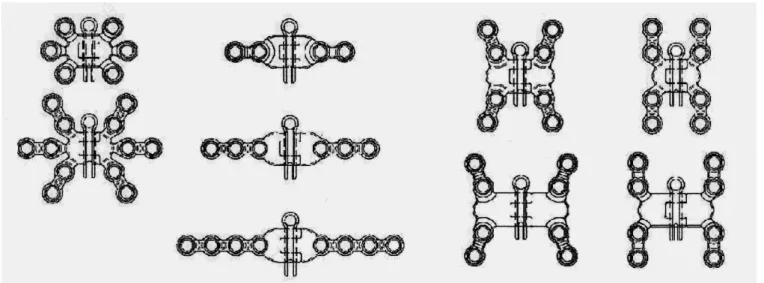
To develop this new device, some materials were produced to assist their implantation, such as: screwdrivers, fasteners, drill bits and stabilizers plates. Plates and screws used for sternal closure have various designs. The current study seeks new board option, with a signiicant difference; it does not need to be removed to proceed with the sternal opening in case of emergent need of opening the bone. The great advantage of this material is its design and the various models of plates, giving the possibility to the surgeon to choose the one that best suits the situation required.
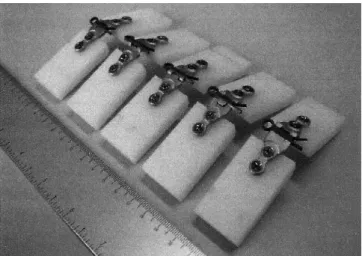
Moreover, the central pin allows opening the sternum, without removing the plate in case of surgical reoperation (Figure 1). It was initially implanted “in vitro” (corpse) by assessing resistance and strength of the plates (Figure 2). It was performed the opening and closing of the corpses sternum with test pins and proper sternal ixation. After these tests the material was made available to perform procedures in patients with coronary artery disease who required surgical treatment. The study was submitted to the Research Ethics INTRODUCTION
Cardiac surgery has been performed mostly through median sternotomy since 1957, when Julian re-introduced the technique previously established by Milton. in 1897 [1]. In 2001, approximately 760,000 surgeries with this type of incision were performed in the United States [2]. Despite its good exposure, it is not free of complications. Mediastinitis is a serious complication that appears with a mortality of about 15% [3]. At Institute Dante Pazzanese of Cardiology, about 1,600 cardiac surgeries are performed per year with an infection rate of 6%. Some methods to prevent mediastinitis after sternotomy included prophylactic methods like antibiotics and special techniques to skin closure, but little has been studied in the prevention of the infectious process by better bone ixation [4]. The current technique for sternal osteosynthesis is to ix the bone with steel wire that involves and approaches both sides of the bone. This procedure, usual in our country, enables the progression of the process of the upper mediastinum into the mediastinal cavity, which is ineffective for the ixation of the sternum [5].
Several new ixing methods have been studied for more rigid ixation of the sternum, among them the use of plates and screws without maintaining contact between the upper portion of the sternum and mediastinal cavity. These boards used
Abbreviations, acronyms & symbols
BMI Body mass index
CONEP Comissão Nacional de Ética em Pesquisa/ National Committee for Research Ethics
EuroSCORE European System for Cardiac Operative Risk Evaluation STS Society of Thoracic Surgeons
Committee under number 3823, approved by the institutional Ethics Committee and CONEP (Comissão Nacional de Ética em Pesquisa/ National Committee for Research Ethics) on 02/03/2010.
After obtaining written informed consent we selected ten patients with coronary artery disease, which had no signiicant risk factors for mediastinitis. It was excluded patients with some comorbidities: 1) Obesity [body mass index (BMI) greater than 30], 2) Elderly (over 75 years), 3) Chronic kidney disease, 4) Diabetes mellitus, 5) Chronic pulmonary obstructive disease, 6) Bilateral internal thoracic artery used as a graft. Underwent surgery ten patients, aged between 46 and 66 years, mean 56 years. The mean body mass index was 27.5 kg/m², ranging 25-30. The patients’ past medical history were restricted to hypertension (present in all). Regarding the context of heart disease, three patients had acute myocardial infarction during the past 30 days, and the remaining patients had stable angina. The study showed low-risk score to all patients; assessed both by EuroSCORE and the Society of Thoracic Surgeons (STS). The irst between zero and three that corresponds to a likelihood of death around 1.43%, and 1.68% to the second.
All surgeries were performed in patients requiring coronary artery bypass grafting with cardiopulmonary bypass with myocardial protection achieved by intermittent aortic clamping. The mean duration of cardiopulmonary bypass was 71.7 minutes (55-80), while the anoxic time ranged between 45 and 65 minutes, averaging 52.4 minutes. The surgeries were performed as the same way as it always be done to all coronary surgeries performed in the institution. Only the time of sternal closure was changed, with the combination of metal wires and plates. Plates were selected according to the preference of the surgeon; steel wires used in “X” or in simply wrap, reinforcing bone ixation. The following table shows how the plates and screws were employed (Table 1).
RESULTS
Revascularization of the affected vessels by coronary artery disease was performed using the left internal thoracic artery to the left anterior descending artery and saphenous vein grafts to the other vessels, the average number of bridges performed per patient was 3.2 bridges, with cases ranging three to four bridges. Sternal closure, as already reported, was performed with plates and screws (Table 1) according to the preference of the surgical team to maintain greater stability of the sternum. Patients were followed-up, without presenting any complications regarding coronary artery disease, all being asymptomatic at the standpoint. The median follow-up was 5.7 months. Regarding the closure, only one patient had wound dehiscence requiring surgical reoperation. This patient had an instable bone ixation with apparent poor coaptation of the bone plate. One of the bolts was not tight. We opted to remove the material synthesis, culture collection and closure with steel wires. Cultures were negative, patient improved after resuture and discharge was possible after a week. This patient is in the Fig. 2 - "In vitro”- straight plate undergone endurance test
Fig. 3 – Fixing the sternum - Image of the plates little star and “H” narrow
Table 1. How to use the plates.
Patient
1 2 3 4 5 6 7 8 9 10
Plate
"H" narrow plate 4 X 4 / Straight plate 2 X 2 "H" narrow plate 4 X 4 / Star plate 3 X 3 "H" narrow plate 4 X 4 / Star plate 3 X 3 "H" narrow plate 4 X 4 / Star plate 3 X 3 "H" narrow plate 4 X 4 / Star plate 3 X 3 "H" narrow plate 4 X 4 / Star plate 3 X 3 "H" narrow plate 4 X 4 / Star plate 3 X 3 "H" narrow plate 4 X 4 / Star plate 3 X 3 "H" narrow plate 4 X 4 / Star plate 3 X 3 "H" narrow plate 4 X 4 / Star plate 3 X 3
"X" shaped steel wire
REFERENCES
1. Dalton ML, Connally SR, Sealy WC. Julian’s reintroduction of Milton’s operation. Ann Thorac Surg. 1992;53(3):532-3.
2. American Heart Association Statistics for Cardiac Procedures 2001; Available from: www.americanheart.org
3. Gummert JF, Barten MJ, Hans C, Kluge M, Doll N, Walther T, et al. Mediastinits and cardiac surgery: an updated risk factors analysis in 10,373 consecutive adult patients. Thorac Cardiovasc Surg. 2002;50(2):87-91.
ifth month post-resuture with favorable clinical course without further complications. The other patients had no complications regarding the synthesis material, which was effective and safe for the ixation of the sternum (Figures 3 and 4).
DISCUSSION
Cardiac surgery has had a major breakthrough in recent years, but the access road trough median sternotomy and its synthesis are almost the same with standard steel wire that is adopted by most centers. This has a non-negligible rate of failure from fatigue of the wires, bone fractures, allowing sternal instability, dehiscence, infection, leading patients to hospitalization and prolonged antibiotic therapy with procedures dressings, debridement and resutures [3,7-9]. Taking as a basis for a good sternal healing and stability, material synthesis with titanium plates and screws is an alternative technique, already in wide use in orthopedic procedures and other cardiac surgery centers primarily in Europe, with proven results [6,8,10,11].
The present study, using plates and screws with domestic technology for its manufacture, aimed to prove the safety and practicality of the method, a lower risk of bleeding than with sternotomy wires and runtime similar after the initial learning curve. Being an initial treatment of new implantable devices, the study began with the evaluation “in vitro” of plates and screws. All models were tested with regard to static bending test (plates) and static test of irmness (screws). The plates are made using a titanium alloy ASTM F 136, as well as screws. All “in vitro” of this innovative material showed great resistance to bending of the plate, as well as secure ixation of the screws.
We proved good results in monitoring all patients in this series from the immediate postoperative period until the Fig. 4 - Postoperative radiological picture - Chest X-ray
sixth month postoperatively, clinically and with chest x-ray. Two steel wires in the upper and lower ends, had to be used only to join the loorboards and facilitate the ixation of the bone plate. An important issue tested was the easy removal of the “pin” that joins the two sides of the plate for a possible need of reoperation postoperatively. This fact allows not to remove the plate already ixed to the bone, and streamlines the reoperation. It also provides that these patients can be re-operated on another center that does not have special materials for implantation and removal of plates and screws. The card can be opened and closed again using a Kelly clamp.
We had one case requiring reoperation even in the initial stage of the series, probably due to deiciency in the technique of ixing the board. A difference was observed in the absence of fractures and preserving the integrity of the sternum. Another important issue that emerges is the cost of the method, not assessed in this series. Based on the good results, we planned a randomized study, between standard closing with steel wires and synthesis with plates and screws, with larger number of patients at high risk of postoperative sternal dehiscence, where in addition to compare the results of two techniques, we will look at the cost effectiveness of the method.
CONCLUSION
The use of ENGIMPLAN boards proved to be safe and effective in sternal closure. Further randomized studies should be done comparing the employment of this material and other techniques like steel wires in patients at high risk for mediastinitis to bring conirmations about the safety of this form of sternal closure.
Authors’ roles & responsibilities
4. Krischak GD, Janousek A, Wolf S, Augat P, Kinzl L, Claes LE. Effects of one-plane and two-plane external ixation on sheep osteotomy healing and complications. Clin Biomech (Briston Avon). 2002;17(6):470-6.
5. Trumble DR, McGregor WE, Magovern JA. Validation of a bone analog model for studies of sternal closure. Ann Thorac Surg. 2002;74(3):739-44.
6. Song DH, Lohman RF, Renucci JD, Jeevanandam V, Raman J. Primary sternal plating in high-risk patients prevents mediastinitis. Eur J Cardiothorac Surg. 2004;26(2):367-72.
7. Jutley RS, Shepherd DE, Hukins DW, Jeffrey RR. Preliminary evaluation of the Sternum Screw: a novel method for improved sternal closure to prevent dehiscence. Cardiovasc Surg. 2003;11(1):85-9.
8. Schimmer C, Reents W, Berneder S, Eigel P, Sezer O, Scheld H, et al. Prevention of sternal dehiscence and infection in high-risk patients: a prospective randomized multicenter trial. Ann Thorac Surg. 2008;86(6):1897-904.
9. Casha AR, Gauci M, Yang L, Saleh M, Kay PH, Cooper GJ. Fatigue testing median sternotomy closure. Eur J Cardiothorac Surg. 2001;19(3):249-53.
10. Hirose H, Yamane K, Youdelman BA, Bogar L, Diehl JT. Rigid sternal ixation improves postoperative recovery. Open Cardiovasc Med J. 2011;5:148-52.