Short-term immunosuppressive therapy does not affect the
density of the pre-existing bone around titanium implants
placed in rabbits
Terapia imunossupressora não afeta a densidade do osso
preexistente ao redor de implantes de titânio inseridos em coelhos
Poliana Mendes Duarte*
Getúlio Rocha Nogueira Filho** Enilson Antônio Sallum** Antonio Wilson Sallum***
Francisco Humberto Nociti Júnior**
ABSTRACT:The aim of this study was to evaluate the influence of the administration and withdrawal of cyclosporin A/nifedipine on the bone density in a lateral area adjacent to implants placed in rabbits. Two screw-type titanium im-plants were placed bilaterally in twenty-eight New Zealand rabbits. The animals were assigned to one of the following groups and received daily subcutaneous injections for 14 days: Groups A and C: vehicle (dimethyl sulfoxide); Groups B and D: CsA (10 mg/kg) plus nifedipine (50 mg/kg). The animals in Groups A and B were sacrificed 14 days postopera-tively and, in Groups C and D, 42 days postoperapostopera-tively. After sacrifice, the tibiae were removed and undecalcified sec-tions were obtained. Bone density was obtained in a 500mm-wide zone lateral to the implant surface. Intergroup anal-ysis showed no significant difference (p > 0.05) in the degree of bone density between control and test groups either on day 14 or on day 42. Thus, it appears that a short-term immunosuppressive therapy may not present a negative influ-ence on the density of the pre-existing bone around titanium implants placed in rabbits.
DESCRIPTORS:Titanium; Osseointegration; Cyclosporine; Nifedipine; Dental implants.
RESUMO:O objetivo deste trabalho foi avaliar a influência da administração de ciclosporina A (CsA)/nifedipina e sua interrupção na densidade óssea em uma região lateral à superfície de implantes de titânio inseridos em coelhos. Dois implantes de titânio comercialmente puros foram inseridos bilateralmente em vinte e oito coelhos. Os animais foram aleatoriamente divididos em um dos seguintes grupos experimentais, recebendo injeções diárias subcutâneas por 14 dias: Grupos A e C: veículo (dimetil sulfóxido); Grupos B e D: CsA (10 mg/kg) e nifedipina (50 mg/kg). Os animais pertencentes aos Grupos A/B e C/D foram sacrificados 14 e 42 dias após a colocação dos implantes, respectivamente. Após o sacrifício, as tíbias foram removidas para a obtenção de secções não-descalcificadas. A densidade óssea foi ob-tida em uma zona de 500mm lateral à superfície do implante através de análise histométrica. A análise intergrupo não revelou diferenças para os grupos teste e controle em 14 e 42 dias (p > 0,05). Dentro dos limites deste estudo podemos concluir que a associação CsA/nifedipina, administrada em um curto prazo, não apresenta uma influência negativa na densidade do osso preexistente ao redor de implantes de titânio inseridos em coelhos.
DESCRITORES:Titânio; Osseointegração; Ciclosporina; Nifedipina; Implantes dentários.
*PhD Student; **Assistant Professors; ***Chairman, Professor – Department of Prosthodontics and Periodontics, Division of Perio-dontics, School of Dentistry of Piracicaba, State University of Campinas.
INTRODUCTION
The use of titanium endosseous dental im-plants has been described for the treatment of edentulous or partially edentulous patients in or-der to restore function and esthetics3,24. However,
the success of osseointegration depends on the state of the host bone and its healing capacity. Thus, some local and systemic conditions may
im-pair bone healing or may interfere with the mainte-nance of osseointegration10
.
the treatment of autoimmune disorders such as psoriasis, atopic dermatitis, rheumatoid arthritis and uveits17
. Its major effect on the immune sys-tem is inhibition of T-cell action and proliferation, thereby altering the T-cell-mediated immune re-sponse. It has also been shown to affect a subset of B cells22
.
In addition to its effects on the immune system, CsA has also been associated with deleterious ef-fects upon bone metabolism16
. Nevertheless, de-spite its many therapeutic uses and its efficient prevention of organ rejection25
, side-effects are not infrequent. Post-transplantation osteoporosis4,15
, bone formation and bone resorption are well-rec-ognized side-effects5,6,14,19,27
. Moreover, CsA admin-istration has been associated with gingival over-growth and nephrotoxicity17
.
In order to control hypertension and reduce cyclosporin-induced nephrotoxicity11
, patients are frequently given nifedipine, a calcium-channel blocker (CCBs). The effect of CCBs appears not to be limited only to the cardiovascular system, but also to calcium metabolism. Similarly to CsA, nifedipine has demonstrated some effects on bone metabolism9,29
.
Therefore, the present study was designed to evaluate, by histological analysis, whether short-term CsA/nifedipine administration influences bone density, in a pre-existing bone area, around titanium implants placed in the tibiae of rabbits. In addition, the study also aims to investigate whether recovery is possible following administra-tion withdrawal.
MATERIAL AND METHODS
Animals
Twenty-eight male New Zealand rabbits (3,000 to 3,500 g), aged 9 to 12 months, were used in the entire study. The animals were kept in individual cages with access to food and water ad libitum.
Prior to the surgical procedures, all animals were allowed to acclimatize to the laboratory environ-ment for a period of 7 days. The protocol was ap-proved by the University of Campinas Institutional Animal Care and Use Committee.
Implant surgery
At the beginning of the experiment, general an-esthesia was performed using intramuscular ad-ministration of ketamine (0.5 ml/kg). The tibiae skin was shaved and disinfected with iodine surgi-cal soap. A 30 mm incision was made and the bone
surface of the tibiae surgically exposed by blunt dissection. Under profuse saline solution irriga-tion, unicortical implant beds were drilled and two screw-shaped commercially available pure tita-nium implants (7.0 mm in length and 3.75 mm in diameter) were placed bilaterally until the screw thread had been completely introduced into the bone cortex. Finally, soft tissues were replaced and sutured. Postoperatively, the animals received an-tibiotic (Pentabiótico®
, Wyeth-Whitehall Ltda., São Paulo, SP, Brazil) given as a single intramuscular injection.
Experimental design
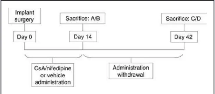
Immediately after implant placement, the ani-mals were randomly assigned to one of the follow-ing four treatment groups (7 animals/group) and received daily subcutaneous injections for 14 days: Groups A and C were injected with a vehi-cle (dimethyl sulfoxide) and groups B and D re-ceived CsA (Sandimmun®
, Novartis Pharma AG, Switzerland) (10 mg/kg) plus nifedipine (Sigma, St. Louis, MO, USA) (50 mg/kg). Groups A and B were sacrificed 14 days postoperatively and Groups C and D were sacrificed 42 days postoper-atively (Figure 1).
Histometric procedures
After sacrifice, the tibiae were removed and fixed in 4% neutral formalin for 48 hours. Undecalcified sections were prepared as previ-ously described7, i.e. the blocks were dehydrated
by using an ascending series of ethanol (60-100%) and embedded in glycolmethacrylate (Technovit 7200â
; Heraeus Kulzer GmbH, Wehrheim, Ger-many). Subsequently, sections (20-30 mm) were obtained and stained as follows: (1) the slide-con-taining specimen was placed in a vessel conslide-con-taining Stevenel’s blue preheated to 60°C, and maintained at this temperature for 15 minutes; (2) the
men was rinsed in distilled water at 60°C and air dried; (3) a small amount of alizarin red stain was placed onto the specimen surface at room temper-ature for 5 minutes. The specimen was then washed thoroughly in running distilled water to re-move excess stain and air-dried. Bone density (BD), i.e., the proportion of mineralized matrix, in a 500 mm-wide zone lateral to the implant surface, was separately recorded for both sides of the im-plant (Image-Proâ
; Media Cybernetics, Silver Spring, MD, USA).
Statistical analysis
The hypothesis that the CsA/nifedipine associ-ation had no influence on the bone density was tested by an intergroup analysis (Group Aversus
Group B and Group C versus Group D) using
Mann-Whitney’s test (a= 0.05). In addition, to test the hypothesis that this immunossupressant ther-apy does not influence body weight during the ex-perimental period, the Student’s t test was used
(a= 0.05).
RESULTS
Clinical observations
On day 14, all animals showed some weight loss. However, the animals that received immunosuppressive therapy demonstrated a sig-nificantly greater weight loss (–0.41±0.20 kg) than the control group (–0.16± 0.10 kg) (p = 0.01). On day 42, after withdrawal of the immunosup-pressive therapy, the control group presented a weight gain (0.27±0.18 kg) and the test group still presented some weight loss (–0.04±0.37 kg);
how-GRAPH 1 -Mean and standard deviation of bone density in a 500mm-wide zone lateral to the implant surface bet-ween test and control groups on days 14 and 42.
FIGURE 2 -Photomicrographs illustrating the histologi-cal aspects observed in a 500mm-wide zone lateral to the implant surface. FiguresAandBillustrate test groups at 14 and 42 days, respectively. (Stevenel’s blue and aliza-rin red stain; 15.75 X).
ever, there was no statistical difference between the groups (p = 0.07). Due to the sample variabil-ity, these data should be considered with caution.
Histometric results
Intergroup analysis did not show statistical dif-ference (p > 0.05) between test and control groups regarding bone density either on day 14, during the CsA/nifedipine administration, or on day 42, after the withdrawal of immunosuppressive ther-apy. Graph 1 illustrates the results observed for all the experimental groups. Figure 2 illustrates the histological aspects observed in the test group on days 14 and 42.
DISCUSSION
High success rates have been reported for im-plant-supported prostheses in fully and partially edentulous patients3,24. However, among other
fac-tors, the medical status of the patients has been associated with biological failures of dental implants10
. Immunosuppression is a therapeutic regimen for several diseases17
, including lifelong medication with drug associations such as CsA/nifedipine in order to counteract rejection of transplanted tissues. Recently, the significance of CsA on alveolar bone has received some atten-tion14
. Thus, with the growth in implant demand, use of immunosuppressive therapy before or dur-ing implantation should be considered.
around the implant, the present study aimed to evaluate the influence of an immunosuppressive therapy and its withdrawal on the proportion of mineralized bone (bone density) on a 500mm-wide zone lateral to the implant inserted in rabbits. The results of the present study demonstrated that short-term administration of CsA/nifedipine may not affect the pre-existing bone adjacent to the im-plant surface.
Increased bone remodeling and trabecular bone loss have been observed in CsA-exposed ani-mals8,13,21. Increased osteoblastic and decreased
osteoclastic activity in patients undergoing CsA treatment have also been reported23. The precise
mechanisms underlying the CsA effect on bone metabolism are still unknown. However, it may be suggested that CsA affects the immune system by selectively influencing the lymphokine-monokine cascade and it has been recognized that cytokines, such as interleukin-1, are among a number of products of the lymphokine-monokine cascade that affect bone metabolism20. Although in vitro
studies indicate that CsA inhibits bone resorp-tion27, it has been observed that, in vivo, CsA
ad-ministration results in a severe high turnover osteopenic state2,4, which is dependent on both the
dose and duration of the therapy26.
The subcutaneous route, used in this study, has been suggested as the route of choice to pro-vide a more consistent cycle of CsA availability than any other route. Blood levels of CsA between 100 and 400 ng/ml have been shown to be suffi-cient to promote immunosuppression in hu-mans30. Likewise, in animals, blood levels between
100 and 400 ng/ml have been considered to be effective1. The dosage of CsA used in the present
study (10 mg/kg, S.Q.) has been shown to produce blood levels sufficient to induce immunosup-pression in rodents12.
The most sensitive index of systemic CsA side-effects appears to be a decrease in renal
func-tion. Calcium-channel blockers (CCB), such as nifedipine, have been successfully utilized for the control of CsA-induced toxicity as well as hyper-tension11. A number of clinical and experimental
studies have suggested that the effects of CCBs are not only limited to the cardiovascular system but might also involve skeletal calcium metabolism, due to the presence of calcium channels in bone cells9,28,29
. Though the mechanisms of the effects of CCBs on bone remain obscure, some explanations may be proposed. Calcium antagonists inhibit cal-cium intracellular influx and there is no doubt that calcium plays an essential role in the mechanism for controlling bone resorption29
. Osteoclast activ-ity depends upon calcium concentration, and CCBs can lead to a rapid increase of cytosolic free calcium with effects on osteoclast morphology and a marked dose-dependent fall in the cell spread area and osteoclast bone resorption29. Clinically, it
has been reported that, in elderly patients treated for coronary heart disease over 2 years, nifedipine caused a temporary increase in bone mineral density18
.
CONCLUSION
In conclusion, the results of the present study suggest that short-term administration of CsA, as-sociated with nifedipine, may not influence bone density in a lateral zone to the titanium implant surface inserted in rabbits. However, further stud-ies should be considered in order to address the long-term consequences of maintenance dosage schedules of CsA/nifedipine on bone mineral me-tabolism around titanium implants.
ACKNOWLEDGMENT
The authors greatly appreciated the assistance of AS Technology, for supplying the implants.
REFERENCES
1. Borel JF. Pharmacology of cyclosporine (Sandimmune) IV. Pharmacological propertiesin vivo. Pharmacol Rev 1989; 421:259-371.
2. Bourbigot B, Moal MC, Cledes J. Bone histology in renal transplant patients receiving cyclosporin. Lancet 1988; 1:1048.
3. Branemark PI. Osseointegration and its experimental background. J Prosthet Dent 1983;50:390-410.
4. Cueto-Manzano AM, Konel S, Hutchison AL, Crowley V, France MW, Freemont AJ,et al. Bone loss in long-term re-nal transplantation: histopathology and densitometry analysis. Kidney Int 1999;55:2021-9.
6. Del Pozo E, Lippuner K, Ruch W, Casez JP, Payne T, Mac-Kenzie A,et al. Different effects of cyclosporin A on bone re-modeling in young and adult rats. Bone 1995;16:271S-5S. 7. Donath K, Breuner GA. A method for the study of
undecalcified bones and teeth with attached soft tissue. J Oral Pathol 1992;11:318-26.
8. Duarte PM, Nogueira Filho GR, Sallum EA, Toledo S, Sallum AW, Nociti Jr FH. The effect of an immu-nosuppressive therapy and its withdrawal on bone healing around titanium implants. A histometric study in rabbits. J Periodontol 2001;72:1391-7.
9. Duriez J, Flautre B, Blary MC, Hardouin P. Effects of the calcium channel blocker nifedipine on epiphyseal growth plate and bone turnover: a study in rabbit. Calcif Tissue Int 1993;52:120-4.
10. Esposito M, Hirsch JM, Lekholm U, Thomsen P. Biological factors contributing to failures of osseointegrated oral im-plants (II). Etiopathogenesis. Eur J Oral Sci 1998; 106:721-64.
11. Feehally J, Walls J, Mistry N, Horsburgh T, Taylor J, Veitch PS. Does nifedipine ameliorate cyclosporin A nephro-toxicity? Br Med J 1987;295:310.
12. Fischer RJ, Klinge B. Clinical and histological evaluation of ligature-induced periodontal breakdown in domestic fer-rets immunosuppressed by cyclosporin-A. J Clin Perio-dontol 1994;21:240-9.
13. Fu E, Hsieh YD, Nieh S, Wikesjo UM, Liu D. Effects of cyclosporin-A on bone: an experimental study in the rat. J Periodontol 1999;70:189-94.
14. Fu E, Hsieh YD, Shen EC, Nieh S, Mao TK, Chiang CY. Cyclosporin-induced gingival overgrowth at the newly formed edentulous ridge in rats: a morphological and histometric evaluation. J Periodontol 2001;72:889-94. 15. Guo C-Y, Johnson A, Locke TJ, Eastell R. Mechanisms of
bone loss after cardiac transplantation. Bone 1998; 22:267-71.
16. Jowell PS, Epstein S, Fallon MD, Reinhardt TA, Ismail F. 1,25-dihydroxyvitamin D3 modulates glucocorticoid-in-duced alteration in serum bone Gla protein and bone histomorphometry. Endocrinology 1987;120:531-6. 17. Kahan BD. Cyclosporin. N Engl J Med 1989; 321:1725-38. 18. Kapitan-Malinowska B, Talalaj M,
Marcinowska-Sucho-wierska E. The influence of calcium channels blockers on Ca-P-Mg homeostasis and bone mass in patients with
cor-onary heart disease and hypertension. Osteoporos Int 1996;6(Suppl 1):184.
19. Kawana K, Takahashi M, Kushida K, Hoshino H, Sakata S, Inoue T. The effect of cyclosporin A administration on bone metabolism in the rat evaluated by biochemical markers. J Endocrinol Invest 1996;19:499-504.
20. Keown PA, Stiller CR. Cyclosporine: a doubled-edged sword. Hosp Pract 1987;22:207.
21. Klein L, Lemel MS, Wolfe MS, Shaffer J. Cyclosporin A does not affect the absolute rate of cortical bone resorption at the organ level in the growing rat. Calcif Tissue Int 1994;55:295-301.
22. Lafferty KJ, Boel JF, Hodgkin P. Cyclosporin A (CsA): mod-els for mechanism of action. Transplant Pro (Suppl) 1983;15:2230-41.
23. Li XQ, Stenvenson S, Klein L, Davy DT, Shaffer JW, Goldberg VM. Differential patterns of incorporation and re-modeling among various types of bone grafts. Acta Anat 1991;140:236-40.
24. Lindquist LW, Carlsson GE. Long terms effects on chewing with mandibular fixed prostheses on osseointegrated im-plants. Acta Odontol Scand 1985;43:39-45.
25. Miller LW, Pennington DG, McBride LR. Long-term effect of cyclosporine in cardiac transplantation. Transplant Proc 1990; 22:15-20.
26. Movsowitz C, Epstein S, Fallon M, Ohno K, Michi K, Yamaguchi A. Cyclosporin A in vivo produces severe osteopenia in rats: effect of dose and duration of adminis-tration. Endocrinol 1988;123:2571-7.
27. Orcel P, Denne MA, De Vernejoul MC. Cyclosporin-Ain vi-tro decreases bone resorption, osteoclast formation, and the fusion of cells of the monocyte-macrophage lineage. Endocrinol 1991;128:1638-46.
28. Redlich K, Pietschmann P, Stulc T, Peterlik M. Compara-tive study on the effect of calcium channel blockers on basal and parathyroid hormone-induced bone resorption in vitro. Pharmacol Toxicol 1997;80:262-5.
29. Ritchie CK, Maercklein PB, Fitzpatrick LA. Direct effect of calcium channel antagonists on osteoclast function: alter-ations in bone resorption and intracellular calcium con-centrations. Endocrinology 1994;135:996-1003.
30. Seymour RA, Heasman PA. Drugs, disease and the periodontium. Oxford: Oxford University Press; 1992. p. 80-91.