Two Dibenzylbutyrolactol Derivatives and Other Chemical
Constituents from Aristolochia peltato-deltoidea
Ana Paula Freitas da Silvaa, Sebastião Ferreira Palmeira Júniora, Lucia
Maria Conservaa,* and Giselle Maria S. Pinheiro Guilhonb
a
Departamento de Química,Universidade Federal de Alagoas,
57072-970 Maceió - AL, Brazil;
b
Departamento de Química, Universidade Federal do Pará,
66060-060 Belém - PA, Brazil
Do extrato hexânico das partes aéreas de Aristolochia peltato-deltoidea Hoehne (Aristolo-chiaceae) foram isolados duas novas lignanas epiméricas do tipo dibenzilbutirolactol, rel-(8R, 8’S,
9S)-3,4-dimetoxi-3’,4’-metilenodioxi-9β-etoxi- e rel-(8R, 8’S, 9R )-3,4-dimetoxi-3’,4’-metilenodi-oxi-9α-etoxi-lignanas-8.8’,9.O.9’, além da neolignana benzofurânica eupomatenóide-7, α -tocofer-ilquinona, β-sitosterol e estigmasterol. Do extrato clorofórmico foram isoladas duas lignanas dibenzilbutirolactonas diasteroisoméricas: rel-(8R, 8’R)- e rel-(8R, 8’S )-3,4-dimetoxi-3’,4’-meti-lenodioxi-9-oxo-lignanas-8.8’,9.O.9’. A composição química das frações apolares do extrato hexânico também foi analisada por CG/EM. Dentre os componentes detectados, dez foram identi-ficados. As estruturas dos compostos isolados foram elucidadas utilizando-se métodos espec-trométricos.
The hexane extract from the aerial parts of Aristolochia peltato-deltoidea Hoehne (Aristolo-chiaceae) afforded two new epimeric lignans dibenzylbutyrolactol type, rel-(8R, 8’S, 9S )-3,4-di-methoxy-3’,4’-methylenodioxy-9β-ethoxy- and rel-(8R, 8’S, 9R )-3,4-dimethoxy-3’,4’-methyleno-dioxy-9α-ethoxy-lignans-8.8’,9.O.9’, besides a benzofuran neolignan, known as eupomatenoid-7, α-tocopherylquinone, β-sitosterol and stigmasterol. From chloroform extract were isolated two diastereomeric dibenzylbutyrolactone lignans: rel-(8R, 8’R)- and rel-(8R, 8’S )-3,4-dimethoxy-3’,4’-methylenodioxy-9-oxo-lignans-8.8’,9.O.9’. Chemical composition analysis by GC/MS of the non polar fractions from hexane extract also was carried out and ten components were identified. The structures of the isolated compounds were elucidated utilizing spectrometric methods.
Keywords: Aristolochia peltato-deltoidea, Aristolochiaceae, benzofuran neolignan,
dibenzylbutyrolactol lignans, α-tocopherylquinone
Introduction
The genus Aristolochia (Aristolochiaceae) is found in wide areas from the tropics to temperate zones and consists of about 300 species1. In Brazil, their genetic diversity has been about 90 species2. Some species from this genus has been known to possess some medicinal properties3-4. The specie Aristolochia peltato-deltoidea Hoehne, known as ‘‘jarrinha’’, is originated from South America5 and no chemical or biological studies on this plant have been reported.Hexane and chloroform extracts of the dried aerial parts of a specimen of this plant after chromatographic
fractionations afforded two epimeric dibenzylbutyrolactol lignans (1 and 2), two diastereomeric dibenzylbutyrolac-tone lignans (3 and 4), a benzofuran neolignan (5), pre-viously isolated from A. taliscana6, along with
α-tocopherylquinone (6), not reported from any
Aristolo-chia species so far, β-sitosterol and stigmasterol. Analysis by GC/MS of the non polar fractions from hexane extract resulted in the identification of phytol, farnesol, sphatu-lenol, hedycaryol, α-eudesmol, δ-selinene, 9-aristolen-1α -ol, caryophyllene oxide, and methyl and ethyl esters of hexadecanoic and nonanoic acids, respectively.
Results and Discussion
Compounds 1 (major component) and 2 were isolated as a mixture whose separation was not achieved by silica gel chromatography. Low resolution mass spectra, [M] at
m/z 400, and comparative analysis of the 13C-NMR proton noise-decoupled and DEPT spectra suggested for both compounds molecular formula of C23H28O6, revealing that the two compounds were isomerics. The IR spectrum indi-cated the presence of absorption for aromatic ring, ether linkage and methylenedioxy groups and no absorption was observed for carbonyl or hydroxyl functions.
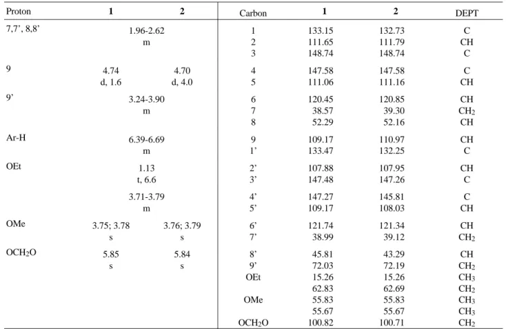
The NMR spectra of 1 and 2 (Table 1) showed signals for methoxyl [1: δH 3.75 and 3.78 (s each); δC 55.83and 55.67 (CH3 each); 2: δH 3.76; 3.79 (s each); δC 55.83and 55.67 (CH3 each)] and methylenedioxy groups [1: δH 5.85 (s); δC 100.82 (CH2); 2: δH 5.84 (s) δC 100.71 (CH2)] and also provided evidence of the existence of two dibenzyl-butyrolactol lignans by the presence of multiplets signals for benzylic hydrogens and methine groups (δH 1.96-2.62), oxymethylene of the tetrahydrofuran system (δH 3.24-3.90) and aromatic hydrogens (δH 6.39-6.69). Moreover, the NMR spectra showed characteristic signals for acetalic hydrogens [1: δH 4.74 (d, J = 1.6 Hz), δC 109.17 (CH); 2:
5' 2' 7' 5
7 2
9' 9
8' 8
O H
MeO MeO
O O
R1 R
H
5' 2' 5
2
7' 7
O H
H O
MeO MeO
O O
9
9 8
8' 9'
1 R = H; R1 = OEt
2 R = OEt; R1 = H
3
9' 8' 8 9
9
O H O
MeO MeO
O
O H 7
7' 2
5
2'
5'
2' 1'
6
5
2
O
OMe HO
OMe
4 5
O
O
OH
1'
3'a 7'a 11'a 15'a
15'b 4
1 2 .
+
MeO MeO
H
H
6 3a
R1O RO
O +
+
MeO HO
O O
H O
MeO
+
1a R = R1 = Me
δH 4.70 (d, J = 4 Hz); δC 110.97 (CH)] and for ethoxy groups by the presence of signals for methyl hydrogens [δH 1.13 (t, J = 6.6 Hz); δC 15.26 (CH3) and oxymethylene hydro-gens probably submerged in the signals for methoxyl groups and methylene in the tetrahydrofuran system [1: δC 62.83 (CH2); 2: δC 62.69 (CH2)] as well as the appearance of peaks at m/z 354 in their MS spectra corresponding to the loss of ethanol from molecular ions. Analysis using Dreiding models and observed couplings between H-8 and H-9 [1 (J = 1.6 Hz); 2 (J = 4 Hz)] suggested the relative configurations depicted in formula 1 and 2 for ethoxy groups at C-9 since their couplings were consistent with the
cis- (a dihedral angle of nearly 90°) and
trans-configura-tions (a dihedral angle of nearly 120°), respectively. The presence of two lignans was also discernable from the 13C-NMR spectra (Table 1) which showed the doubling of the signals. Since these spectra furnished different inten-sities signals for both compounds [ratio 2:1 (1):(2)], assign-ments of the chemical shifts for individually lignan were inferred. Noteworthy is the fact that corresponding carbon chemical shifts for lignans 1 and 2 are very similar, the only significative difference being the chemical shifts of C-8’[1 (δC 45.81); 2 (δC 43.29, γ-effect by the oxygen atom of the ethoxy group on C-8’)], reflecting the different configura-tion at the C-9 chiral centre. Considering that 1 and 2 have a cis- and trans-relationship between H-8 and H-9, and
trans- and cis-relationship between H-8’ and the ethoxy
groups at C-9, respectively, is consistent deduced that the relative configurations between H-8 and H-8’ for both lignans are cis. By analogy of the chemical shifts of H-9’ of other 8,8’-trans-dibenzylbutyrolactol lignans containing ethoxy group at C-9 (δH 3.22-4.20)7-8, 1 as well as 2, must be cis-oriented since its chemical shifts of H-9’ (δH 3.24-3.90) revealed at upfield (anisotropic effect by the π-sistem aromatic ring). This fact, suggest that 1 and 2 are epimers. Of additional interest were also the mass spectra which exhibited strong tropilium ions at m/z 135 [1 (100); 2 (71)] and m/z 151 [1 (85); 2 (100)] assignable to methylenedioxy-and dimethoxybenzyl units, respectively. Besides from the base peaks, the MS spectra also showed peaks with differ-ent intensities at m/z 219 [1 (9); 2 (3)], m/z 203 [1 (4); 2 (20)] and m/z 152 [1 (32); 2 (43)] corresponding to the fragments 1a, 1b, 2a and 3a, respectively.Thus, the struc-tures of two epimers were elucidated as rel-(8R, 8’S, 9S)-3,4-dimethoxy-3’,4’-methylenodioxy-9β-ethoxy- (1) and
rel-(8R, 8’S,
9R)-3,4-dimethoxy-3’,4’-methylenodioxy-9α-ethoxy-lignans-8.8’,9.O.9’ (2).
The possibility that the lignans 1 and 2are artifacts is discarded due to the fact that its presence have been con-firmed by comparison with the original hexane extract on co-TLC. Lignans containing β- and α-ethoxy groups at C-9
have been isolated previously from some Piper7-10 and
Dacrydium11-12 species.
The structures of two diastereomeric lignans (3 and
4)13-17, neolignan eupomatenoid-7 (5)6 and α -toco-pherylquinone (6)18-21 were established on the basis of their spectral data and comparison with those of the analogous compounds recorded in previous reports.
Experimental
General experimental procedures
Mp are uncorrected. IR spectrum was obtained as film on a FT-IR/1600 Perkin Elmer spectrofotometer. NMR spectra were measured in a Bruker AC-200 spectrometer at 200 and 50.3 MHz for 1H- and 13C-NMR, respectively. Proton and carbon shifts are reported in δ units (ppm) relative to TMS as the internal standard. Mass spectra were recorded in a Hewlett Packard instrument using electron impact (EI) at 70 eV. Qualitative GC/MS analysis was carried out on GC-5890 (Hewlett Packard) coupled to a Mass Selective Detector (Hewlett Packard MSD-5970) controled by a computer ChemStation 50070 C, utilizing a glass capillary column coated with dimethylsiloxane im-mobilized (12 m x 0.32 mm x 0.25 µm). The column temperature was programmed from 100 to 150 °C at a rate of 5°/min. and Helium was the carrier gas (1mL/min.).
Plant material
Aerial parts of A. peltato-deltoidea Hoehne were col-lected in Ilha de Maçaranduba, Pará State, Brazil, and identified by a specialist from the Museu Paraense Emílio Goeldi (Belém/PA), where a voucher specimen (MG-0147607) was deposited.
Extraction and isolation of the constituents
850 g of dried aerial parts were successively extracted in a Soxhlet apparatus with n-hexane and 90% ethanol. After removal solvents under vacuum, the residues were suspended in 90% and 60% MeOH/H2O solutions and extracted with n-C6H14 and n-C6H14, CHCl3 and EtOAc, respectively.
rechromatogra-phed as described above for furnished a mixture, mp 138-138.8 °C, containing β-sitosterol and stigmasterol (168 mg) after crystalization from MeOH. Fraction 9 (2.6 g) was rechromatographed as described previously and the frac-tions were combined. Some of them were further purified by gel filtration on Sephadex LH-20 with MeOH to yield eupomatenoid-7 (5, 7.9 mg) and α-tocopherylquinone (6, 13 mg) after preparative TLC [silica gel PF-254, n-C6H14 -EtOAc (9:1)] in three consecutives elutions. Finally, frac-tion 10 (3.1 g) was dissolved in MeOH and submitted to centrifugation (1 h/7000 rpm). The portion soluble in MeOH (1.3 g) was permeated on Sephadex LH-20 with MeOH. After chromatographic fractionation on silica gel 60 H [n-C6H14-EtOAc (9:1)] and preparative TLC [silica gel PF-254, n-C6H14-EtOAc (9:1)] in four successives de-velopments afforded a mixture containing lignans 1 and 2 (19.7 mg).
The CHCl3 residue (8.2 g) was suspended in aqueous 5 % NaHCO3 and extracted with CHCl3. The CHCl3 layer was taken to dryness (1.1 g) and fractioned on silica gel column using n-C6H14 with increasing proportions of CHCl3. This procedure resulted in the isolation of a mixture constituted lignans 3 and 4 (38 mg).
Rel-(8R, 8’S, 9S)-3,4-dimethoxy-3’,4’-methylenodioxy-9β-ethoxy- (1) and rel-(8R, 8’S, 9R)- 3,4-dimethoxy-3’,4’-methylenodioxy-9α-ethoxy-lignans-8.8’,9.O.9’ (2)
Yellow oil, IR ν max (cm-1, Film): 2925, 1609, 1511, 1460, 1445, 1367, 1267, 1147, 1058, 932. 1H-NMR (200 MHz, CDCl3, δ): Table 1. 13C-NMR (50.3 MHz, CDCl3,
δ): Table 1. EIMS m/z (rel. int.): 400 (1, 3; 2, 9), 354 (1 and
2, 7), 219 (1, 9; 2, 3), 203 (1, 4; 2, 20), 178 (1, 12; 2, 16),
177 (1, 69; 2, 10), 152 (1, 32; 2, 43), 151 (1, 85; 2,100), 135 (1, 100; 2, 71), 113 (1, 44; 2, 5), 91 (1, 9; 2, 12).
Acknowledgements
This work represents part of the MSc dissertation pre-sented by A.P.F.S. at the Universidade Federal de Alagoas on February 1998.We wish to thank the Conselho Nacional de Desenvolvimento Científico e Tecnológico (CNPq) -Programa de Apoio ao Desenvolvimento Científico Tec-nológico (PADCT II) for the financial support. The authors are also grateful to the Instituto de Química, Universidade de São Paulo, Museu Paraense Emílio Goeldi for acquisi-tion of NMR and MS spectra and Dr Ruth Rufino do Nascimento for analysis by GC/MS.
Table 1. NMR data for compounds 1 and 2. 1H (200 MHz, CDCl3); 13C (50.3 MHz, CDCl3). Chemical shifts (δ) expressed in ppm from internal TMS or residual undeuterated solvent, coupling constants (J) in Hz.
Proton 1 2 Carbon 1 2 DEPT
7,7’, 8,8’ 1.96-2.62 m 1 2 3 133.15 111.65 148.74 132.73 111.79 148.74 C CH C 9 4.74 d, 1.6 4.70 d, 4.0 4 5 147.58 111.06 147.58 111.16 C CH 9’ 3.24-3.90 m 6 7 8 120.45 38.57 52.29 120.85 39.30 52.16 CH CH2 CH Ar-H 6.39-6.69 m 9 1’ 109.17 133.47 110.97 132.25 CH C OEt 1.13 t, 6.6 2’ 3’ 107.88 147.48 107.95 147.26 CH C 3.71-3.79 m 4’ 5’ 147.27 109.17 145.81 108.03 C CH
OMe 3.75; 3.78
s 3.76; 3.79 s 6’ 7’ 121.74 38.99 121.34 39.12 CH CH2
OCH2O 5.85
s 5.84 s 8’ 9’ OEt OMe OCH2O
References
1. Mizuno, M.; Oka, M.; Iinuma, M.; Tanaka, T. J. Nat.
Prod. 1990, 53, 179.
2. Leitão, G.G.; Kaplan, M.A.C. Rev. Bras. Farm. 1992,
73, 65.
3. Wu, T.; Ou, L.; Teng, C. Phytochemistry 1994, 36, 1063.
4. Priestap, H.A. Phytochemistry 1987, 26, 519. 5. Hoehne, F.B. Flora Brasílica 1942, 15, 102.
6. Enriquez, R.G.; Chavez, M.A.; Reynolds, W.F. J. Nat.
Prod. 1984, 47, 896.
7. Badheka, L.P.; Prabhu, B.R.; Mulchandani, N.B.
Phy-tochemistry 1987, 26, 2033.
8. Koul, S.K.; Taneja, S.C.; Dhar, K.L.; Atal, C.K.
Phy-tochemistry 1984, 23, 2099.
9. Koul, S.K.; Taneja, S.C.; Dhar, K.L.; Atal, C.K.
Phy-tochemistry 1993, 32, 478.
10. Parmar, V.S.; Jain, S.C.; Birsht, K.S.; Jain, R.; Taneja, P.; Jha, A.; Tyagi, O.D.; Prasad, A.K.; Wengel, J.; Olsen, C.E.; Boll, P.M. Phytochemistry 1997, 46, 597. 11. Cambie, R.C.; Parnell, J.C. Tetrahedron Letters 1979,
12, 1085.
12. Cambie, R.C.; Pang, G.T.M.; Parnell, J.C.; Rodrigo, R.; Weston, R.J. Aust. J. Chem. 1979, 32, 2741. 13. Sheriha, G.M.; Abouamer, K.; Elshtaiwi, B.Z.;
Ashour, A.S.; Abed, F.A.; Alhallaq, H.H.
Phytochem-istry 1987, 26, 3339.
14. Estévez-Braun, A.; Estévez-Reyes, R.; González, A. G. Phytochemistry 1996, 43, 885.
15. Lopes, L.M.X.; Yoshida, M.; Gottlieb, O.R.
Phyto-chemistry 1983, 22, 1516.
16. McDoniel, P.B.; Cole, J.R. J. Pharm. Sci. 1972, 61, 1992.
17. Rücker, G.; Langmann, B.; Siqueira, N.S. Planta Med.
1981, 41, 143.
18. Schudel, P.; Mayer, H.; Metzger, J.; Riiegg, R.; Isler, O. Helv. Chim. Acta 1963, 46, 333.
19. Teresa, J.P.; Urones, J.G.; Marcos, I.S.; Ferreras, J.F.; Bertelloni, A.M.L.; Barcala, P.B. Phytochemistry
1987, 26, 1481.
20. Urones, J.G.; Marcos, I.S.; Cubillo, L., Garrido, N.M.; Basabe, P. Phytochemistry 1990, 29, 2228.
21. Mayer, H.; Schudel, P.; Riiegg, R.; Isler, O. Helv.
Chim. Acta 1963, 46, 650.
Received: May 19, 1998