Myocardial Bridging: Therapeutic and Clinical Development
Aline Braz Pereira, Danilo Spricigo Peressoni Castro, Emanuela Todeschini Menegotto, Wladimir Maia do Amaral,
Gustavo Spricigo Peressoni Castro
Universidade do Extremo Sul Catarinense, Hospital São José de Criciúma SC, Florianópolis, SC – Brazil
Abstract
Background: The myocardial bridge constitutes one of the main differential diagnoses of coronary artery disease. However, it remains an underdiagnosed condition and its physiopathological mechanisms and therapeutics are yet to be elucidated.
Objectives: To analyze and describe the clinical and therapeutic evolution of patients with an angiographic diagnosis of myocardial bridge, comparing the data with that in the current literature, in order to clarify the patients’ clinical profile and prognosis.
Methods: The results of coronary angiographies carried out from 2003 to 2007 in a Laboratory of Hemodynamics were reviewed; the analysis of patients’ files was carried out and selected patients were interviewed.
Results: The frequency of myocardial bridge diagnosis was 3.6%. The mean age of patients was 56.8 years (SD = 11.83; CI = 0.73). The anterior descending artery was affected in isolation in 100% of the cases. After the selection, the analysis and interview of 31 patients were carried out. There was no correlation between symptoms and degree of angiographic narrowing observed in the studied patients. The drug treatment included the use of beta-blockers, calcium-channel antagonists, platelet antiaggregants and/or nitrates and resulted in clinical improvement in 30%, absence of alterations in the clinical picture in 60% and symptom worsening in 10% of the patients. One patient presented sudden death; two patients underwent angioplasty followed by significant clinical improvement and none of the patients underwent surgical procedures.
Conclusion: Most of the patients with myocardial bridge have a good prognosis, but in the long term, there are not enough data, obtained from a large sample of symptomatic patients, to draw definitive conclusions. (Arq Bras Cardiol 2010;94(2): 175-181)
Key Words: Coronary artery disease/therapy; myocardial bridging; angioplasty, transluminal, percutaneous coronary; cohort studies.
Mailing address: Aline Braz Pereira •
Rua Coronel Carvalho, 33 - Centro - 89240-000 - São Francisco do Sul, SC - Brazil
E-mail: [email protected]
Manuscript received May 13, 2009; revised manuscript received May 20, 2009; accepted August 06, 2009.
Introduction
The myocardial bridge (MB) is a congenital anomaly of the coronary arteries that usually affect the left anterior descending artery (ADA), when one or more myocardial bundles cross or involve a segment of the epicardial coronary artery, which crosses the intramural portion of the myocardium, below the muscular bridge. The MB constitutes one of the main differential diagnoses of coronary artery disease (CAD) and can manifest as typical or atypical angina pectoris and, more rarely, as acute myocardial infarction (AMI) or sudden death.
It is a relatively common and usually benign pathology among the general population, affecting mainly patients at low risk for CAD; however, when symptomatic, it can manifest
as unstable or stable angina, cardiac arrhythmias (ventricular tachycardia and supraventricular tachycardia), AMI and sudden death, with the latter two being rare1.
It remains underdiagnosed due to the fact that only a minority of patients present symptoms as well as the lack of availability and, consequently, the restricted use of more accurate diagnostic methods and therefore, its physiopathological mechanisms and therapeutics have not been fully elucidated.
The present study aimed at analyzing the clinical and therapeutic evolution of patients with an angiographic diagnosis of myocardial bridge, from 2003 to 2007, comparing the data with those in the current literature, in order to elucidate the clinical profile and the evolution of these patients.
Methods
Table 1 - Degree of narrowing of the anterior descending artery at the coronary angiography
Degree of narrowing Patient % (nº)
Grade 1: < 49% 50.41 (62)
Grade 2: 50%-74% 36.59 (45)
Grade 3: > 75% 13.01 (16)
Total 100 (123)
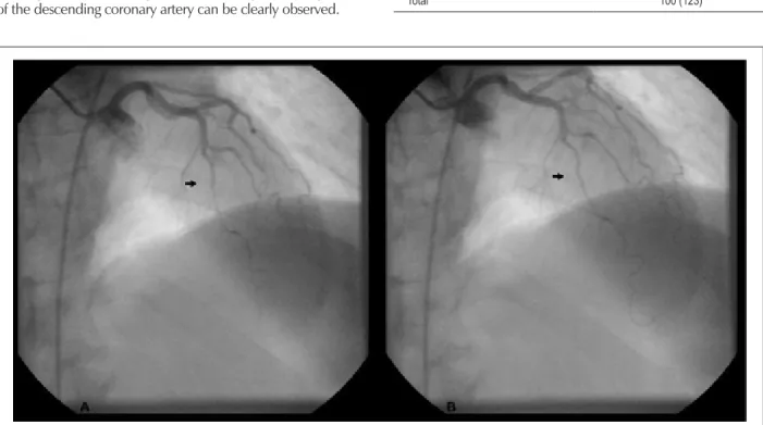
Figure 1 -Coronary angiography performed in a 56-year-old female patient that presented compression of the left anterior descending artery, with an 80% decrease in
vessel lumen diameter (arrow) at systole (A) and persistence of the phenomenon at diastole (B), with slight vessel dilatation (arrow). materials used in the study. The study was carried out through
the analysis of medical files and by applying a questionnaire to a group of selected patients.
The study was carried out in a Laboratory of Hemodynamics after being approved by the Ethics Committee of the institution. The patients’ confidentiality and the use of the data for scientific research purposes only were warranted. After the pre-selection of the medical files, the patients or their tutors were contacted, the study objective was explained to them and the Free and Informed Consent Form was signed by all participants, which had been previously approved by the Scientific Committee in Research and Ethics of the Institution.
Patients aged 20 to 75 years that had been submitted to a coronary angiography from 2003 to 2007, due to suspected and undergoing investigation for probable CAD, in whom the angiographic results showed the presence of MB, were evaluated. Patients who did not present associated cardiac pathologies capable of producing angina symptoms were also assessed: cardiac valvular disease or myocardiopathies that had been previously diagnosed or were diagnosed at the coronary angiography and the presence of CAD at the coronary angiography.
Patients with neurological pathologies that prevented the correct use of the questionnaire and those who available data were incomplete or outdated, which prevented the contact for the interview, were excluded.
Although the patients were included in the study based on the coronary angiographies carried out before the study, all of them were carried out according to the protocol of the Laboratory of Hemodynamics of the institution, following the Judkins technique, in which the phenomenon of systolic compression of the descending coronary artery can be clearly observed.
This protocol did not include the intracoronary administration of nitroglycerin.
Results
The results of 3,375 coronary angiographies, carried out from 2003 to 2007, were assessed, of which 123 presented the phenomenon of systolic constriction of the left ADA (Figure 1), with a diagnosis of MB. The frequency of diagnoses of MB in coronary angiographies performed within this period was 3.6%. The mean age of patients was 56.8 years (SD=11.83; CI=0.73). All patients presented only one affected coronary artery and the left ADA was the affected one in 100% of the cases. Thirty-four patients (27.6%) presented CAD that affected the proximal segment of the bridge at different degrees. The involvement of the left ADA at the coronary angiography was classified as Grade 1 (≤ 49%), grade 2 (50-74%) and grade 3 (≥ 75%) (Table 1).
Table 2 -Degree of narrowing of the anterior descending artery at the coronary angiography of the patients selected for the study
Degree of narrowing Patient % (nº) Grade 1: < 49% 61.29 (19)
Grade 2: 50%-74% 29.03 (09)
Grade 3: > 75% 9.68 (03)
Total 100 (31)
Table 3 - Canadian Cardiovascular Society Grading Scale for Angina Pectoris
Class I – Usual physical activity, e.g. walking or climbing stairs, does not cause angina; angina is evoked by strenuous and/or rapid work or recreation.
Class II – Slight limitation of ordinary activities, e.g. after walking 2 blocks, climbing one light of steps, under normal circumstances, after meals, in the cold, wind, in the morning, or when under emotional stress
Class III – Marked limitation of ordinary activities, e.g. walking 1-2 blocks or climbing stairs under normal circumstances.
Class IV – Inability to carry out any physical activity without discomfort–angina may be present at rest ..
Source: Stable Angina Guideline. Arquivos Brasileiros de Cardiologia, 2004. Modiied from Campeau, Circulation, 19761. whom one presented a neurological pathology that prevented
the interview to be carried out and in 10 cases, the interview could not be carried out as the available data were incomplete or outdated, which prevented the contact with these patients, resulting in the analysis and interview of 31 patients.
The mean age at the symptom onset among the studied patients was 45.9 years (CI=11.7; SD=4.2) and in 50% of them, the diagnosis of MB took ≥ one year. The analysis of the degree of narrowing of the left ADA was performed (Table 2). The patients’ symptoms were classified according to the classification proposed by the Canadian Cardiovascular Society Grading Scale for angina pectoris, before and after the treatment (Table 3).
According to this classification, patients with grade 1 of systolic narrowing at the coronary angiography presented the classifications CCS-I (31.6%), II (31.6%) and III (31.6%). Patients with grade 2 of narrowing were classified as I (11.1%), II (11.1%), III (66.7%) and IV (11.1%). Patients with grade 3 were classified as II (33.3%), III (33.3%) and IV (33.3%).
A patient who died due to sudden death was excluded from the analysis of symptoms; this patient presented grade 1 systolic narrowing of the ADA. Two patients that underwent angioplasty presented grade 1 narrowing and were both classified as CCS-III; they were later reclassified as classes I and II after the procedure.
Based on these data, one concludes that there is no correlation between symptom classification and the degree of angiographic narrowing observed in the studied patients. However, the intensity of these symptoms also depends on the number, thickness, location and length of the MB, which vary from patient to patient and can even vary in a same patient, from one evaluation to another. The adverse events reported by the patients were: ischemia and acute coronary syndrome, AMI and sudden death (Chart 1).
There was a significant variation regarding the time of treatment performed and the therapeutic regimens used by
the patients. The drug treatment included the use of beta-blockers, calcium-channel antagonists, platelet antiaggregants and/or nitrates. Two patients underwent angioplasty and none of the patients was submitted to surgical procedure. Nitrates were used by 43.3% of the patients, associated or not to other drugs, in spite of reports of the worsening of symptoms and clinical picture in the literature. However, it was not possible to assert the real impact of these medications on the clinical outcome of these patients.
Of the patients that presented CCS-I classification before treatment, all remained at the same classification after the treatment. Of the patients that presented CCS-II classification before the treatment, 37.5% remained at the same class after the treatment, 50% became class I, with clinical improvement and 12.5% presented clinical worsening and became class III. Of the patients that presented CCS-III classification before treatment, 53.8% remained at the same classification after the treatment, 30.8% presented improvement to classes I or II and 15.4%, presented symptom worsening and became class IV. Of the patients that presented CCS-IV classification before the treatment, 50% remained at the same classification after the treatment and the other 50% presented clinical improvement and became class III. This group included two patients with classification III that underwent PCI and maintained the same drug treatment, presenting clinical improvement.
In conclusion, 30% of the patients presented clinical improvement, 60% did not present alterations in the clinical picture and 10% presented symptom worsening, in spite of the treatment (Chart 2).
Discussion
The myocardial bridge is a congenital anomaly2,3 that
results from the failure in the synchronic development of the myocardium and the coronary branches4, in which a segment
of one epicardial coronary artery is involved by a cardiac muscle bundle, called “tunneled segment”, crossing the intramural portion of the myocardium, below the myocardial muscle bridge5. The frequency of MB diagnoses in this study
was 3.6%. Study data have shown a significant variation regarding the frequency, incidence and prevalence rates and the angiographic results. The angiographic prevalence of myocardial bridges is < 5%6-8, a fact attributed to thin
myocardial bridges that cause little compression to the tunneled segment and are present in most cases8.
Chart 1 - Adverse events reported by the patients with myocardial bridge.
Ischemia and Acute coronary syndrome
Acute myocardial infarction
Sudden death
No complications
that the area below the MB is spared in atherosclerotic disease, whereas the proximal area is prone to the development of atheroclerosis9,10. In the present study, 27.6% of the patients
presented involvement of the proximal segment of the bridge due to varying degrees of CAD. Disorders in blood flow contribute to the development of atherosclerosis in the proximal segment of the bridge11, modulating the production
of vasoactive substances by the endothelial cells, affecting vascular cell functions such as the thrombogenic potential, blood flow regulation and vascular tonus12,13.
The typical angiographic finding of the MB is the systolic reduction of the epicardial coronary artery diameter and the persistence of this reduction during diastole14. Its transient
nature and the dynamics of the obstruction help the differential diagnosis of fixed coronary stenoses. The left ADA is the most commonly affected artery15 and 100% of the patients
in the present study presented the isolated form of left ADA involvement. However, other arteries can be involved, such as the left circumflex artery and the right coronary artery4. The
quantification of the involvement of the coronary artery was carried out according to Noble et al6, in which the narrowing
of the ADA during systole was classified as: Grade 1 (≤ 49%); Grade 2 (50%-74%); and Grade 3 (≥ 75%).
In some cases, when the angiographic results disclose normal coronary arteries, the use of provocative tests through pre-load decrease and adrenergic stimulation with nitroglycerin and orciprenaline, respectively, can enhance the systolic compression of the tunneled segment, thus establishing the diagnosis in up to 40% of the cases7,16. Such procedures
were not carried out in the present study.
The clinical diagnosis of MB must be considered in patients with angina symptoms, in the absence of risk factors or evidence of ischemia17. Although it is a malformation present
since birth, symptoms onset does not occur until the third decade of life. The mean age of symptom onset presented
by the patients was 45.9 years (CI=11.7; SD=4.2). The late symptom manifestation can be explained by the systolic tension increase in the myocardial wall, as a consequence of the heart growth due to the elevation in the left ventricular diastolic-end pressure, through the association with arterial hypertension (myocardial hypertrophy) and with the eventual decrease in the coronary flow due to atherosclerotic processes that occur later18.
A considerable number of patients with an angiographic diagnosis of MB present associated cardiac valvular, muscular or atherosclerotic disease, which independently affects the clinical presentation and the response to treatment6. Such
patients were excluded from the present study.
It has been well established the association between MB and complications such as ischemia and acute coronary syndrome19, reported by 56% of the studied patients; AMI20
affecting 40% of the patients and sudden death21, which
was the cause of death of one patient in the present study. Other complications are: coronary spasm22; ventricular
septum rupture23, supraventricular paroxysmal tachycardia24;
ventricular tachycardia25 and exercise-induced atrioventricular
block26, which were not reported by the patients in the
present study.
The drug therapy is the first-choice treatment for symptomatic patients with MB. Interventions are reserved for patients with angina that is refractory to drug treatment. The drug treatment consists in the use of negative inotropic and chronotropic agents, such as adrenergic receptor blockers or calcium-channel blockers and the use of anti-platelet agents, with the objective of relieving symptoms and signs of myocardial ischemia and reducing the risk of future cardiac adverse events27. Treatment duration is not well clarified
among the several studies.
Chart 2 - Individualized categorization according to the Canadian Cardiovascular Society Angina Classiication, before and after treatment. Clinical classiication
Patients Before treatment
After treatment
used by the patients, which prevented the individualized analysis of each drug class.
The use of beta-blockers results in decreased heart rate, increased diastolic time and reduced contractility and systolic compression of the vessel, with a consequent return to normal of the ST-segment alterations at the ECG, in addition to the improvement of clinical symptoms of angina and signs of ischemia28,29.
The use of calcium-channel antagonists can be particularly useful when there is a contraindication for the use of blockers or, first choice, when there is a suspected coronary vasospasm30. The use of nitrates must be avoided, as while
they improve cardiac contractility, they worsen the degree of systolic narrowing of the coronary artery, which can aggravate symptoms16,31,32. In spite of the presented studies, the nitrate
was used in 43.3% of the patients in association or not with other drugs. However, the real impact of the use of these drugs on the clinical outcome of these patients cannot be established.
Stent implants prevent the phasic compression of the coronary lumen, eliminate the abnormalities of the diastolic flow and the maximum elevation of the intracoronary systolic pressure, normalizing the clinical symptoms13. After the stent
implant, the systolic compression of the anterior descending artery disappears and the luminal diameter increases as well as the transversal section of the artery and the coronary flow reserve. However, according to Haager et al33, analyses after
7 weeks showed moderate to severe stenosis of the stents in 45% of the patients, which required a new intervention in 36% of the patients.
The two-year clinical and angiographic follow-up showed good results, with improvement of the angina symptoms and absence of cardiac events33. However, long-term favorable
results have been described by other authors34,35. In the
present study, two patients with grade 1 angiographic systolic narrowing underwent angioplasty, presenting significant symptom improvement, free of adverse events, after one and two years of follow-up.
Before the current era of percutaneous coronary intervention, the surgical myotomy was considered the treatment of choice for patients with persistent symptoms, despite the intensive drug therapy27,36. The surgical decompression of the tunneled
artery results in the disappearance of the angina symptoms and ischemia in patients with severe systolic compression of the ADA37. Another alternative is the myocardial revascularization
surgery, with the anastomosis of the internal mammary artery to the left ADA33. None of the patients in the present study
underwent surgical procedures.
The long-term prognosis of MB is good, regardless of the severity of the systolic narrowing of the luminal diameter of the coronary artery38.
In this study, the analysis of 31 patients showed that only one patient died, which was attributed to sudden death due to myocardial bridge; angioplasty was performed in two patients, with long-term significant clinical improvement; and 96.7% of the patients underwent clinical pharmacological therapy. In another study, during a 43-month follow-up of a group of 35 patients with MB, a patient died due to sudden cardiac death; 20% of the patients persisted with CCS I-II angina classification; one patient needed percutaneous revascularization for symptom improvement and 63% of the patients had kept the same drug therapy by the end of the study39.
Limitations
References
1. Sociedade Brasileira de Cardiologia. Diretrizes de doença coronariana crônica e angina estável. Arq Bras Cardiol. 2004; 83 (2): 2-43.
2. Angelini P, Velasco JA, Flamm S. Coronary anomalies: incidence, pathophysiology, and clinical relevance. Circulation. 2002; 105: 2449-54.
3. Angelini P, Trivellato M, Donis J, Leachman RD. Myocardial bridges: a review. Prog Cardiovasc Dis. 1983; 26: 75-88.
4. Polacek P. Relation of myocardial bridges and loops on the coronary arteries to coronary occlusions. Am Heart J. 1961; 61: 44-52.
5. Alegria JR, Herrmann J, Holmes DR, Lerman AJ, Rihal CS. Myocardial bridging. Eur Heart J. 2005; 26 (12): 1159-68.
6. Noble J, Bourassa MG, Petitclerc R, Dyrda I. Myocardial bridging and milking effect of the left anterior descending artery: normal variant or obstruction? Am J Cardiol. 1976; 37 (37): 993-9.
7. Diefenbach C, Erbel R, Treese N, Bollenbach E, Meyer J. Häufigkeit von myokardbrucken nach adrenerger stimulation und nachlastsenkung bei patienten mit angina pectoris, aber unauffälligen koronararterien. Z Kardiol. 1994; 83: 809-15.
8. Garcia JF, Villalon AM, Chavero EP. Significado clinico de las bandas musculares en las arterias coronaries. Arch Inst Cardiol Méx. 1983; 53: 413-20.
9. Ishii T, Asuwa N, Masuda S, Ishikawa Y. The effects of a myocardial bridge on coronary atherosclerosis and ischemia. J Pathol. 1998; 185: 4-9.
10. Ishikawa Y, Ishii T, Asuwa N, Masuda S. Absence of atherosclerosis evolution in the coronary arterial segment covered by myocardial tissue in cholesterol-fed rabbits. Virchows Arch. 1997; 430: 163-71.
11. Ge J, Eebel R, Görge G, Haude M, Meyer J. High wall shear stress proximal to myocardial bridging and atherosclerosis: intracoronary ultrasound and pressure measurements. Br Heart J. 1995; 73 (5): 462-5.
12. Zoghi M, Duygu H, Nalbantgil S, Kirilmaz B, Turk U, Ozerkan F, et al. Impaired endothelial function in patients with myocardial bridge. Echocardiography. 2006; 23 (7): 577-81.
13. Klues HG, Schwarz ER, vom Dahl J, Reffelmann T, Reul H, Potthast K, et al. Disturbed intracoronary hemodynamics in myocardial bridging: early normalization by intracoronary stent placement. Circulation. 1997; 96:
2905-13.
14. Schwarz ER, Klues HG, vom Dahl J, Klein I, Krebs W, Hanrath P. Functional characteristics of myocardial bridging: a combined angiographic and intracoronary doppler flow study. Eur Heart J. 1997; 18 (3): 434-42.
15. Ferreira AGJ, Trotter SE, König BJ, Décourt LV, Fox K, Olsen EG. Myocardial bridges: morphological and functional aspects. Br Heart J. 1991; 66 (5): 364-7.
16. Hongo Y, Tada H, Ito K, Yasumura Y, Miyatake K, Yamagishi M. Augmentation of vessel squeezing at coronary-myocardial bridge by nitroglycerin: study by quantitative coronary angiography and intravascular ultrasound. Am Heart J. 1999; 138: 345-50.
17. Kulan K, Kulan C, Tuncer C, Komsuoğlu B, Telatar M. Myocardial perfusion scintigraphy in a myocardial bridging of coronary artery. Clin Nucl Med. 1996; 21 (11): 888-9.
18. Maia GAS, Alfieri RG, Chalela WA, Moffa PJ, Pastore CA, Del Nero Junior E. Ponte miocárdica. Arq Bras Cardiol. 1987; 48: 389-93.
19. Tauth J, Sullebarger T. Myocardial infarction associated with myocardial bridging: case history and review of the literature. Cathet Cardiovasc Diagn. 1997; 40 (4): 364-7.
20. Bestetti RB, Costa RS, Zucolotto S, Oliveira JS. Fatal outcome associated with autopsy proven myocardial bridging of the left anterior descending coronary artery. Eur Heart J. 1989; 10 (6): 573-6.
21. Bestetti RB, Costa RS, Kazava DK, Oliveira JS. Can isolated myocardial bridging of the left anterior descending coronary artery be associated with sudden death during exercise? Acta Cardiol. 1991; 46 (1): 27-30.
22. Sakuma M, Kamishirado H, Inoue T, Ichihara M, Takayanagi K, Hayashi T. Acute myocardial infarction associated with myocardial bridge and coronary artery vasospasm. Int J Clin Pract. 2002; 56 (9): 721-2.
23. Tio RA, Ebels T. Ventricular septal rupture caused by myocardial bridging. Ann Thorac Surg. 2001; 72 (4): 1369-70.
24. Morales AR, Romanelli R, Boucek RJ. The mural left anterior descending coronary artery, strenuous exercise and sudden death. Circulation. 1980; 62 (2): 230-7.
25. Feld H, Guadanino V, Hollander G, Greengart S, Lichstein E, Shani J. Exercise-MB. For this retrospective study, patients were selected from
a database of a Laboratory of Hemodynamics that functions as a reference center in the region, which does not statistically reflect the data of the local population. As the patients were selected based on the data of the coronary angiographies previously carried out during a 5-year interval, it was not possible to carry out a follow-up and perform uniform noninvasive diagnostic tests in all patients, as well as confirm the reported complications, which were solely based on the report of the interviewed patients.
It was only possible to indicate the number of patients in whom these tests were performed and the type of test that was carried out, as well as their results. Finally, the criteria on which the decision of maintaining the long-term treatment were based are out of the scope of the present study, being influenced by the cardiologist’s criterion.
Conclusion
Myocardial bridges affect individuals with angina symptoms in the absence of risk factors or evidence of ischemia, in which
symptom onset does not take place before the third decade of life. Most patients with myocardial bridges have a good prognosis, but in the long term, there are not enough data obtained from a large number of symptomatic patients that have presented an elevated degree of systolic and diastolic compression and ischemia evidence for definitive conclusions to be drawn.
Potential Conflict of Interest
No potential conflict of interest relevant to this article was reported.
Sources of Funding
There were no external funding sources for this study.
Study Association
induced ventricular tachycardia in association with a myocardial bridge. Chest. 1991; 99: 1295-6.
26. Den Dulk K, Brugada P, Braat S, Heddle B, Wellens HJ. Myocardial bridging as a cause of paroxysmal atrioventricular block. J Am Coll Cardiol. 1983; 1: 965-9 .
27. Bourassa MG, Butnaru A, Lesperance J, Tardif JC. Symptomatic myocardial bridges: overview of ischemic mechanisms and current diagnostic and treatment strategies. J Am Coll Cardiol. 2003; 41 (3): 351-9.
28. Schwarz ER, Klues HG, vom Dahl J, Klein I, Krebs W, Hanrath P. Functional, angiographic and intracoronary Doppler flow characteristics in symptomatic patients with myocardial bridging: effect of short-term intravenous beta-blocker medication. J Am Coll Cardiol. 1996; 27 (7): 1637-45.
29. Nair CK, Dang B, Heintz MH, Sketch MH. Myocardial bridges: effect of propanolol on systolic compression. Can J Cardiol. 1986; 2: 218-21.
30. Kracoff OH, Osvyshcher I, Gueron M. Malignant course of a benign anomaly: myocardial bridging. Chest. 1987; 92 (6): 1113-5.
31. Ishimori T, Raizner AE, Chahine RA, Awdeh M, Luchi RJ. Myocardial bridges in man: clinical correlations and angiographic accentuation with nitroglycerin. Cathet Cardiovasc Diagn. 1977; 3: 59-65.
32. Carvalho VB, Macruz R, Décourt LV, Arie S, Manrique R, Mello SC, et al. Hemodynamic determinants of coronary constriction in human myocardial bridges. Am Heart J. 1984; 108: 73-80.
33. Haager PK, Schwarz ER, vom Dahl J, Klues HG, Reffelmann T, Hanrath P. Long-term angiographic and clinical follow-up in patients with stent implantation for symptomatic myocardial bridging. Heart. 2000; 84 (4): 403-8.
34. Bayes A, Marti V, Auge JM. Coronary stenting for symptomatic myocardial bridging. Heart. 1998; 80: 102-3.
35. Prendergast BD, Kerr F, Starkey IR. Normalisation of abnormal coronary fractional flow reserve associated with myocardial bridging using an intra-coronary stent. Heart. 2000; 83: 705-7.
36. Katznelson Y, Petchenko P, Knobel B, Cohen AJ, Kishon Y, Schachner A. Myocardial bridging: surgical technique and operative results. Mil Med. 1996; 161: 248-50.
37. Grondin P, Bourassa MG, Noble J, Petitclerc R, Dydra I. Successful course after supraarterial myotomy for myocardial bridging and milking effect of the left anterior descending artery. Ann Thorac Surg. 1977; 24 (5): 422-9.
38. Juilliere Y, Berder V, Suty-Selton C, Buffet P, Danchin N, Cherrier F. Isolated myocardial bridges with angiographic milking of the left anterior descending coronary artery: a long-term follow-up study. Am Heart J. 1995; 129 (4): 663-5.