Contents lists available atScienceDirect
Separation and Purification Technology
j o u r n a l h o m e p a g e :w w w . e l s e v i e r . c o m / l o c a t e / s e p p u r
Isolation and analysis of bioactive isoflavonoids and chalcone from a new type of
Brazilian propolis
Tatiane Luiza C. Oldoni
a, Ingridy S.R. Cabral
a, Marisa A.B. Regitano d’Arce
a, Pedro L. Rosalen
b,
Masaharu Ikegaki
c, Andrea M. Nascimento
a, Severino M. Alencar
a,∗aDepartment of Agri-Food Industry, Food and Nutrition, “Luiz de Queiroz” College of Agriculture, University of Sao Paulo (USP), P.O. Box. 9, 13418-900, Piracicaba, SP, Brazil bDepartment of Physiological Sciences, Piracicaba Dental School, University of Campinas (UNICAMP), P.O. Box 52, 13414-903, Piracicaba, SP, Brazil
cFaculty of Pharmaceutical Sciences, Federal University of Alfenas, Av. Gabriel Monteiro da Silva, 700, 37130-000, Alfenas, MG, Brazil
a r t i c l e
i n f o
Article history: Received 9 August 2010 Received in revised form 26 November 2010 Accepted 6 December 2010
Keywords: Isoflavonoids Chalcone Antioxidant activity Antimicrobial activity Brazilian propolis
a b s t r a c t
Activity-directed fractionation and purification processes were employed to identify isoflavonoids with antioxidant and antimicrobial activities from Brazilian red propolis. Crude propolis was extracted with ethanol (80%, v/v) and fractioned by liquid–liquid extraction technique using hexane and chloroform. Since chloroform fraction showed strong antioxidant and antimicrobial activities it was purified and isolated using various chromatographic techniques. Comparing our spectral data (UV, NMR, and mass spectrometry) with values found in the literature, we identified two bioactive isoflavonoids (vestitol and neovestitol), together with one chalcone (isoliquiritigenin). Vestitol presented higher antioxidant activity against-carotene consumption than neovestitol. The antimicrobial activity of these three compounds againstStaphylococcus aureus,Streptococcus mutans, andActinomyces naeslundiiwas evaluated and we concluded that isoliquiritigenin was the most active one with lower MIC, ranging from 15.6 to 62.5g/mL. Our results showed that Brazilian red propolis has biologically active isoflavonoids that may be used as a mild antioxidant and antimicrobial for food preservation.
© 2010 Elsevier B.V. All rights reserved.
1. Introduction
Flavonoids and the isomeric isoflavonoids are important constituents in vegetables used for human consumption [1,2]. Isoflavonoids are plant secondary metabolites with multiple biological and pharmacological effects, such as antioxidant, antibacterial, antiviral, anti-inflammatory, etc. [3,4]. They occur widely in legumes but have also been found in a considerable num-ber of non-legumes. Reynaud et al.[5]listed 35 non-leguminous families that produce isoflavones to which[6]added an additional 17 families. According to recent epidemiological investigations, isoflavonoids abundant in certain plant foods are more benefi-cial for human health than flavonoids[7]. Dietary consumption of foods and food additives containing isoflavone phytoestrogens has been associated with several beneficial properties to human health, such as prevention of coronary heart disease and osteo-porosis, reduction of menopausal symptoms, and prevention of distinct cancer forms (e.g. breast, prostate, and colon cancer)
[8,9].
∗Corresponding author. Tel.: +55 19 34294150; fax: ++55 19 34029437. E-mail address:[email protected](S.M. Alencar).
In our previous studies, we reported the crude extract antiox-idant and antimicrobial activities, the botanical origin of Brazilian red propolis, as well as the presence of the isoflavones, suggest-ing new biological potentialities for this novel type of propolis
[10–12]. Nevertheless, the presence of isoflavonoids has never been reported in other types of Brazilian propolis. Trusheva et al.[13]
isolated phenolic compounds from Brazilian red propolis; how-ever, the authors did not use the bioassay-guided fractionation and found only the isoflavonoids isosativan and medicarpin with antimicrobial and antioxidant activities.
Propolis, a natural resinous product collected by honeybees from buds and exudates of various plant sources, has been used empirically as a traditional remedy in folk medicine for cen-turies[14]. It is well known for its potential benefits to human health and for presenting valuable biological activities such as antioxidant [10,15], antibacterial[16], anticariogenic[17], anti-inflammatory[18], and anticancer[19]properties. It has recently gained popularity as a healthy food supplement and has been extensively used in foods and beverages in different parts of the world, where it is claimed to improve health and prevent ailments such as inflammation, heart diseases, diabetes, and even cancer
[14].
The present study aimed at evaluating the antioxidant and antimicrobial activities of Brazilian red propolis extracts
1383-5866/$ – see front matter© 2010 Elsevier B.V. All rights reserved.
and fractions and, subsequently, isolating isoflavonoids through bioassay-guided fractionation techniques.
2. Materials and methods
2.1. Materials
Silica gel 60 and Brain Heart Infusion Agar (BHI) were purchased form Merck (Darmstadt, Germany); 2,2-diphenyl-1-picrylhydrazyl hydrate (DPPH), (±)␣-tocopherol (VE), trans--carotene, Tween 40, and chlorhexidine from Sigma Co. (St. Louis, MO); linoleic acid from Agros Organics (Geel, Belgium); sodium carbonate, butylated hydroxytoluene (BHT), and butylated hydroxyanisole (BHA) stan-dards from Synth (Diadema, SP, Brazil); resazurin from Sigma Co. (St. Louis, MO); and Sephadex LH-20 from Amersham Pharmacia (Uppsala, Sweden). All the solvents used for chromatography were of high performance liquid chromatography (HPLC) grade and all the other chemicals were of analytical-reagent grade.
A total of 11 crude propolis samples were obtained from colonies ofApis melliferacollected from March, 2007 to January, 2008, in an apiary located in the municipality of Marechal Deodoro, state of Alagoas, in the Northern Region of Brazil. The samples with the same chemical profile by HPLC were cleaned, ground after addi-tion of liquid nitrogen, weighed, mixed, and stored at−18◦C until analysis.
2.2. Extraction and isolation of bioactive compounds
The representative propolis sample (100 g) was extracted with ethanol 80% (450 mL) in water bath at 70◦C for 30 min and after filtration yielded the ethanolic extract of propolis (EEP). The EEP was further fractioned by liquid–liquid extraction with hexane and chloroform yielding 11.4 g of hexanic fraction (Hex-fr) and 30 g of chloroform fraction (Chlo-fr), respectively. The active Chlo-fr (30 g) was subjected to open dry column chromatography on nor-mal phase silica gel (particle size: 0.0063–0.2 mm; pore size: 60 ˚A; pore volume:∼0.8 cm3/g; and specific surface area: 500 m2/g) and
eluted with a solvent mixture of chloroform/ethyl acetate (70:30, v/v) to afford seven major subfractions. The subfractions obtained were monitored by thin layer chromatography (TLC) using the anisaldehyde reagent (4-methoxy-benzaldehyde, acetic acid, sul-phuric acid – 1.0:48.5:0.5), followed by incubation at 100◦C for 5 min. Fluorescent substances were visualized under ultra violet (UV) light at the wavelengths of 254 nm and 366 nm[10]. Sub-fractions 1, 6, and 7 showed no activity and negligible activities were found in subfractions 2, 4, and 5. The most bioactive sub-fraction (3) was chromatographed over a Sephadex LH-20 column (5 cm×30 cm) using methanol to yield three bioactive subfractions. Active subfractions 3.2 and 3.3 were purified by semi-preparative reverse-phase HPLC [Shimadzu PREP-ODS (H) 250×20 mm col-umn eluted with a gradient starting with CH3OH:H2O (65:35) to
CH3OH:H2O (95:5) in 35 min, flow rate 3 mL/min] and yielded three
active compounds.
2.3. Chromatography with mass spectrometry (GC–MS)
Aliquots of 400L (10 mg/mL) of each compound were trans-ferred to vials, received 1 mL of CH2N2solution for methylation, and
were maintained in ice bath for 4 h to complete the methylation reaction. Samples of the methylated solutions were analyzed by GC–MS using a capillary column CBP5 (30 m×0.25 mm×0.25m) installed in a Hewlett-Packard 5890 Series II instrument interfaced with a HP-5971 mass selective detector operated in scanning mode (m/z40–400). GC–MS analysis was previously programmed from 50◦C (0.3 min hold) to 285◦C (15 min hold) at a rate of 6◦C/min.
Samples were injected with an auto injector using a splitless injec-tion technique (0.6L injection volume) and the carrier gas (He) flow was set at 1.0 mL/min. The GC–MS peaks were identified by comparison with data from the literature and the profiles from the Wiley 138 and Nist 98 libraries[12].
2.4. Nuclear magnetic resonance (NMR)
The NMR spectra, obtained in CD3OD using TMS as internal
standard, were recorded in a Brucker DPX 500 MHz spectrometer, operating at 500 MHz for1H. The chemical shifts are expressed in
ı(parts per million) and the coupling constants (J) in Hz.
All the compounds, already described in the literature, were identified by comparison of their spectral data (UV, nuclear mag-netic resonance – NMR, and mass spectrometry) with reported values.
Vestitol (1): UVmax(EtOH): 208, 280 (sh); EIMS (70 eV)m/z(rel.
int.): 300 [M+], 272 (38), 150 (100), 137 (32), 123 (11).1H- and 13C-NMR data were consistent with those previously reported[20].
Neovestitol (2): UVmax (EtOH): 211, (sh); EIMS (70 eV)m/z(rel. int.): 300 [M+], 272 (38), 150 (16,7), 137 (100), 121 (15).1H- and 13C-NMR data were consistent with those previously reported[21].
Isoliquiritigenin (3):1H- and13C-NMR data were in agreement
with the reported literature values[22].
2.5. Antioxidant activity on linoleic acid oxidation
This analysis was carried out using the method of[23] with some modifications. We dissolved 10 mg of-carotene in 100 mL of chloroform, an aliquot of 3 mL was added to 40 mg of linoleic acid and 400 mg of Tween 40, and the chloroform was removed under a stream of nitrogen gas. Oxygenated distilled water (100 mL) was added and thoroughly mixed. Aliquots of 3 mL of the  -carotene/linoleic acid emulsion were mixed with 50L of test samples and incubated in water bath at 50◦C. The antioxidant activ-ity was expressed as percent inhibition relative to the control after 120 min incubation using the equation:
AA=[(DRcontrol−DRsample)] DRcontrol ×100
AA = antioxidant activity; DRcontrol= degradation rate of 
-carotene without addition of antioxidant; DRsample= degradation
rate of-carotene with addition of antioxidant.
The degradation rates were calculated according to first order kinetics:
DR=natural log
At Ax×
1t
At= initial absorbance at 470 nm andt= 0; Ax= absorbance at
470 nm andt= 120 min.
Test samples were evaluated at the final concentration of 90g/mL and VE and BHT were used as the reference samples. All analyses were carried out in triplicate.
2.6. Antimicrobial activity
The antimicrobial activity was determined by minimum inhibitory concentration (MIC) and minimum bactericidal con-centration (MBC) in accordance with the Clinical and Laboratory Standards Institute [24], using the following bacterial strains: Staphylococcus aureus ATCC 25923, Streptococcus mutansIngbritt 1600, andActinomyces naeslundii ATCC 12104. To determine MIC, the starting inoculum was 5×10⁵CFU/mL. The technique was
1
Extract, fractions, subfractions or compounds with antioxidant activity 2
Extract, fractions, subfractions or compounds with antimicrobial activity
Crude propolis
(
1
00
g)
1,2
Ethanolic Extract of Propolis (EEP)
(5
8 g)
Extracted with ethanol 80% at 70 oC, 30 min
Extraction liquid-liquid with hexane and chloroform
Hexane fraction (Hex-fr)
(
11.
4
g)
Aqueous
phase
1,2
Chloroform fraction (Chlo
-fr)
(3
0
g)
Dry column chromatograph on silica gel (SiO2)(chloroform/ethyl acetate (70:30, v/v)
Subfr
. 3.1
1,2Subfr 3.3
.
Rechromatography onsemi-prep HPLC
Subfr
.
7
Subfr
.
6
Subfr
.5
Subfr
.
4
1,2
Subfr.
3
Subfr
.
2
Subfr.1
Rechromatography on Sephadex LH-20
Rechromatography onsemi-prep HPLC
1,2
Compound 1 (200mg)
1,2
Compound 3 (100mg) 1
Compound 2 (40mg)
1,2
Subfr 3.2
.
Fig. 1.Procedure for bioguided isolation of compounds from Brazilian red propolis.
used chlorhexidine 0.12% (w/v) as positive control and 80% ethanol (v/v), employed to solubilize the samples, as negative control. The microplates were incubated at 37◦C for 24 h and, after incubation, 30L of resazurin (0.01%, w/v) was added to identify the wells presenting bacterial growth. The wells presenting no changes in color were considered free of viable bacteria and those presenting changes in color were considered positive for bacterial growth.
To determine MBC, aliquots of 10L of all incubated wells pre-senting concentrations higher than the MIC were subcultured on BHI agar. The MBC was defined as the lowest concentration that allowed no visible growth on the agar. All the analyses were per-formed in six replicates.
3. Results and discussion
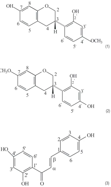
In this study, the antimicrobial and antioxidant activities of the EEP, Hex-fr, Chlo-fr, and isolated compounds from Brazilian red propolis were investigated. Bioguided purification of the EEP using a series of chromatographic separations (Fig. 1) resulted in the isolation of three compounds: 200 mg of vestitol (1), 40 mg of neovestitol (2), and 100 mg of isoliquiritigenin (3) (Fig. 2). The structures of the compounds were identified by their spectroscopic data (1H NMR,13C NMR, UV, and mass spectrometry) measurement
and by comparison with published values. The structure of these compounds has never been reported in other 12 types of Brazilian propolis[25].
3.1. Antioxidant activity
The first goal of this work was to detect and isolate compounds from Brazilian red propolis with potential antioxidant capacity. We performed analyses for the detection of antioxidant activity at every step of isolation of compounds from the EEP. The analyses using the -carotene–linoleic acid model system led us to the three purified compounds (vestitol, neovestitol, and isoliquiritigenin) that were obtained from subfractions presenting antioxidant activity (Fig. 2). The-carotene–linoleic acid method has been considered an efficient technique to detect and evaluate antioxidants from propo-lis and other natural sources by several investigative groups
[23,26,27]. The consumption of-carotene is related to the ther-mally induced formation of linoleic acid hydroperoxides. After the isolation and identification of the three flavonoids compounds, a comparison of their activities in the-carotene–linoleic acid sys-tem was performed. Their antioxidant activity was compared with that of the EEP, fractions, BHT, and vitamin E, a major natural lipid-soluble antioxidant[28].
OH
O
OH
H
OCH
32 ,
3
,4
,5
,6
,2
3
4
5
6
7
8
(1)
CH
3O
O
OH
OH
H
2 ,
3
,4
,5
,6
,2
3
4
5
6
7
8
(2)
HO
O
OH
OH
1
2
3
4
5
6
α
β
1'
6'
5'
4'
3'
2'
(3)
Fig. 2.Structures of the bioactive compounds isolated from Brazilian red propolis: (1) vestitol, (2) neovestitol, and (3) isoliquiritigenin.
Fig. 3.Antioxidant activity of the ethanolic extract of propolis (EEP), hexanic fraction (Hex-fr), chloroform fraction (Chlo-fr), vestitol (1), neovestitol (2), isoliquir-itigenin (3),␣-tocopherol (VE), and butylated hydroxytoluene (BHT). Measurements were performed in triplicate and the standard deviations are indicated.
but all of them are highly important antioxidant compounds found in the EEP. In fact, isoflavonoids have been considered the most abundant and effective antioxidant compounds found in soybean
[27]. The confirmed occurrence of isoflavonoids in this novel type of Brazilian propolis also suggests the presence of other biological activities in addition to the antioxidant activity detected in this study.
Ahn et al. [30] reported the antioxidant activity of propolis from various areas of Korea. The EEP from Cheongju presented stronger antioxidant activity (∼40%) than those from other regions. EEP from Muju also had high antioxidant activity (∼28%) evalu-ated by-carotene–linoleic acid system. In the study mentioned, the contents of both total polyphenol and flavonoid were high, indicating a correlation between total polyphenol content and antioxidant activity. Flavonoids have been reported to be the most abundant and most effective antioxidant in propolis[31–33]. In our study, two isoflavonoids and one chalcone were important com-pounds responsible for the antioxidant activity in Brazilian red propolis.
3.2. Antimicrobial activity
The antimicrobial activity of EEP, fractions and compounds iso-lated were tested againstS. aureusATCC 25923,S. mutansIngbritt 1600, andA. naeslundiiATCC 12104 (Table 1). Chlo-fr presented MIC values ranging from 31.2 to 62.5g/mL forS. aureusand from 62.5 to 125g/mL forS. mutansandA. naeslundii, the latter ones lower than those found for the Hex-fr and EEP. As to MBC, Chlo-fr was as effective as the EEP forS. aureusandS. mutans, and forA. naeslundiiit proved to be more bioactive than the EEP, presenting values ranging from 62.5 to 125g/mL.
These results are similar to those found by[10], when evaluating MIC and MBC of red propolis collected in the state of Alagoas and its Hex-fr and Chlo-fr usingS. aureus ATCC 25923 andS. mutans UA159. The authors attributed the high antimicrobial activity of red propolis and its Chlo-fr to their high content in phenolic com-pounds, respectively 232 mg/g and 324 mg/g, the highest values ever found in Brazilian propolis samples[10,29]. In this work we were able to isolate and identify the phenolic compounds respon-sible for antimicrobial activity.
Chlo-fr was found to be the principal source of bioactive com-pounds during the bioguided fractionation and isolation process since it presented the best antibacterial activity among the frac-tions. After isolation, we observed that two compounds showed strong activity against all bacterial strains tested (Table 1). Vesti-tol presented MIC ranging from 31.2 to 62.5g/mL, showing no distinction among the microorganisms assessed. Isoliquiriti-genin was more potent than vestitol and exhibited MIC values ranging from 15.6 to 31.2g/mL for the three bacterial strains used.
Isoliquiritigenin presented the best MBC values, which ranged from 31.2 to 62.5g/mL forS. aureusandS. mutans.No isolated compound showed antibacterial effect against A. naeslundii. As reported by[34], the strong antibacterial activity of propolis may be due to the synergistic effect among flavonoids, hydroxy-acids, and sesquiterpens. Therefore, in the present study we also evaluated the synergistic effect among the isolated compounds. The solution was prepared by mixing vestitol and isoliquiritigenin at a concen-tration of 1:1 and the final concenconcen-tration in the tests ranged from 125 to 1.8g/mL.
Table 1
Minimum inhibitory concentration (MIC) and minimum bactericidal concentration (MBC) of the ethanolic extract of propolis (EEP), hexanic fraction (Hex-fr), chloroform fraction (Chlo-fr), and bioactive compounds isolated from Brazilian red propolis.
Treatment S. aureus S. mutans A. naeslundii
MIC (g/mL) MBC (g/mL) MIC (g/mL) MBC (g/mL) MIC (g/mL) MBC (g/mL)
EEP 62.5–125 250–500 62.5–125 250–500 125–250 125–250
Hex-fr 62.5–125 250–500 125–250 500–1000 250–500 500–1000
Chlo-fr 31.2–62.5 250–500 62.5–125 250–500 62.5–125 62.5–125
Vestitol (1) 31.2–62.5 62.5–125 31.2–62.5 125–250 31.2–62.5 nd
Isoliquiritigenin (3) 15.6–31.2 31.2–62.5 15.6–31.2 31.2–62.5 15.6–31.2 nd
1 + 3 [1:1] 31.2–62.5 nd 62.5–125 nd 31.2–62.5 nd
nd: Activity not detected at the concentration tested. Neovestitiol (2) did not show antimicrobial activity.
them separately. This demonstrates that these compounds are not able to potentialize each other’s activity for microbial control, since their antibacterial activity was lower when they were employed together than when they were tested separately.
Koo et al.[16]analyzed MIC and MBC of 30 compounds from Brazilian propolis against S. mutans GS-5 and UA159 as well as Streptococcus sobrinus 6715. All the flavanones tested inhib-ited bacterial growth and, among them, pinocembrine proved to be the most efficient compound (MIC 64g/mL), although pinobanksin-3-acetate also inhibited the growth of the bacte-rial strains tested (MIC 157g/mL). The concentrations reported by those authors are higher than the ones we found in this study for the flavonoids isolated from Brazilian red propolis that inhibited the growth ofS. mutans(Table 1). Among all the com-pounds evaluated by [16], tt-farnesol was the most efficient (MIC 28g/mL), presenting antibacterial activity similar to that observed for the isoliquiritigenin isolated from Brazilian red propo-lis reported herein, which exhibited MIC ranging from 15.6 to 31.2g/mL.
Pepeljnjak and Kosalec[35] assessed the antibacterial activ-ity of galangin isolated from three samples of propolis collected in Croatia against resistant strains ofS. aureus,Enterococcusspp. andPseudomonas aeruginosaand found MIC values ranging from 1600 to 2400g/mL. The high concentration of galangin necessary to inhibit bacterial growth was already expected by the authors since the strains employed showed resistance to several types of antibiotics.
Melliou et al.[36]evaluated the antibacterial effect of␣-pinene, a compound found in large amounts in propolis collected in Greece, againstS. aureusATCC 25923. Although this compound is widely known for its strong antibacterial activity[29], it presented MIC value of 6500g/mL. This concentration was significantly higher than that required to inhibit the growth ofS. aureususing vestitol and isoliquiritigenin isolated from Brazilian red propolis. In fact, isoflavonoids and chalcones, possess several biological properties such as marked antimicrobial activity[4].
4. Conclusions
The bio-assay guided isolation of active compounds from Brazil-ian red propolis resulted in the identification of two isoflavonoids (vestitol and neovestitol) and one chalcone (isoliquiritigenin). We assessed the activity of the isolated compounds, by measuring their ability to inhibit-carotene consumption, and observed that vesti-tol is a potent antioxidant. Furthermore, isoliquiritigenin presented the most active antimicrobial activity among the compounds tested with no synergistic effect.
The identification of these bioactive isoflavonoids in Brazilian red propolis is important to improve the development and produc-tion of this natural product, and in the future, may be used as a mild antioxidant and antimicrobial for food preservation.
Acknowledgments
The authors thank FAPESP (Grants 04/08635-6, 08/51048-6 and 08/58492-8) for financial support, and SEBRAE/AL and beekeepers for providing the propolis samples.
References
[1] J. Kuhnau, The flavonoids. A class of semi-essential food components: their role in human nutrition, World Rev. Nutr. Diet. 24 (1976) 117–191.
[2] T.J. Key, M. Thorogood, P.N. Appleby, M.L. Burr, Dietary habits and mortality in 11,000 vegetarians and health conscious people: results of a 17-year follow up, Br. Med. J. 313 (1996) 775–779.
[3] T. Cornwell, W. Cohick, I. Raskin, Dietary phytoestrogens and health, Phyto-chemistry 65 (2004) 995–1016.
[4] S.G. Dastidar, A. Manna, K.A. Kumar, K. Mazumdar, N.K. Dutta, A.N. Chakrabarty, N. Motohashi, Y. Shirataki, Studies on the antibacterial potentiality of isoflavones, Int. J. Antimicrob. Agents 23 (2004) 99–102.
[5] J. Reynaud, D. Guilet, R. Terreux, M. Lussignol, N. Walchshofer, Isoflavonoids in non-leguminous families: an update, Nat. Prod. Rep. 22 (2005) 504–515. [6] Z. Macková, R. Koblovská, O. Lapc¸ík, Distribution of isoflavonoids in
non-leguminous taxa—an update, Phytochemistry 67 (2006) 849–855.
[7] R. Koblovská, Z. Macková, M. Vítková, L. Kokoska, B. Klejdus, O. Lapcík, Isoflavones in the Rutaceae family: twenty selected representatives of the gen-era Citrus, Fortunella, Poncirus, Ruta and Severinia, Phytochem. Anal. 19 (2008) 64–70.
[8] M. Lof, E. Weiderpass, Epidemiologic evidence suggests that dietary phytoe-strogen intake is associated with reduced risk of breast, endometrial, and prostate cancers, Nutr. Res. 26 (2006) 609–619.
[9] M. Cotterchio, B.A. Boucher, M. Manno, S. Gallinger, A. Okey, P. Harper, Dietary phytoestrogen intake is associated with reduced colorectal cancer risk, J. Nutr. 136 (2006) 3046–3053.
[10] S.M. Alencar, T.L.C. Oldoni, M.L. Castro, I.S.R. Cabral, C.M. Costa-Neto, J.A. Cury, P.L. Rosalen, M. Ikegaki, Chemical composition and biological activity of a new type of Brazilian propolis: red propolis, J. Ethnopharmacol. 113 (2007) 278–283. [11] I.S.R. Cabral, T.L.C. Oldoni, A. Prado, M. Ikegaki, P.L. Rosalen, S.M. Alencar, Composic¸ão fenólica, atividade antibacteriana e antioxidante da própolis ver-melha brasileira, Quim. Nova 32 (2009) 1523–1527.
[12] B.B. Silva, P.L. Rosalen, J.A. Cury, M. Ikegaki, V.C. Souza, A. Esteves, S.M. Alen-car, Chemical composition and botanical origin of red propolis, a new type of Brazilian propolis, Evid. Based Complement. Altern. Med. 5 (2008) 313–316. [13] B. Trusheva, M. Popova, V. Bankova, S. Simova, M.C. Marcucci, P.L. Miorin, F.R.
Pasin, I. Tsvetkova, Bioactive constituents of Brazilian red propolis, Evid. Based Complement. Altern. Med. 3 (2006) 249–254.
[14] A.H. Banskota, Y. Tezuka, S. Kadota, Recent progress in pharmacological research of propolis, Phytother. Res. 15 (2001) 561–571.
[15] S. Kumazawa, R. Ueda, T. Hamasaka, S. Fukumoto, T. Fujimoto, T. Nakayama, Antioxidant prenylated flavonoids from propolis collected in Okinawa, Japan, J. Agric. Food Chem. 55 (2007) 7722–7725.
[16] H. Koo, P.L. Rosalen, J.A. Cury, Y.K. Park, W.H. Bowen, Effects of compounds found in propolis onStreptococcus mutansgrowth and on glucosyltransferase activity, Antimicrob. Agents Chemother. 46 (2002) 1302–1309.
[17] M. Hayacibara, H. Koo, P.L. Rosalen, S. Duarte, E.M. Franco, W.H. Bowen, M. Ikegaki, J.A. Cury, In vitro and in vivo effects of isolated fractions of Brazilian propolis on caries development, J. Ethnopharmacol. 101 (2005) 110–115. [18] N. Paulino, C. Teixeira, R. Martins, A. Scremin, V.M. Dirsch, A.M. Vollmar,
S.R. Abreu, S.L. Castro, M.C. Marcucci, Evaluation of the analgesic and anti-inflammatory effects of a Brazilian green propolis, Planta Med. 72 (2006) 899–906.
[19] S. Awale, F. Li, H. Onozuka, H. Esumi, Y. Tezuka, S. Kadota, Constituents of Brazilian red propolis and their preferential cytotoxic activity against human pancreatic PANC-1 cancer cell line in nutrient-deprived condition, Bioorg. Med. Chem. 16 (2008) 181–189.
[21] J.L. Ingham, Isoflavonoid phytoalexins from leaflets ofDalbergia sericea, Z. Naturforsch. C 34 (1979) 630–631.
[22] A.F. Barrero, M.M. Herrador, P. Arteaga, I. Rodriguez-Garcia, M. Garcia-Moreno, Resorcinol derivatives and flavonoids of Ononis natrix subspecies ramosissima, J. Nat. Prod. 60 (1997) 65–68.
[23] M.R. Ahn, S. Kumazawa, Y. Usui, J. Nakamura, M. Matsuka, F. Zhu, T. Nakayama, Antioxidant activity and constituents of propolis collected in various areas of China, Food Chem. 101 (2007) 1383–1392.
[24] CLSI, Clinical and Laboratory Standards Institute, Methods for determining bac-tericidal activity of antimicrobial agents, Tentative standard M26-T. Wayne: National Committee for Clinical Laboratory Standards (2007).
[25] Y.K. Park, M. Ikegaki, S.M. Alencar, F.F. Moura, Evaluation of brazilian propolis by both physicochemical methods and biological activity, Honey Bee Sci. 21 (2000) 85–90.
[26] V.W. Bowry, R. Stocker, Tocopherol-mediated peroxidation. The prooxidant effect of vitamin E on the radical-initiated oxidation of human low-density lipoprotein, J. Am. Chem. Soc. 115 (1993) 6029–6044.
[27] C.H. Lee, L. Yang, J.Z. Xu, S.Y.V. Yeung, Y. Huang, Z.Y. Chen, Relative antioxi-dant activity of soybean isoflavones and their glycosides, Food Chem. 90 (2005) 735–741.
[28] M.S. Taga, E.E. Miller, D.E. Pratt, Chia seeds as a source of natural lipid antioxi-dants, J. Am. Oil Chem. Soc. 61 (1984) 928–931.
[29] S. Kumazawa, T. Hamasaka, T. Nakayama, Antioxidant activity of propolis of various geographic origins, Food Chem. 84 (2004) 329–339.
[30] M.R. Ahn, S. Kumazawa, T. Hamasaka, K.S. Bang, T. Nakayama, Antioxidant activ-ity and constituents of propolis collected in various areas of Korea, J. Agric. Food. Chem. 52 (2004) 7286–7292.
[31] J.S. Bonvehi, F.V.V. Coll, Phenolic composition of propolis from China and South America, Z. Naturforsch C 49 (1994) 712–718.
[32] M.I. Isla, M.I. Nieva Moreno, A.R. Sampietro, M.A. Vattuone, Antioxidant activity of Argentine propolis extracts, J. Ethnopharmacol. 76 (2001) 165–170. [33] I. Martos, M. Cossentini, F. Ferreres, F.A. Tomas-Barberan, Flavonoid
composi-tion of Tunisian honeys and propolis, J. Agric. Food Chem. 45 (1997) 2824–2829. [34] M.C. Marcucci, Propolis: chemical composition, biological properties and
ther-apeutic activity, Apidologie 26 (1995) 83–99.
[35] S Pepeljnjak, I. Kosalec, Galangin expresses bactericidal activity against multiple-resistant bacteria: MRSA, Enterococcus spp. and Pseudomonas aeruginosa, FEMS Microbiol. Lett. 240 (2004) 111–116.