Humoral and cellular immune responses in dogs with inapparent
natural
Leishmania infantum
infection
q
W. Coura-Vital
a,b, M.J. Marques
c, R.C. Giunchetti
a,d, A. Teixeira-Carvalho
d, N.D. Moreira
a,
J. Vitoriano-Souza
a, P.M. Vieira
a, C.M. Carneiro
a, R. Corrêa-Oliveira
e, O.A. Martins-Filho
d,
M. Carneiro
b, A.B. Reis
a,e,⇑
a
Laboratório de Imunopatologia, Núcleo de Pesquisas em Ciências Biológicas/NUPEB, Universidade Federal de Ouro Preto, Ouro Preto, Minas Gerais, Brazil
bLaboratório de Epidemiologia de Doenças Infecciosas e Parasitárias, Departamento de Parasitologia, Universidade Federal de Minas Gerais, Belo Horizonte, Minas Gerais, Brazil cLaboratório de Parasitologia Básica, Departamento de Ciências Biológicas, Universidade Federal de Alfenas, Alfenas, Minas Gerais, Brazil
dLaboratório de Biomarcadores de Diagnóstico e Monitoração, Centro de Pesquisas René Rachou, Fundação Oswaldo Cruz, Belo Horizonte, Minas Gerais, Brazil eLaboratório de Imunologia Celular e Molecular, Centro de Pesquisas René Rachou, Fundação Oswaldo Cruz, Belo Horizonte, Minas Gerais, Brazil
a r t i c l e
i n f o
Article history:
Accepted 7 April 2011
Keywords:
Canine visceral leishmaniasis Asymptomatic dogs Immune response
a b s t r a c t
Molecular analysis, serology and immunophenotyping for T lymphocytes and their subsets, B lympho-cytes and monolympho-cytes were performed on dogs naturally infected withLeishmania infantum. Animals were categorised as asymptomatic dogs I (AD-I), with negative serology and positive molecular results, and asymptomatic dogs II (AD-II), with positive serology and positive molecular results, and these were com-pared to symptomatic dogs (SD) and control dogs (CD).
AD-I exhibited immunophenotypic features similar to those of CD, including isotype profiles and con-centrations of monocytes. Similar biomarkers were found in AD-II and SD, such as, higher levels of immu-noglobulins IgG, IgG2, IgM and IgA and higher concentrations of eosinophils. High frequencies of T lymphocytes and CD4+T cells were observed in both AD-I and AD-II compared to SD, whereas CD8+T cells were higher only in AD-II compared with SD. Analysis of B lymphocytes revealed an increased frequency of this cell type only in AD-II animals compared with SD. Asymptomatic dogs appear to have a dichoto-mous infection spectrum that can influence the humoral and cellular immunological status during canine visceral leishmaniasis.
Ó2011 Elsevier Ltd. All rights reserved.
Introduction
Leishmaniasis is endemic in 88 countries in tropical and
sub-tropical regions of the Old and New World, with more than 350
million people exposed to the infection (
Desjeux, 2001
). The
esti-mated incidence is 2 million new cases per year, with 0.5 million
being visceral leishmaniasis (VL) (
Desjeux, 2004
). Infected dogs
have a high cutaneous parasite density and represent the main
domestic reservoir of
Leishmania infantum,
contributing to the
propagation of the disease (
Deane and Deane, 1962
). In Brazil over
the past 10 years, more than 2 million dogs were screened and
more than 160,000 seropositive dogs were eliminated; however,
the incidence of human VL has not been reduced to an acceptable
level (
Lemos et al., 2008; Romero and Boelaert, 2010
).
Canine visceral leishmaniasis (CVL) can be categorised into
three distinct clinical forms on the basis of major features observed
in seropositive infected dogs, which can be classified as
asymptom-atic dogs (AD), oligosymptomasymptom-atic dogs and symptomasymptom-atic dogs (SD)
dogs (
Mancianti et al., 1988; Reis et al., 2009
).
L. infantum
-infected
dogs also include animals in which the presence of parasites is
con-firmed through direct methods (such as PCR) and which have
low-titre anti-
Leishmania
antibodies (
Paltrinieri et al., 2010
).
Oliva et al. (2006)
identified subpatent infection with
L.
infan-tum
in asymptomatic dogs that were intermittently positive by
nested PCR but became negative for long periods of time.
Seropos-itive asymptomatic dogs are important sources of amastigotes for
infection of phlebotomines that contribute to the transmission of
L. infantum
(
Marzochi et al., 1985; Molina et al., 1994; da
Costa-Val et al., 2007
). In endemic areas, 10–62% of apparently healthy
and/or seronegative dogs were positive for
Leishmania
by PCR
(
Martin-Sanchez et al., 2001; Solano-Gallego et al., 2001; Lachaud
et al., 2002; Andrade et al., 2006
). Thus, asymptomatic dogs may
play a role in the transmission of
Leishmania
parasites but cannot
be detected by conventional serological tests, such as the indirect
fluorescent antibody test (IFAT) and enzyme-linked
immunosor-bent assay (ELISA).
1090-0233/$ - see front matterÓ2011 Elsevier Ltd. All rights reserved. doi:10.1016/j.tvjl.2011.04.005
qThis article is available only in the online version of this issue of
The Veterinary Journalathttp://www.sciencedirect.com/science/journal/10900233
⇑
Corresponding author. Tel.: +55 21 31 35591694.E-mail address:alexreis@nupeb.ufop.br(A.B. Reis).
Contents lists available at
ScienceDirect
The Veterinary Journal
The aim of this study was to use the humoral and cellular
im-mune response to explore the dichotomy in dogs with
asymptom-atic and symptomasymptom-atic infection with
L. infantum
and to identify
features that could be used to identify resistant and susceptible
profiles.
Materials and methods
Dogs and experimental design
Forty-one mongrel dogs of either gender from the endemic area of Belo Hori-zonte, Minas Gerais, Brazil, were selected on the basis of serological tests (IFAT and ELISA, Biomanguinhos/Fiocruz) forLeishmaniaspp. Dogs with an IFAT titre <1/40 were considered to be seronegative and dogs with an IFAT titreP1/40 were
considered to be seropositive and infected withLeishmaniaspp. Positive infection was confirmed by ELISA and PCR in at least one skin sample (Degrave et al., 1994) and the species ofLeishmaniaresponsible was determined by restriction frag-ment length polymorphism–PCR (Volpini et al., 2004).
The study was conducted from June 2008 to August 2009 after approval by the ethical committees for the use of experimental animals of the Federal University of Ouro Preto (CETEA/UFOP 032/2007), Federal University of Minas Gerais (CETEA/ UFMG 020/2007) and the Municipal Health Secretariat of Belo Horizonte City Coun-cil, Minas Gerais State, Brazil (CEP-SMSA/PBH 001/2008).
Selection and clinical classification of dogs
Dogs were selected and clinically classified according to the presence/absence of clinical signs: (1) asymptomatic, with no signs suggestive of disease; (2) symp-tomatic, with characteristic clinical signs of visceral leishmaniasis, such as opaque bristles, severe loss of weight, onychogryphosis, cutaneous lesions, apathy and keratoconjunctivitis; and (3) control dogs, classified according to negative serolog-ical and molecular results and absence of clinserolog-ical signs.
Haematology
Peripheral blood (5 mL) from the brachiocephalic vein was collected into tubes containing ethylene diamine tetraacetic acid (EDTA) at a final concentration of 1 mg/mL. Erythrocytes and leucocytes were quantified using an automatic cell counter (Model 2800 Vet, Mindray). Differential leucocyte counts were performed by examination of at least 200 leucocytes in Giemsa-stained blood smears by light microscopy.
ELISA for immunoglobulin isotype profile
ELISAs were performed to determine the anti-Leishmaniaimmunoglobulin pat-tern using solubleL. infantum(MHOM/BR/1972/BH46) promastigotes antigen (SLA) (Rosário et al., 2005; Reis et al., 2006c). The protein concentration was quantified by the Lowry method, adjusted to 1 mg/mL and samples were stored at 70°C. Ninety-six well microplates (MaxiSorp, Nalge Nunc) were coated with SLA at a con-centration of 2
l
g/well, left overnight at 4–8°C and then washed. Serum samples were added to the wells at a dilution of 1:80, followed by washes after the addition of goat anti-dog IgG1 (anti-heavy chain specific) conjugated with peroxidase, IgM (anti-l
chain specific), IgA (anti-a
chain specific) and IgE (anti-e
chain specific) orsheep anti-dog IgG and IgG2 (both anti-heavy chain specific) (Bethyl Laboratories). Wells were washed and then the substrate and chromogen (O-phenylenediamine, Sigma–Aldrich) were added. The absorbance was read on an automatic ELISA micro-plate reader (EL 800G PC, Bio-Tek) at 492 nm. The concentrations of conjugate were determined by a block titration method with positive and negative standard sera. The conjugates IgG1, IgM, IgA, IgE were used at a dilution of 1:1,000, anti-IgG was used at a dilution of 1:8,000 and anti-anti-IgG2 was used at a dilution of 1:16,000.
Immunophenotyping by flow cytometry
Immunophenotyping of peripheral blood by flow cytometry was performed as described byReis et al. (2005). After a pre-fixation step, erythrocytes were lysed in 1 mL EDTA-treated whole blood by the slow addition of 13 mL fluorescence-acti-vated cell sorter (FACS) lysing solution (Becton Dickinson), followed by incubation for 10 min at room temperature (RT). After centrifugation (450gfor 10 min at RT), the pellet was resuspended in 500 mL phosphate-buffered saline (PBS) supple-mented with 10% fetal bovine serum (PBS–10% FBS). Using 96-well U-bottom plates (Limbro Biomedicals), 30
l
L prefixed leucocyte suspension were incubated at RT for30 min in the dark with 30
l
L monoclonal antibodies (mAbs) diluted previously inPBS–10% FBS.
The mAbs used in the study were diluted purified anti-canine CD5 (1:800, rat IgG2a, clone YKIX3223), anti-canine CD4 (1:1,000, rat IgG2a, clone YKIX3029) and anti-canine CD8 (1:800, rat IgG1, clone YCATE559) (Serotec). Undiluted fluo-rescein isothiocyanate (FITC)-labelled mouse anti-human CD21 (5
l
L, mouseIgG1, clone IOBla; Immunotech) and diluted phycoerythrin (PE)/Cy-5-conjugated mouse anti-human CD14 (50
l
L, 1:200, mouse IgG2a, clone TÜK4; Serotec) werealso used in direct immunofluorescence procedures. Cells were also incubated in the same conditions in the presence of 60
l
L of diluted FITC-conjugated sheepanti-rat IgG polyclonal antibody (1:100 or 1:200; Serotec).
Before flow cytometric data collection and analysis were performed, labelled cells were fixed for 30 min with 200
l
L FACS fix solution (10.0 g/Lparaformalde-hyde, 10.2 g/L sodium cacodylate, 6.6 g/L sodium chloride; pH 7.2). Flow cytometric measurements were performed on a FACScan (Becton Dickinson) and analysed using CellQuest software (10,000 events acquired per sample). The results were ex-pressed in absolute counts (cell number/mm3) through the product of the percent-age of positive cells (CD5+, CD4+, CD8+and CD21+) within gated lymphocytes by absolute lymphocyte counts. Absolute counts for T lymphocyte subsets were also calculated as the sum of absolute values of CD4+plus CD8+cells. The absolute counts for monocytes were the products of CD14+cells within ungated leucocytes by the total leucocyte counts.
Statistical analysis
Statistical analysis was performed using GraphPad Prism 5.0. The normality of the data was assessed using the Kolmogorov–Smirnoff test. Considering the non-parametric nature of all data sets, Kruskal–Wallis tests were used to investigate dif-ferences between the four groups, followed by Dunn’s test for pairwise compari-sons. Differences were considered to be significant atP< 0.05.
Results
Reclassification according to serological, molecular and clinical
features
Dogs with no clinical signs and negative serological and
molec-ular results were included in the control group (CD,
n
= 7).
Seroneg-ative dogs without clinical signs but positive molecular results for
L. infantum
were classified as asymptomatic dogs I (AD-I,
n
= 8).
Dogs with positive serology and molecular results for
L. infantum
but no clinical signs were classified as asymptomatic dogs II
(AD-II,
n
= 10). Dogs with clinical signs and positive serological and
molecular results were classified as symptomatic dogs (SD)
(
n
= 16) (
Table 1
).
Haematology
There were lower concentrations of eosinophils in AD-II and SD
compared with AD-I and CD and lower concentrations of
lympho-cytes in SD compared with AD-II (
Table 2
). Symptomatic
L.
infan-tum
infection was associated with decreased erythrocyte counts
and haematocrits in AD-II compared with AD-I and CD (
P
< 0.05)
and decreased haemoglobin concentrations in SD compared to
other groups (
P <
0.05).
Anti-Leishmania immunoglobulin isotypes
Concentrations of IgG, IgG1, IgG2, IgM, IgA and IgE in AD-I were
similar to CD (
Fig. 1
). There were increased IgG, IgG2, IgM and IgA
concentrations in AD-II and SD compared with AD-I and CD
Table 1
Serological and molecular status of dogs categorised according to clinical status with and without natural infection byLeishmania infantum.
Groups Serodiagnosis Molecular diagnosis Total IFAT and ELISA PCR–RFLP
Positive Negative Positive Negative
Control (CD) 0 7 0 7 7
Asymptomatic I (AD-I) 0 8 8 0 8
Asymptomatic II (AD-II) 10 0 10 0 10
Symptomatic (SD) 16 0 16 0 16
Total 26 15 34 7 41
(
P
< 0.05). Concentrations of IgE were lower in AD-I compared with
SD (
P
< 0.05). There were no significant differences in
concentra-tions of IgG1 between groups.
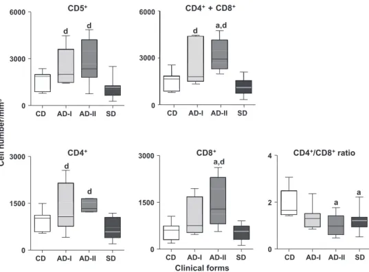
Immunophenotyping of circulating I lymphocytes and their subsets
AD-I and AD-II had increased concentrations of T lymphocytes
compared with SD (
P
< 0.05). AD-I and AD-II had higher counts of
CD4
+T lymphocytes than SD, whereas AD-II, but not AD-I, had
higher counts of CD8
+T lymphocytes than CD and SD. There was
a decreased ratio of CD4
+:CD8
+in AD-II and SD as compared with
CD (
Fig. 2
).
Immunophenotyping of circulating B lymphocytes and monocytes
AD-II and CD had higher concentrations of CD21
+B
lympho-cytes than SD (
Fig. 3
). AD-II and SD had lower concentrations of
monocytes than AD-I and CD.
Discussion
In the current study, we identified lower concentrations of
eosinophils in AD-II and SD compared with AD-I and CD. Decreased
concentrations of eosinophils have been identified previously in
Leishmania
-infected dogs with clinical signs (
Reis et al., 2006a
),
as well as in dogs with medium and high parasite densities in
spleen and skin (
Guerra et al., 2009
).
AD-II and SD had higher levels of IgG, IgG2, IgM and IgA than
AD-I or CD, whereas AD-I and CD had similar immunoglobulin
iso-type profiles. These findings suggest that AD-I are in the initial
phase of CVL, whereas AD-II are unable to control the parasitism,
as indicated by intense polyclonal activation (
Reis et al., 2006c;
Teixeira Neto et al., 2010
). None of these isotypes can confer
pro-tective immunity against
L. infantum
.
High concentrations of IgG2 are associated with clinical signs in
dogs with leishmaniasis (
Bourdoiseau et al., 1997; Cavaliero et al.,
1999; Boceta et al., 2000; Solano-Gallego et al., 2000; Cardoso
et al., 2007
), whereas high concentrations of IgG1 are present in
asymptomatic carriers (
Reis et al., 2006c
). Although IgM usually
is considered to be an immunological marker for the acute phase
of parasitic disease, it has been demonstrated that this
immuno-globulin also can be detected during the chronic phases of CVL
(
Genaro et al., 1992; Reis et al., 2006c; Rodriguez-Cortes et al.,
2007
).
Reis et al. (2006c)
suggest that IgA reactivity is a marker
for tissue parasite density in CVL. SD had higher concentrations
of IgE than AD-I. Increased levels of IgE have been associated with
severe forms of the disease and elevated tissue parasite densities,
as well as with the T helper type 2 immune response profile (
Reis
et al., 2006c; Guerra et al., 2009
).
Table 2
Haematological parameters of naturally infected withLeishmania infantumand uninfected dogs.
Haematological parameter Clinical groups
CD AD-I AD-II SD
Erythrocytes (million/mm3) 6.8 ± 0.4 6.7 ± 1.0 5.8 ± 0.7 4.2 ± 1.3a,b
Haemoglobin (g%) 17.0 ± 0.6 14.8 ± 2.3 14.1 ± 2.3 9.8 ± 2.9a,b,c
Haematocrit (%) 49.1 ± 2.0 44.0 ± 6.7 40.7 ± 7.1 28.6 ± 8.2a,b
Leucocytes (103/mm3) 13.4 ± 3.5 12.0 ± 2.6 13.0 ± 4.5 10.2 ± 3.9
Granulocytes 10.1 ± 3.6 8.2 ± 3.3 7.4 ± 3.1 8.0 ± 3.2
Neutrophils 7.9 ± 3.8 6.6 ± 3.1 6.5 ± 2.7 7.2 ± 3.0
Eosinophils 2.1 ± 0.8 1.7 ± 0.6 0.5 ± 0.3a,b 0.5 ± 0.4a,b
Lymphocytes 2.1 ± 0.8 3.0 ± 1.9 4.5 ± 3.3 1.7 ± 1.0c
Monocytes 1.1 ± 0.6 1.0 ± 0.5 1.0 ± 0.6 0.5 ± 0.4a,b
Results are shown as the average values ± standard deviation.
CD, Control dogs; AD-I, Asymptomatic dogs I; AD-II, Asymptomatic dogs II; SD, Symptomatic dogs. a Statistically significant differences compared with CD.
b Statistically significant differences compared with AD-I. c Statistically significant differences compared with AD-II.
IgG
IgM
0.8 0.8
a,b
a,b
a,b
a,b
a,b
,
0.4 0.4
CD AD-I AD-II SD 0.0
CD AD-I AD-II SD 0.0
IgG1
IgA
a,b
b
0.8 0.8
s
ity
den
s
tical
Op
t
CD AD-I AD-II SD CD AD-I AD-II SD
IgG2
IgE
b
0.8 1.0
a,b
a,b
0.4 0.5
d
CD AD-I AD-II SD 0.0
CD AD-I AD-II SD 0.0
Clinical forms
0.4
0.0 0.0
0.4
b
a,b
AD-I and AD-II had similar total concentrations of CD5
+T
lym-phocytes, with the main contribution being from CD4
+T
lympho-cytes, whereas CD8
+T lymphocytes were only high in AD-II. The
lower CD4
+:CD8
+ratio in AD-II and SD suggests a distinct cellular
immune response after seroconversion and disease progression.
A more intense parasite load in AD-II and SD may augment the
pro-duction of CD8
+T lymphocytes and polyclonal immunoglobulin
activation, resulting in increased concentrations of IgG. Progressive
disease in CVL is associated with suppression of cell-mediated
immunity (
Pinelli et al., 1994; Reis et al., 2009, 2010
). Increased
levels of CD8
+T lymphocytes are evident in asymptomatic dogs
with a low parasite load (
Reis et al., 2006b; Guerra et al., 2009
).
The absolute number of CD8
+T lymphocytes could be used to
discriminate between AD-I and AD-II. However, further studies
are require to confirm this hypothesis.
Concentrations of CD21
+B lymphocytes were increased in AD-II
but decreased in SD, as described previously (
Reis et al., 2006b,
2009; Giunchetti et al., 2008
). Analysis of lymphoid organs from
dogs naturally infected with
L. infantum
revealed hyperplasia of B
cells, mainly plasma cells, associated with increased
concentra-tions of anti-
Leishmania
antibodies (
Martinez-Moreno et al., 1993
).
AD-I and CD had an increased frequency of CD14
+monocytes
compared with AD-II and SD, suggesting that higher counts of
CD14
+monocytes in AD-I could be important in the control of
tis-sue parasite load and the establishment of resistance mechanisms
during ongoing CVL.
Conclusions
AD-I had elevated counts of circulating eosinophils, CD14
+monocytes and T lymphocytes, particularly CD4
+T lymphocytes,
whereas AD-II had higher counts of T lymphocytes mainly due to
increased CD8
+T lymphocytes and accompanied by a decreased
CD4
+:CD8
+ratio. AD-I exhibit a resistance phenotype, whereas
AD-II exhibit a susceptibility phenotype.
Conflict of interest statement
None of the authors of this paper has a financial or personal
relationship with other people or organisations that could
inappro-priately influence the content of the paper.
Acknowledgements
This work was supported by Fundação de Amparo à Pesquisa do
Estado de Minas Gerais, Brazil (FAPEMIG Grant: CBB –
APQ-3073-4.01/07), Programa de Pesquisa para o SUS
(PPSUS/MS/CNPq/FAP-6000 6000
d
6000 6000
d
d
d
3000 3000
CD AD-I AD-II SD 0
m
3
0
CD AD-I AD-II SD CD AD I AD II SD
/m
m
CD AD I AD II SD
ber
/
CD8
+n
um
CD4
+CD4
d
3000a,d
3000 4
CD4
+/CD8
+ratio
e
ll
n
d
a,d
C
e
d
d
1500 1500
a
a
21500 1500 2
a
0 0 0
CD AD-I AD-II SD
0 0
Clinical forms
CD AD-I AD-II SD CD AD-I AD-II SD
a,d
CD5
+CD4
CD4
++
CD8
+Fig. 2.Immunophenotypic profile of peripheral blood leucocytes in dogs naturally infected withLeishmania infantum, categorised according to their clinical status as asymptomatic dogs I (AD-I), asymptomatic dogs II (AD-II) and symptomatic dogs (SD). Uninfected dogs were used as a control group (CD). The results are expressed as absolute cell counts and cell ratio in box plot format highlighting the gap of 50% of data set measurement and the median and maximum and minimum values.a,dSignificant differences atP< 0.05 compared with CD and SD, respectively.
CD21
+800 1500
mm
3
d
ber/m
numb
400 750
a
a,b
a b
Cell n
a,b
a,b
C
0
CD AD-I AD-II SD CD AD-I AD-II SD 0
Clinical forms
CD14
+EMIG/SES-MG/Grant CBB-APQ-00356-10), Conselho Nacional de
Pesquisa (CNPq Grant: 472554/2007-7) and Departamento de
Ciência e Tecnologia do Ministério da Saúde (DECIT/MS/CNPq/BR/
Grant: 576062/2008-1). ATC, CMC, RCO, OAMF, MC and ABR are
thankful to CNPq for the PQ fellowship program.
References
Andrade, H.M., Reis, A.B., dos Santos, S.L., Volpini, A.C., Marques, M.J., Romanha, A.J., 2006. Use of PCR–RFLP to identifyLeishmaniaspecies in naturally-infected dogs. Veterinary Parasitology 140, 231–238.
Boceta, C., Alonso, C., Jimenez-Ruiz, A., 2000. Leucine rich repeats are the main epitopes in Leishmania infantum PSA during canine and human visceral leishmaniasis. Parasite Immunology 22, 55–62.
Bourdoiseau, G., Bonnefont, C., Hoareau, E., Boehringer, C., Stolle, T., Chabanne, L., 1997. Specific IgG1 and IgG2 antibody and lymphocyte subset levels in naturallyLeishmania infantum-infected treated and untreated dogs. Veterinary Immunology and Immunopathology 59, 21–30.
Cardoso, L., Schallig, H.D., Cordeiro-da-Silva, A., Cabral, M., Alunda, J.M., Rodrigues, M., 2007. Anti-Leishmaniahumoral and cellular immune responses in naturally infected symptomatic and asymptomatic dogs. Veterinary Immunology and Immunopathology 117, 35–41.
Cavaliero, T., Arnold, P., Mathis, A., Glaus, T., Hofmann-Lehmann, R., Deplazes, P., 1999. Clinical, serologic, and parasitologic follow-up after long-term allopurinol therapy of dogs naturally infected with Leishmania infantum. Journal of Veterinary Internal Medicine 13, 330–334.
da Costa-Val, A.P., Cavalcanti, R.R., de Figueiredo Gontijo, N., Michalick, M.S., Alexander, B., Williams, P., Melo, M.N., 2007. Canine visceral leishmaniasis: Relationships between clinical status, humoral immune response, haematology andLutzomyia(Lutzomyia)longipalpisinfectivity. The Veterinary Journal 174, 636–643.
Deane, L.M., Deane, M.P., 1962. Visceral leishmaniasis in Brazil. Geographical distribution and transmission. Revista do Instituto de Medicina Tropical de São Paulo 4, 149–212.
Degrave, W., Fernandes, O., Campbell, D., Bozza, M., Lopes, U., 1994. Use of molecular probes and PCR for detection and typing ofLeishmania– a mini-review. Memorias do Instituto Oswaldo Cruz 89, 463–469.
Desjeux, P., 2001. Worldwide increasing risk factors for leishmaniasis. Medical Microbiology and Immunology 190, 77–79.
Desjeux, P., 2004. Leishmaniasis: Current situation and new perspectives. Comparative Immunology, Microbiology and Infectious Diseases 27, 305–318. Genaro, O., Raso, P., da Costa, C.A., Carvalho, M.D., do Amaral, F., Botelho, A.C.,
Williams, P., Dias, M., Mayrink, W., 1992. Montenegro skin tests in dogs experimentally infected withLeishmania(Viannia)braziliensis. Memorias do Instituto Oswaldo Cruz 87, 163–164.
Giunchetti, R.C., Martins-Filho, O.A., Carneiro, C.M., Mayrink, W., Marques, M.J., Tafuri, W.L., Correa-Oliveira, R., Reis, A.B., 2008. Histopathology, parasite density and cell phenotypes of the popliteal lymph node in canine visceral leishmaniasis. Veterinary Immunology and Immunopathology 121, 23–33. Guerra, L.L., Teixeira-Carvalho, A., Giunchetti, R.C., Martins-Filho, O.A., Reis, A.B.,
Correa-Oliveira, R., 2009. Evaluation of the influence of tissue parasite density on hematological and phenotypic cellular parameters of circulating leukocytes and splenocytes during ongoing canine visceral leishmaniasis. Parasitology Research 104, 611–622.
Lachaud, L., Chabbert, E., Dubessay, P., Dereure, J., Lamothe, J., Dedet, J.P., Bastien, P., 2002. Value of two PCR methods for the diagnosis of canine visceral leishmaniasis and the detection of asymptomatic carriers. Parasitology 125, 197–207.
Lemos, E.M., Laurenti, M.D., Moreira, M.A., Reis, A.B., Giunchetti, R.C., Raychaudhuri, S., Dietze, R., 2008. Canine visceral leishmaniasis: Performance of a rapid diagnostic test (Kalazar Detect) in dogs with and without signs of the disease. Acta Tropica 107, 205–207.
Mancianti, F., Gramiccia, M., Gradoni, L., Pieri, S., 1988. Studies on canine leishmaniasis control. 1. Evolution of infection of different clinical forms of canine leishmaniasis following antimonial treatment. Transactions of the Royal Society of Tropical Medicine and Hygiene 82, 566–567.
Martin-Sanchez, J., Lopez-Lopez, M.C., Acedo-Sanchez, C., Castro-Fajardo, J.J., Pineda, J.A., Morillas-Marquez, F., 2001. Diagnosis of infections with Leishmania infantumusing PCR–ELISA. Parasitology 122, 607–615.
Martinez-Moreno, A., Martinez-Cruz, M.S., Blanco, A., Hernandez-Rodriguez, S., 1993. Immunological and histological study of T- and B-lymphocyte activity in canine visceral leishmaniosis. Veterinary Parasitology 51, 49–59.
Marzochi, M.C., Coutinho, S.G., De Souza, W.J., De Toledo, L.M., Grimaldi Jr., G., Momen, H., Pacheco Rda, S., Sabroza, P.C., De Souza, M.A., Rangel Jr., F.B., Tramontano, N.C., 1985. Canine visceral leishmaniasis in Rio de Janeiro, Brazil. Clinical, parasitological, therapeutical and epidemiological findings (1977– 1983). Memorias do Instituto Oswaldo Cruz 80, 349–357.
Molina, R., Amela, C., Nieto, J., San-Andres, M., Gonzalez, F., Castillo, J.A., Lucientes, J., Alvar, J., 1994. Infectivity of dogs naturally infected withLeishmania infantumto colonizedPhlebotomus perniciosus. Transactions of the Royal Society of Tropical Medicine and Hygiene 88, 491–493.
Oliva, G., Scalone, A., Foglia Manzillo, V., Gramiccia, M., Pagano, A., Di Muccio, T., Gradoni, L., 2006. Incidence and time course ofLeishmania infantuminfections examined by parasitological, serologic, and nested-PCR techniques in a cohort of naive dogs exposed to three consecutive transmission seasons. Journal of Clinical Microbiology 44, 1318–1322.
Paltrinieri, S., Solano-Gallego, L., Fondati, A., Lubas, G., Gradoni, L., Castagnaro, M., Crotti, A., Maroli, M., Oliva, G., Roura, X., Zatelli, A., Zini, E., 2010. Guidelines for diagnosis and clinical classification of leishmaniasis in dogs. Journal of the American Veterinary Medical Association 236, 1184–1191.
Pinelli, E., Killick-Kendrick, R., Wagenaar, J., Bernadina, W., del Real, G., Ruitenberg, J., 1994. Cellular and humoral immune responses in dogs experimentally and naturally infected withLeishmania infantum. European Journal of Immunology 62, 229–235.
Reis, A.B., Carneiro, C.M., Carvalho, M.G., Teixeira-Carvalho, A., Giunchetti, R.C., Mayrink, W., Genaro, O., Correa-Oliveira, R., Martins-Filho, O.A., 2005. Establishment of a microplate assay for flow cytometric assessment and it is use for the evaluation of age-related phenotypic changes in canine whole blood leukocytes. Veterinary Immunology and Immunopathology 103, 173–185. Reis, A.B., Martins-Filho, O.A., Teixeira-Carvalho, A., Carvalho, M.G., Mayrink, W.,
Franca-Silva, J.C., Giunchetti, R.C., Genaro, O., Correa-Oliveira, R., 2006a. Parasite density and impaired biochemical/hematological status are associated with severe clinical aspects of canine visceral leishmaniasis. Research in Veterinary Science 81, 68–75.
Reis, A.B., Teixeira-Carvalho, A., Giunchetti, R.C., Guerra, L.L., Carvalho, M.G., Mayrink, W., Genaro, O., Correa-Oliveira, R., Martins-Filho, O.A., 2006b. Phenotypic features of circulating leucocytes as immunological markers for clinical status and bone marrow parasite density in dogs naturally infected by
Leishmania chagasi. Clinical and Experimental Immunology 146, 303–311. Reis, A.B., Teixeira-Carvalho, A., Vale, A.M., Marques, M.J., Giunchetti, R.C., Mayrink,
W., Guerra, L.L., Andrade, R.A., Correa-Oliveira, R., Martins-Filho, O.A., 2006c. Isotype patterns of immunoglobulins: Hallmarks for clinical status and tissue parasite density in Brazilian dogs naturally infected byLeishmania(Leishmania)
chagasi. Veterinary Immunology and Immunopathology 112, 102–116. Reis, A.B., Martins-Filho, O.A., Teixeira-Carvalho, A., Giunchetti, R.C., Carneiro, C.M.,
Mayrink, W., Tafuri, W.L., Correa-Oliveira, R., 2009. Systemic and compartmentalized immune response in canine visceral leishmaniasis. Veterinary Immunology and Immunopathology 128, 87–95.
Reis, A.B., Giunchetti, R.C., Carrillo, E., Martins-Filho, O.A., Moreno, J., 2010. Immunity toLeishmaniaand the rational search for vaccines against canine leishmaniasis. Trends in Parasitology 169, 240–249.
Rodriguez-Cortes, A., Fernandez-Bellon, H., Ramis, A., Ferrer, L., Alberola, J., Solano-Gallego, L., 2007.Leishmania-specific isotype levels and their relationship with specific cell-mediated immunity parameters in canine leishmaniasis. Veterinary Immunology and Immunopathology 116, 190–198.
Romero, G.A., Boelaert, M., 2010. Control of visceral leishmaniasis in Latin America – a systematic review. PLoS Neglected Tropical Diseases 4, e584.
Rosário, E.Y., Genaro, O., Franca-Silva, J.C., da Costa, R.T., Mayrink, W., Reis, A.B., Carneiro, M., 2005. Evaluation of enzyme-linked immunosorbent assay using crude Leishmania and recombinant antigens as a diagnostic marker for canine visceral leishmaniasis. Memórias do Instituto Oswaldo Cruz 100, 197–203. Solano-Gallego, L., Llull, J., Ramos, G., Riera, C., Arboix, M., Alberola, J., Ferrer, L.,
2000. The Ibizian hound presents a predominantly cellular immune response against naturalLeishmaniainfection. Veterinary Parasitology 90, 37–45. Solano-Gallego, L., Morell, P., Arboix, M., Alberola, J., Ferrer, L., 2001. Prevalence of
Leishmania infantuminfection in dogs living in an area of canine leishmaniasis endemicity using PCR on several tissues and serology. Journal of Clinical Microbiology 39, 560–563.
Teixeira Neto, R.G., Giunchetti, R.C., Carneiro, C.M., Vitor, R.W., Coura-Vital, W., Quaresma, P.F., Ker, H.G., de Melo, L.A., Gontijo, C.M., Reis, A.B., 2010. Relationship ofLeishmania-specific IgG levels and IgG avidity with parasite density and clinical signs in canine leishmaniasis. Veterinary Parasitology 169, 248–257.
Volpini, A.C., Passos, V.M., Oliveira, G.C., Romanha, A.J., 2004. PCR-RFLP to identify