Corresponding author: Herintha Coeto Neitzke-Abreu.
e-mail: [email protected]
Received 31 January 2018
Accepted 5 June 2018
Short Communication
PCR sensitivity of peripheral blood of dogs co-infected with
Leishmania
spp. and
Ehrlichia
spp. in
endemic area of Brazil
Ana Paula Stefanello da Silveira
[1], Victor Bruno Duarte Vieira
[1], Leticia Surian Batalini
[2],
Silvia Barbosa do Carmo
[3], Elisabete Friozi
[3], Eduardo José de Arruda
[4],
Manoel Sebastião da Costa Lima Junior
[5]and Herintha Coeto Neitzke-Abreu
[1],[6][1]. Programa de Pós-Graduação em Ciências da Saúde, Universidade Federal da Grande Dourados, MS, Brasil. [2]. Graduação de Medicina, Universidade Federal da Grande Dourados, Dourados, MS, Brasil.
[3]. Centro de Controle de Zoonozes de Campo Grande, MS, Brasil.
[4]. Faculdade de Ciências Exatas e Tecnologia, Universidade Federal da Grande Dourados, Dourados, MS, Brasil. [5]. Fundação Oswaldo Cruz - Instituto Aggeu Magalhães, Recife, PE, Brasil.
[6]. Faculdade de Ciências da Saúde, Universidade Federal da Grande Dourados, Dourados, MS, Brasil.
Abstract
Introduction: Peripheral blood of 400 dogs infected with Leishmania and Ehrlichia were analyzed using polymerase chain reaction (PCR), and clinical signs were characterized. Methods: PCR and parasitological tests were conducted. Results: PCR was positive for Leishmania in 84.75%, and parasitological tests showed that 63.25% and 31.75% were positive for Leishmania and Ehrlichia, respectively. All animals showed more than three clinical signs. PCR results were negative for Leishmania in 15.25% of the samples. Conclusions: Conventional PCR of peripheral blood can be used for diagnosing canine visceral leishmaniasis in
combination with other techniques, especially in uncertain cases that need species identification.
Keywords: Canine visceral leishmaniasis. PCR. Sensitivity. Peripheral blood.
Leishmaniasis is an endemic protozoonosis found in 97 countries. It accounts for about 20 to 30 thousand deaths, with more than 350 million people at risk of infection. In Brazil, leishmaniasis is endemic in 22 states; visceral leishmaniasis (VL) is mainly caused by Leishmania infantum (Leishmania chagasi), and American tegumentary leishmaniasis1 by Leishmania braziliensis and Leishmania amazonensis.
Visceral canine leishmaniasis (VCL) is a chronic and progressive zoonosis with extreme relevance. It causes high mortality in humans in the endemic regions, which can be attributed to the high number of contagious dogs and intense parasitism. Thus, dogs are the most important reservoirs in the urban areas.
VCL diagnosis and correct identification of the species are
important, especially in the regions with different species, in
order to know the epidemiological profile and to create strategies
for treatment and control. Several techniques are used for this
purpose, however the sensitivity or specificity are not enough
to distinguish the Leishmania species2.
Molecular biology techniques are increasingly being used to diagnose and identify the Leishmania species, and to avoid possible cross-reaction with other diseases in the serological tests. We aimed to evaluate the sensitivity of PCR (polymerase chain reaction) analysis of peripheral blood samples of dogs infected with Leishmania and Ehrlichia and to characterize the clinical signs of the animals.
We used 400 samples, collected in 2016 by the Control Center of Zoonozes (CCZ) of Campo Grande City (Mato Grosso do Sul State, Brazil). Dogs were referred to CCZ for euthanasia because they were positive for VCL based on the immunochromatographic DPP™ rapid test and subsequent
confirmation by ELISA, the serological tests recommended
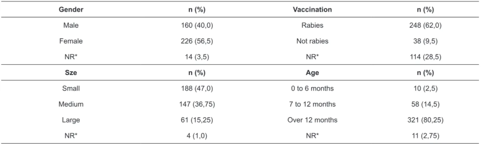
TABLE 1: Gender, size, vaccination, and age distributions of the 400 sampled dogs.
Gender n (%) Vaccination n (%)
Male 160 (40,0) Rabies 248 (62,0) Female 226 (56,5) Not rabies 38 (9,5)
NR* 14 (3,5) NR* 114 (28,5)
Sze n (%) Age n (%)
Small 188 (47,0) 0 to 6 months 10 (2,5) Medium 147 (36,75) 7 to 12 months 58 (14,5)
Large 61 (15,25) Over 12 months 321 (80,25)
NR* 4 (1,0) NR* 11 (2,75)
*NR: it was not rated (no information).
From 300 μL of peripheral blood, DNA was extracted
according to Araújo et al.3. The DNA pellet was hydrated with 50 μL of TE buffer (10 mM Tris, 1 mM EDTA, pH 8.0), and frozen at −20°C.
Initially, LEISH-1 and LEISH-2 primers4 were used to amplify the kDNA (kinetoplast) of L. (L.) infantum. The negative samples were subjected to PCR using 13A and 13B primers5 to amplify the kDNA of the genus Leishmania. The reaction
(25 μL) mixture was composed of 0.4 μM of each primer
(Sigma), 1.5 mM MgCl2, 0.2 mM dNTP (Invitrogen), 1.5 U Taq DNA Polymerase (Phoneutria), 1× enzyme buffer, and 2
μL of DNA. Amplification was performed in a thermocycler (BIORAD, T100 Thermal Cycler) at 95°C/5 min, followed by 35 cycles each at: 95°C/30 sec, 57.5°C (LEISH-1/LEISH-2 primers) and 61°C (13A/13B primers) for 30 sec each, 72°C/30 sec, and finally 72°C/10 min. Three positive controls [DNA of
L. (V.) braziliensis, L. (L.) amazonensis, and L. (L.) infantum] and a negative (water) control were used.
Samples with negative PCR results for Leishmania were
subjected to PCR with internal canine control. Specific primers for β-actin Forward (5′-CTTCTACAACGAGCTGCGCG-3′)
and Reverse (5'-TCATGAGGTAGTCGGTCAGG-3') were used
(GenBank: NM_001195845.1). The reaction (15 μL) mixture
was composed of 0.4 mM of each primer (Sigma), 1.0 mM MgCl2, 0.2 mM dNTP (Invitrogen), 1.5 U Taq DNA Polymerase
(Phoneutria), 1× enzyme buffer, and 1 μL of DNA. Amplification was performed in the thermocycler at 95°C/5 min, followed by 35 cycles each at: 95°C/30 sec, 54.9°C/30 sec, 72°C/1 min, and finally at 72°C/10 min. A negative control (water) was used.
For electrophoresis, 8 μL of the amplified product was loaded
on agarose gel stained with ethidium bromide. Presence of bands
was identified in a transilluminator (Loccus).
Peripheral blood smears prepared using glass slides were stained with Giemsa for studying Ehrlichia and Leishmania (parasitological test).
The 400 analyzed dogs (Table 1) belonged to different ages, races, sizes, and genders; some of them had been given
the anti-rabies vaccine and majority (76.0%) were bred. Some
of this information was not found because the animal files were
not dated; however, biological samples from such animals were also analyzed.
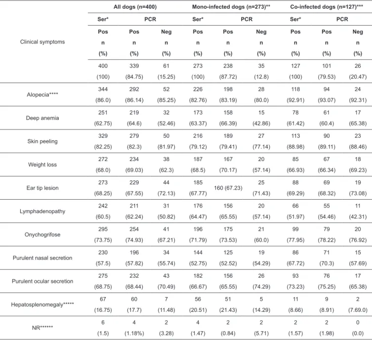
The PCR results were positive for 84.75% of the samples. Among the samples subjected to parasitological test, 63.25% were positive for Leishmania and 31.75% for Ehrlichia. Among the former, 38.94% were negative for Leishmania and 29.79% were positive for Ehrlichia, based on the parasitological test. Among the samples negative for Leishmania (15.25%) according to PCR analysis, the parasitological test showed that 75.41% were positivity for Leishmania and 42.62% for Ehrlichia (Table 2).
All the animals showed more than three VCL-compatible clinical signs. Among the mono-infected dogs, the proportions of samples that were positive for Leishmania were 87.18% and 56.78% according to the PCR and parasitological test, respectively. Among the co-infected dogs, 79.53% and 77.17% were positive for Leishmania based on the PCR and parasitological analyses, respectively (Table 2).
Many samples were identified as positive based on PCR analysis as compared to those identified by serological testing.
PCR has been used for the diagnosis of diseases, and for epidemiological studies; it is the most sensitive technique.
The analyzed animals had previously been diagnosed as serologically positive for VCL by CCZ using the DPPTM method and ELISA. According to Hirschmann6, this method showed a sensitivity of 30% and a specificity of 94.8%, and the best results can be obtained by a combination of DPPTM/RIFI and standard gold ELISA. Although serological tests are more sensitive than molecular tests, many factors such as quantities of serum antibodies that are higher than the quantity
of parasite DNA, may affect the specificity. Therefore, PCR is
a reliable methodological alternative, due to high sensitivity
and specificity.
TABLE 2: Individual breakup of the number of dogs experiencing each of the main clinical symptoms; serological and PCR test results of the peripheral blood samples of dogs; and their correlation.
Clinical symptoms
All dogs (n=400) Mono-infected dogs (n=273)** Co-infected dogs (n=127)***
Ser* PCR Ser* PCR Ser* PCR
Pos n (%) Pos n (%) Neg n (%) Pos n (%) Pos n (%) Neg n (%) Pos n (%) Pos n (%) Neg n (%) 400 (100) 339 (84.75) 61 (15.25) 273 (100) 238 (87.72) 35 (12.8) 127 (100) 101 (79.53) 26 (20.47) Alopecia**** 344 (86.0) 292 (86.14) 52 (85.25) 226 (82.76) 198 (83.19) 28 (80.0) 118 (92.91) 94 (93.07) 24 (92.31)
Deep anemia 251 (62.75) 219 (64.6) 32 (52.46) 173 (63.37) 158 (66.39) 15 (42.86) 78 (61.42) 61 (60.4) 17 (65.38)
Skin peeling 329 (82.25) 279 (82.3) 50 (81.97) 216 (79.12) 189 (79.41) 27 (77.14) 113 (88.98) 90 (89.11) 23 (88.46)
Weight loss 272 (68.0) 234 (69.03) 38 (62.3) 187 (68.5) 167 (70.17) 20 (57.14) 85 (66.93) 67 (66.34) 18 (69.23)
Ear tip lesion 273 (68.25) 229 (67.55) 44 (72.13) 185
(67.77) 160 (67.23) 25 (71.43) 88 (69.29) 69 (68.32) 19 (73.08) Lymphadenopathy 242 (60.5) 211 (62.24) 31 (50.82) 176 (64.47) 156 (65.55) 20 (57.14) 66 (51.97) 55 (54.46) 11 (42.31) Onychogrifose 295 (73.75) 254 (74.93) 41 (67.21) 196 (71.79) 175 (73.53) 21 (60.0) 99 (77.95) 79 (78.22) 20 (76.92)
Purulent nasal secretion 230 (57.5) 196 (57.82) 34 (55.74) 144 (52.75) 125 (52.52) 19 (54.29) 86 (67.72) 71 (70.3) 15 (57.69)
Purulent ocular secretion 275 (68.75) 232 (68.44) 43 (70.49) 182 (66.67) 156 (65.55) 26 (74.29) 93 (73.23) 76 (75.25) 17 (65.38) Hepatosplenomegaly***** 67 (16.75) 60 (17.7) 7 (11.48) 56 (20.51) 51 (21.43) 5 (14.29) 11 (8.66) 9 (8.91) 2 (7.69.0) NR****** 6 (1.5) 4 (1.18%) 2 (3.28) 4 (1.47) 2 (0.84) 2 (5.71) 2 (1.57) 2 (1.98) 0 (0.0)
*Ser: serological tests; **Mono-infected dogs: serological positive for Leishmania and parasitological negative for Ehrlichia; ***Co-infected dogs: serological positive for Leishmania and parasitological positive for Ehrlichia; ****Alopecia: generalize or local; *****Hepatosplenomegaly: although it was not one of the most frequent signs, it was frequent in VCL; ******NR: it was not rated (no information); PCR: polymerase chain reaction.
for Leishmania, 42.62% were positive for Ehrlichia. Clinical signs of the dogs that were PCR-negative for leishmaniasis may indicate Ehrlichia infection, because both the parasites show similar clinical signs7. Failed serological tests have been reported as false-positives due to cross-reactions with other pathologies, and due to chronic and old infections7,8. The Bio-Manguinhos ELISA kits use the promastigotes of Leishmania major, the species that causes cutaneous leishmaniasis, as the antigen, which generates false results. Although cross-reactivity in animals infected with Ehrlichia is a limitation of the Bio-Manguinhos kits, the combination
of ELISA and DPPTM significantly improves sensitivity and specificity9 and overcomes this limitation. Combination of serological methods and molecular tests increases the accuracy of disease detection. Thus, peripheral blood PCR of kDNA can be combined with the serological tests and clinical signs to increase the accuracy of VCL diagnosis.
Based on PCR analysis 15.25% of the animals were negative for Leishmania, despite clinical signs of VCL. The
sensitivity and specificity PCR can be affected by the choice of
One of the important advantages of using peripheral blood is that the collection is less invasive than from bone marrow, lymph node, and spleen aspirates, and the samples do not require special processing. However, the parasite concentration of the peripheral blood is lower than that of the bone marrow, lymph nodes, and spleen. Another disadvantage of peripheral blood is the presence of inhibitors that affect PCR sensitivity11.
The choice of the primers is important, because it can
influence the sensitivity of the technique, in addition to allowing
the differentiation of subgenera and species. The combination of two pairs of primers increased PCR sensitivity and indicated the possibility of the presence of other animal-infecting Leishmania
species. Recently LEISH1/LEISH2 primers amplified L. (V.) braziliensis, contrary to the possibility of identifying only L. (L.) infantum; however, infection by the former is low in dogs
and thus needs another method of identification12.
All the control samples were positive for Leishmania in the PCR analysis. The DNA extraction method interferes with PCR sensitivity. Extracting DNA from the blood samples was
efficient. The PCR analysis showed that all the samples were positive for the canine β-actin gene, representing the absence
of PCR inhibitors and excellent DNA integrity.
PCR is essential for the detection and identification of
the protozoan involved in the advancement of clinical signs. In addition to monitoring the parasite load, the possibility of analyzing different clinical samples with high sensitivity and
specificity makes PCR an undoubtedly advantageous method
when compared to the traditional diagnostic methods.
Clinical signs of leishmaniasis can vary because the disease shows several pathological mechanisms. Among the several clinical signs and symptoms, skin lesions, generalized lymphadenomegaly, progressive weight loss, muscular atrophy, decreased appetite, lethargy, splenomegaly, ocular lesions, epistaxis, onychogryphosis, vomiting, and diarrhea are prominent. All the dogs studied showed more than three signs. Alopecia and skin peeling, the classical dermatological alterations of leishmaniasis, were observed in most of the dogs; the dermatitis varied in extent and severity.
Anemia was observed in 62.75% of the animals. This is commonly caused by chronic kidney disease or because reduction of erythropoiesis in chronic diseases, which is aggravated by blood loss, immunosuppression, or destruction of blood cells. Co-infection with Ehrlichia also contributed to anemia.
Weight loss was observed in 68.0% of the animals. This finding is in agreement with other studies13,14 and can be explained by albuminuria triggered by protein imbalance and gastric mucosal involvement.
Ear tip lesions were observed in 68.25% of the animals. Lesions are more frequently located at the ear tip, muzzle, face, and ears, because they are more exposed.
Lymphadenopathy was detected in 60.5% of the animals, which was also described in other studies15. The increase in cell numbers of the phagocytic mononuclear system when the infection is installed, explains this.
Onychogryphosis, the most striking features of VCL, was detected in majority of the animals (73.75%). The parasite can stimulate the nail matrix to grow, and the apathy of animal decreases its movements; therefore, there is no natural nail wear.
Purulent ocular secretion occurred in 68.75% of the animals, which may be due to deposition of immunocomplexes and anti-Leishmania antibodies in ocular tissues.
Hepatosplenomegaly, although infrequent in the studied animals, is a common clinical sign, which may occur due to B cell and macrophage production, and proliferation of amastigotes.
Although the analyzed animals had previously been diagnosed with VCL at CCZ, it is important to emphasize that the objective, among other points, is not to question the
adopted methods, but to confirm the positive samples through
standardization of PCR as a complementary method to conventional tests, especially in uncertain cases, as in the case
of asymptomatic animals, which require species identification,
and when serological methods are non-resolute. The samples used in this study to evaluate the sensitivity of conventional
PCR reflect the reality of CCZ routine. The present work is the first step of a more complex study that aims to validate the
complementary molecular test as routine exams.
The clinical picture of mono-infected Leishmania cases does not change with co-infection of Ehrlichia, although the latter may aggravate the clinical signs. In addition, clinical proximity and endemicity between VCL and ehrlichiosis may
make diagnosis difficult. In areas endemic for leishmaniasis and
ehrlichiosis, conventional PCR can be used for the diagnosis of VCL in combination with other traditional techniques that identify co-infection of Ehrlichia spp., and especially in dubious
cases that need species identification.
Acknowledgments
The authors would like to thank CCZ for collecting the biological samples and realizing the parasitological tests.
Conflict of Interest
The authors declare that they have no conflicts of interest.
Financial Support
Financial support was provided by Conselho Nacional de Desenvolvimento Científico e Tecnológico (CNPq) and Fundação de Apoio ao Desenvolvimento do Ensino, Ciência e Tecnologia do Estado de Mato Grosso do Sul (Fundect).
REFERENCES
1. Ministério da Saúde (MS). Secretaria de Vigilância em Saúde. Manual de Recomendações para Diagnóstico, Tratamento e Acompanhamento de Pacientes com a Coinfecção Leishmania-HIV. 1ª edição. Brasília: MS; 2011. 112 p.
2. Nunes RV, Quaresma PF, Rugani JMN, Teixeira-Neto RG, Vahia LS, Silva ES. Molecular characterization of Leishmania infantum
3. Araújo FR, Ramos CAN, Luíz HL, Péres IAHFS, Oliveira RHM, Souza IIF, et al. Avaliação de um protocolo de extração de DNA genômico a partir de sangue total. Embrapa Comunicado Técnico. 2009;120:1-5. 4. Francino O, Altet L, Sánchez-Robert E, Rodriguez A,
Solano-Gallego L, Alberola J, et al. Advantages of real-time PCR assay for diagnosis and monitoring of canine leishmaniosis. Vet Parasitol. 2006;137(3-4):214-21.
5. Rodgers MR, Popper SJ, wirth DF. Amplification of kinetoplast DNA as a tool in the detection and diagnosis of Leishmania. Exp Parasitol. 1990;71(3):267-75.
6. Hirschmann LC, Brod CS, Jradin J, Simon CF, Recuero ALC. Leishmaniose Visceral Canina: Comparação de métodos sorológicos em cães de área indene do Rio Grande do Sul no Brasil. Rev Patol Trop. 2015;44(1):33-44.
7. Sousa VRF, Almeida ABPF. Co-infecção entre leishmaniose visceral e ehrlichiose monocítica em cães de Cuiabá, Mato Grosso. Acta Sci Vet. 2008;36(2):113-7.
8. Faria AR, Andrade HM. Diagnóstico da leishmaniose visceral canina: grandes avanços tecnológicos e baixa aplicação prática. Rev Pan-Amaz Saude. 2012;3(2):47-57.
9. Gomes YM, Paiva Cavalcanti M, Lira RA, Abath FGC, Alves LC. Diagnosis of canine visceral leishmaniasis: biotechnological advances. Vet J. 2008;175(1):45-52.
10. Lachaud L, Chabbert E, Dubessay P, Reynes J, Lamothe J, Bastien P. Comparison of various sample preparation methods for PCR diagnosis of visceral leishmaniasis using peripheral blood. J Clin Microbiol. 2001;39(2):613-7.
11. Reithinger R, Espinoza JC, Courtenay O, Davies CR. Evaluation of PCR as a diagnostic mass-screening tool to detect Leishmania (Viannia) spp. in domestic dogs (Canisfamiliaris). J Cli Microbiol. 2003;41(4):1486-93.
12. Dantas-Torres F, Sales KGS, Siva LG, Otranto D, Figueredo LA. Leishmania-FAST15: A rapid, sensitive and low-cost real-time PCR assay for the detection of Leishmania infantum and Leishmania braziliensis kinetoplast DNA in canine blood samples. Mol Cell Probes. 2017;31:65-9.
13. Almeida MAO, Jesus EEV, Atta MLBS, Alves LC, Berne MEA, Atta AM. Clinical and serological aspects of visceral leishmaniasis in Northeast Brazilian dogs naturally infected with Leishmania chagasi. Vet Parasitol. 2005;127(3-4):227-32.
14. Almeida ABPF, Mendonça AAJ, Sousa VRF. Prevalence and epidemiology of visceral leishmaniasis in dogs and humans in the city Cuiaba, Mato Grosso, Brazil. Cienc Rural. 2010;40(7):1610-5. 15. Mattos JR DG, Pinheiro JM, Menezes RC, Costa DA. Aspectos