Ocular alterations in dogs naturally infected by Leishmania (Leishmania) chagasi
[Alterações oculares em cães infectados naturalmente por Leishmania (Leishmania)chagasi]
F.L.C. Brito1, L.C. Alves1, F.C.L. Maia1, E.S.C. Santos1, J.L. Laus2, I.M.J. Meunier1
1
Universidade Estadual de Maringá – Umuarama, PR
2Universidade Federal Rural de Pernambuco – Recife, Pe 3
Faculdade de Ciências Agrárias e Veterinárias – UNESP Via de Acesso Professor Paulo Donato Castellane, s/n
14884-900 - Jaboticabal, SP
ABSTRACT
Ocular conditions, anti-Leishmania antibodies and total protein of the aqueous humor were studied in dogs naturally infected by Leishmania (Leishmania) chagasi. Fifty dogs were analyzed and assigned into two groups of 25 animals each. All animals were submitted to routine ophthalmic exam. Results showed that 76% of the affected animals presented ocular signs, being uveitis the predominant. The mean of total protein in the aqueous humor of animals with uveitis was higher (P<0.05) when compared to the mean of animals with other ocular signs or no clinical sign. The anti-Leishmania chagasi antibody values in plasma were superior to those found in the aqueous humor (P<0.05).
Keywords: dog, Leishmania (Leishmania) chagasi, aqueous humor, antibodies, protein
RESUMO
Estudaram-se as condições oculares, os anticorpos anti-Leishmania e os valores de proteína total no humor aquoso de cães infectados naturalmente por Leishmania (Leishmania)chagasi. Analisaram-se 50 cães, divididos em dois grupos de 25 animais. Todos os animais foram submetidos a exame oftálmico rotineiro. Os resultados mostraram que 76% dos cães infectados apresentaram sinais oculares, sendo a uveíte a alteração predominante. A média de proteína total no humor aquoso dos animais com uveíte foi maior (P<0,05), que a dos animais com outros sinais oculares ou sem qualquer sinal. Os valores de anticorpos anti-L. chagasi obtidos no plasma foram estatisticamente superiores aos encontrados no humor aquoso (P<0,05).
Palavras-chave: cão, Leishmania (Leishmania) chagasi, humor aquoso, anticorpo, proteína
INTRODUCTION
In Brazil, canine visceral leishmaniasis (CVL), also known as canine calazar, is caused by L. chagasi and affects mainly domestic and wild canides and men. The infection by Leishmania
sp. usually causes a systemic disease with chronic evolution, clinically apparent from three
months to a few years after infection, and its
Recebido em 24 de novembro de 2004 Aceito em 14 de fevereiro de 2006
*Autor para correspondência (corresponding author) E-mail: fabiobrito@click21.com.br
known to be the most frequent alterations, although granulomatous or lymphocytic uveitis, associated to corneal edema and synechiae, have prevailed (Garcia-Alonso et al., 1996; Peña et al., 2000).
Increased level of total plasmatic protein has been observed in CVL. Due to the ocular findings, particularly in the anterior segment of the eye (Puchol and Gonzáles, 1989; Molleda et al., 1993), the aqueous humor plays an important role on the course of the disease (Novales et al., 1994). According to aqueous humor alterations, antibody detection and dosage of protein level revealed an useful method in the diagnosis of ocular affections (Hazel et al., 1985; Garcia-Alonso et al., 1996), despite it is not frequently used in canine leishmaniasis (Blogg and Colles, 1971).
Because of the importance of canine calazar on public health and its association to ophthalmopathies, particularly in endemic regions, this paper aimed to study the ocular conditions, specific antibodies and total protein dosage of the aqueous humor of dogs naturally infected by L. chagasi.
MATERIAL AND METHODS
Fifty dogs of several breeds and ages, males and females, coming from the metropolitan region of Recife and Bezerros cities, Pernambuco, Brazil, were analyzed. All the animals were clinically evaluated as well as submitted to routine ophthalmic examination. The animals were assigned into two groups composed by 25 dogs. The infected group (IG) was formed by animals positive for L. chagasi according to parasitological and serologic exams, presenting or not ocular alterations. The control group (CG) was composed by animals with negative results for L. chagasi according to parasitological and serologic exams, without clinical signs of systemic disease or ocular alterations. Parasitological diagnosis was accomplished after analysis of cutaneous scraps and bone marrow fluid, which was aspirated by punction in the sternum bone, besides the visualization of the amastigotas forms of L. chagasi. The serological diagnosis was done by indirect
immunofluorescence1, as described by Camargo (1977).
Following recommendations of the World Health Organization (World…, 2003), the animals were euthanased. Immediately after, it was performed the paracentesis of the anterior chamber at limbal portion of both eyes to collect the aqueous humor using a 13×4.5mm gauge needle coupled to a 3ml disposable syringe. The samples were divided and disposed into 1ml plastic microtubes, which were identified and stored at the -20ºC. Total protein was determined using the Coomassie blue technique for the analyses of microproteins (Bradford, 1976). The ELISA test was performed according to the instructions of the manufacturer for the diagnosis of canine calazar using S7 recombining peptides. The Goldmann-Witmer coefficient was obtained according to Hill et al. (1995).
For the statistical analysis, the Student (t) test was used to assess differences of protein concentration. Total proteins of the aqueous humor were studied using the analysis of variance. Means were compared using the Tukey test (P<0.05). The Signs test was applied to evaluate the results from the ELISA, with two variables: magnitude of the results and interpretation of positive and negative results using the Pearson’s correlation coefficient.
RESULTS
From the 25 animals naturally infected by L. chagasi, 76% presented ocular lesions at ophthalmic examination. It was observed that, amongst the animals that presented ocular signs, 73.7% had only one type of alteration, while 26.3% showed more than one alteration in the same eye. Unilateral ocular alterations were observed in 10.5% of the dogs, while 89.5% presented bilateral alterations. It was also noted that the anterior chamber was affected in 76% and 4% of the animals presented alterations in both ocular chambers. In some cases, visualization of the inner eye structures was disturbed by opacifications of the aqueous humor.
The evaluation of the occurrence of ocular lesions showed that uveitis was the main alteration. This condition was bilateral in five dogs (5/25). Two dogs (2/25) with uveitis presented hyphema (Fig. 1). Glaucoma secondarily to uveitis was found in one (1/25) dog naturally infected by L. chagasi. Regarding other ocular alterations, 16% of the dogs developed blepharitis. Edema, seborrheic dry exudate and thickening with a fibrotic appearance of the eyelids were also observed in one animal (Fig. 2). The conjunctiva was affected in 40% (10/25) of the animals infected by L. chagasi. The lesion was restricted to the conjuctiva in 20% (2/10) of them (Fig. 3). The
involvement of the conjunctiva and cornea was observed in 20% (5/25) of the animals (Fig. 2), being keratitis the second most frequent finding associated to conjunctivitis. From the animals with keratoconjunctivitis, 8% (2/25) presented the keratoconjunctivitis sicca (Fig. 4). Isolated corneal lesions were observed in 12% (3/25) of the dogs. Two animals presented keratitis and four dogs exhibited superficial corneal ulceration with a focused edema. Coriorretinitis was observed in only one animal. Twenty per cent (5/25) of the animals did not exhibit any ocular problems.
Figure 1. Discrete iris edema with hyphema in dog naturally infected by Leishmania (Leishmania) chagasi, Pernambuco State, Brazil, 2004.
Figure 2. Diffuse blepharedema with periocular alopecia, seborrheic dry exudate and thicknening fibrotic eyelids, keratitis with corneal edema, pigmentation and neovascularization in a dog with visceral leishmaniasis, Pernambuco State, Brazil, 2004.
Figure 3. Conjunctivitis in a dog with visceral leishmaniasis. Note the prominent thickening of the ventral bulbar conjuctiva, Pernambuco State, Brazil, 2004.
The mean protein level was the same for both right and left eyes, (P>0.05). Comparing the means of infected and control groups, a significant increase of the values for the infected IG group was observed (Table 1). When the aqueous humor protein concentration of dogs with CVL were evaluated, according to ocular signs (Table 2), differences between the mean values (P<0.05) were found. The mean of total protein concentration of the aqueous humor in animals with uveitis was higher (P<0.05) when compared to animals with other ophthalmopathies or without ocular alterations. It was also observed a difference (P<0.05) between animals clinically healthy and those with leishmaniasis, regardless of ocular lesion. Dogs presenting canine visceral leishmaniasis had total protein values seven times higher than those found in animals of the control group. It was interesting to note that the highest variability of protein values was observed amongst individuals without ocular signs (Table 2). However no difference was found amongst animals that presented others signs besides uveitis.
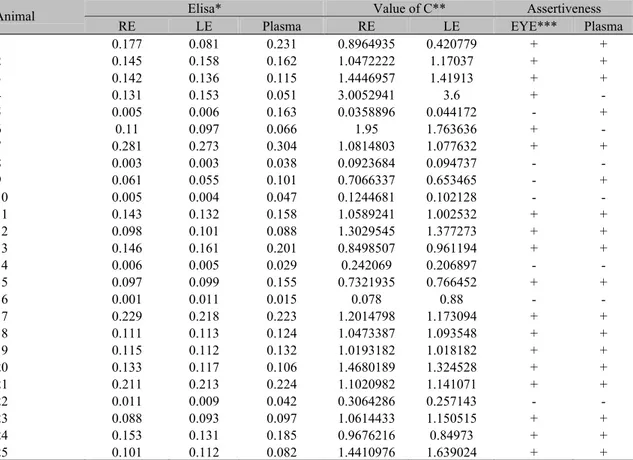
Statistical analysis of the anti-L. chagasi IgG concentration, in the aqueous humor as measured by optical density, showed similarity in both right and left eyes. However values obtained from plasma were superior to those found in the eyes (P<0.05) (Table 3).
The ELISA test using the S7 recombinant peptide detected antibodies anti-L. chagasi, expressed in optical density, in the aqueous humor of naturally infected dogs with high IgG titers. Some of these animals presented anti-L.
chagasi IgG in the aqueous humor,
independently of their presence in the plasma (Table 3).
As the Pearson’s correlation coefficient was evaluated, regarding both right (r=0.828) and left (r=0.748) eyes, a significant positive correlation was observed when all the animals of this study were evaluated. It means that when the level of anti-L. chagasi antibodies increases in the plasma, it will also increase in the aqueous
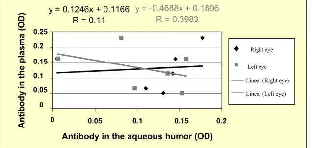
humor (Fig. 5). Both right (r=0.110) and left (r=0.398) eyes of the animals with uveitis were evaluated and it was observed that the higher the level of anti-L. chagasi antibodies in the eye, the lower is its level in the plasma (Fig. 6). Although a significant correlation could not be confirmed.
The Goldman-Witmer coefficient (C) was used to verify the occurrence of local intraocular antibodies production. When C value is greater than one there is local production of intraocular antibodies (Hill et al., 1995). A C value greater than one was observed in 56% of the studied animals. Positive results from the aqueous humor and plasma samples were observed in 72% out of the 25 studied animals, from which two dogs were positive only to the aqueous humor test and another two only to the plasma test (Table 3). Paired analyses of the aqueous humor and plasma confirmed the diagnosis in 80% of the cases.
DISCUSSION
The frequency of the ocular alteration was low when compared to the results obtained by Molleda et al. (1993), reporting ocular alterations in 80.5% of the 41 animals with canine leishmaniasis. In a retrospective study, only 24.4% of the dogs infected by Leishmania infantum presented ocular alterations (Peña et al., 2000). The differences of these results are probably due to the pathogenicity of the
Table 1. Levels of total protein (mg/dl) of the aqueous humor of both right and left eyes of dogs infected by Leishmania (Leishmania) chagasi, Pernambuco State, Brazil, 2004
Infected Group Control Group
Mean Minimum- maximum
values Mean
Minimum- maximum values
Right eye 138.68±58.61a 30-289 26.2±12.98b 10-53
Left eye 149.88±56.27a 66-270 25.52±12.43b 10-51
Mean 144.28±16.24a - 25.86±3.59b -
Means in the same column with common letter do not differ (P>0.05) and means in the same row with no common letter differ (P<0.05) according to the Student test.
Table 2. Levels of total protein (mg/dl) of the aqueous humor of dogs naturally infected by Leishmania (Leishmania) chagasi with and without ocular signs, Pernambuco State, Brazil, 2003
Ocular sign N** Mean Minimum-maximum
values
Uveitis 12 200.67±33.61a 129-289
Other* 26 134.77±16.96b 81-231
Without alteration 12 108.5±32.78b 30-252
Means in the column with no common letters differ (P<0.05) according to Tukey test.
* Other signs: conjunctivitis, keratoconjunctivitis, keratoconjunctivitis sicca, keratitis, ulcerative keratitis. ** Number of eyes used to dose protein.
Table 3. Levels of antibodies to Leishmania chagasi, coefficient of Goldmann-Witmer and assertiveness in the aqueous humor and plasma of dogs naturally infected by Leishmania (Leishmania) chagasi, Pernambuco State, Brazil, 2004
Elisa* Value of C** Assertiveness
Animal
RE LE Plasma RE LE EYE*** Plasma
1 0.177 0.081 0.231 0.8964935 0.420779 + +
2 0.145 0.158 0.162 1.0472222 1.17037 + +
3 0.142 0.136 0.115 1.4446957 1.41913 + +
4 0.131 0.153 0.051 3.0052941 3.6 + -
5 0.005 0.006 0.163 0.0358896 0.044172 - +
6 0.11 0.097 0.066 1.95 1.763636 + -
7 0.281 0.273 0.304 1.0814803 1.077632 + +
8 0.003 0.003 0.038 0.0923684 0.094737 - -
9 0.061 0.055 0.101 0.7066337 0.653465 - +
10 0.005 0.004 0.047 0.1244681 0.102128 - -
11 0.143 0.132 0.158 1.0589241 1.002532 + +
12 0.098 0.101 0.088 1.3029545 1.377273 + +
13 0.146 0.161 0.201 0.8498507 0.961194 + +
14 0.006 0.005 0.029 0.242069 0.206897 - -
15 0.097 0.099 0.155 0.7321935 0.766452 + +
16 0.001 0.011 0.015 0.078 0.88 - -
17 0.229 0.218 0.223 1.2014798 1.173094 + +
18 0.111 0.113 0.124 1.0473387 1.093548 + +
19 0.115 0.112 0.132 1.0193182 1.018182 + +
20 0.133 0.117 0.106 1.4680189 1.324528 + +
21 0.211 0.213 0.224 1.1020982 1.141071 + +
22 0.011 0.009 0.042 0.3064286 0.257143 - -
23 0.088 0.093 0.097 1.0614433 1.150515 + +
24 0.153 0.131 0.185 0.9676216 0.84973 + +
25 0.101 0.112 0.082 1.4410976 1.639024 + +
y = 0.77x + 0.0457
R = 0.7481
y = 0.8219x + 0.0367
R = 0.8280
0 0,05 0,1 0,15 0,2 0,25 0,3 0,35
0 0,05 0,1 0,15 0,2 0,25 0,3
Antibody in the aqueous humor (OD)
A
n
ti
body
i
n
the
pl
a
s
m
a
(O
D
)
Figure 5. Pearson’s correlation coefficient among the level of anti-L. chagasi antibodies measured by optical density (OD) in the plasma and in the aqueous humor of dogs naturally infected by Leishmania (Leishmania) chagasi, using the ELISA/S7, Pernambuco State, Brazil, 2004.
y = 0.1246x + 0.1166
R = 0.11
y = -0.4688x + 0.1806
R = 0.3983
0 0,05 0,1 0,15 0,2 0,25
0
0,05
0,1
0,15
0,2
Antibody in the aqueous humor (OD)
A
n
ti
body
i
n
t
h
e
pla
s
m
a
(
O
D
)
Figure 6. Pearson’s correlation coefficient among the level of anti-Leishmania (Leishmania) chagasi
antibodies measured by optical density (OD) in the plasma and in the aqueous humor of dogs with uveitis, naturally infected by Leishmania (Leishmania) chagasi, using ELISA/S7, Pernambuco State, Brazil, 2004.
♦
Right eye
Left eyeLineal (Right eye)
Lineal (Left eye)
0.05 0.1 0.15 0.2 0.25 0.3 0.3
0.35
0.3
0.25
0.2
0.15
0.1
0.05
0.05 0.1 0.15 0.2 0.05
0.1 0.15 0.2
0
0 0.25
♦
Right eye
Left eyeLineal (Right eye)
Uveitis was the most frequent ophthalmic finding, as observed in other studies (Molleda et al., 1993; Feitosa et al., 2000). In dogs with ocular and periocular alterations associated to CVL, anterior uveitis occurred in a frequency of 42% (Peña et al., 2000), which was a higher rate than that obtained in this study. Nevertheless, up to 70% of the dogs with visceral leishmaniasis from endemic areas may present uveitis. Immune complex deposition on vascular walls may cause uveitis in dogs with leishmaniasis (Pumarola et al., 1991; Garcia-Alonso et al., 1996). Hyphema is also a common finding in dogs with CVL. Eyelid lesions were identified. Blepharitis has been described as one of the most common findings in canine calazar (Ferrer, 1999).
Conjunctival lesions have been frequently identified in CVL dogs. In this study, around 75.6% of dogs were infected by Leishmania spp. In spite of being an isolated finding, conjunctival inflammation was observed in only 34.1% of the animals (Molleda et al., 1993). Keratoconjunctivitis sicca in visceral leishmaniasis may sometimes be due to lacrimal glands inflammation or obstruction of their secretory ducts (Molleda et al., 1993). In spite of the rare isolated involvement of the cornea, similar lesions were also found by other authors (Molleda et al., 1993; Peña et al., 2000).
Unlike other systemic diseases with ocular manifestations, leishmaniasis shows some preference for the anterior chamber of the eye (Puchol and Gonzáles, 1989). In this study, only one animal presented posterior chamber lesion. Ocular conditions were not observed in some of the studied dogs. It is known that the absence of ocular signs may occur in 10-50% of the canine population because of natural resistance to infection (Abranches et al., 1991). Interleukin–2 (IL-2) and tumor necrosis factor alpha (TNF-α) play an important role on protection against clinical disease (Pinelli et al., 1994).
The level of total protein of the aqueous humor of control group animals was within normal values as described by other authors (Hazel et al., 1985; Krohne et al., 1995). However, there are no data for dogs with leishmaniasis. In this study, dogs naturally infected by L. chagasi presented higher aqueous humor total protein values than clinically healthy dogs. Animals naturally infected by Leishmania spp. showed a higher total protein level than clinically healthy animals,
which is in agreement with Novalis et al. (1994). The mean, of the total protein of dogs presenting conjunctivis, keratoconjunctivitis, keratoconjunctivitis sicca, keratitis, ulcerative keratitis were 134.77±16.96mg/dl, which was statistically different from those found in the control group. Previous research comparing the mean protein values of the aqueous humor of healthy and Leishmania infantum infected dogs did not find significant differences (Novalis et al., 1994). In this study, the total protein mean value verified in the aqueous humor of dogs presenting uveitis and visceral leishmaniasis was 200.67±33.61mg/dl. Similar values (231.8mg/dl) were also found by other authors (Novalis et al., 1994).
The severity in which the aqueous humor barrier is broken may increase with protein concentration (Dernouchamps, 1982). This might be due to iris and ciliary vessel dilation (Krohne and Vestre, 1987), due to the presence of the parasite or deposition of immune complexes in the iris stroma (Molleda et al., 1993). Increase of total protein may also be correlated to the great amount of plamocytes at the ciliary body (Brito et al., 2004).
The recombinant ELISA technique used in this study was effective. Previous studies using ELISA showed a high titer of anti- Leishmania
IgG in the aqueous humor of dogs presenting uveitis (Alonso et al., 1995; Garcia-Alonso et al., 1996). Antibody detection in the aqueous humor of dogs presenting uveitis, despite of the level of antibody in the serum, may be related to the uveitis found in sick animals (Garcia-Alonso et al., 1996). In this study, specific antibodies were found in the aqueous humor of infected dogs presenting uveitis, but not in the plasma.
CONCLUSIONS
Uveitis and other ophthalmopathies, mainly of the anterior chamber of the eye, must be considered as a differential diagnosis in visceral leishmaniasis of animals from the endemic areas. The procedure for dosage of total protein of the aqueous humor is easy to perform and the values obtained in the infected animals may be used as a complementary exam for dogs infected by Leishmania (Leishmania) chagasi. It can be still admitted that intraocular immunogenic factors are associated to the uveitis in dogs with visceral leishmaniasis. Canine visceral leishmaniasis diagnosis may be aided by the use of the ELISA technique, using the S7 recombinant peptide being worth for the aqueous humor.
REFERENCES
ABRANCHES, P.; SANTOS-GOMES, G.; RACHAMIN, N. et al. An experimental model for canine visceral leishmaniasis. Parasite. Immunol., v.13, p.537-550, 1991. BLOGG, J.R.; COLES, E.H. Clinicopathological aspects of canine aqueous humour proteins. Res. Vet. Sci., v.12, p.95-100, 1971.
BRADFORD, M.M. A rapid and sensitive method for the quantitation of microgram quantities of protein utilizing the principal of protein-dye binding. Anal. Bioch., v.72, p.248-254, 1976.
BRITO, F.L.C.; ALVES, L.C.; ORTIZ, J.P.D. et al. Uveitis associated by Leishmania chagasi in dog from Olinda City, Pernambuco, Brazil. Ciên. Rural, v.34, p.925-929, 2004. CARMAGO, M.E. Cross-reactivity in immunofluorescense for Tryposoma and Leishmania antibodies. Am. J. Trop. Med. Hyg., v.18, p.500-505, 1977.
DERNOUCHAMPS, J. The proteins of the aqueous humor.
Doc. Ophthalmol., v.53, p.1993-248, 1982.
FEITOSA, M.M.; IKEDA, F.A.; LUVIZOTTO, M.C. et al. Aspectos clínicos de cães com leishmaniose visceral no município de Araçatuba – São Paulo (Brasil). Clin. Vet., v.28, p.36-42, 2000.
FERRER, L. Clinical aspects of canine leishmaniasis. In: INTERNATIONAL CANINE LEISHMANIASIS FORUM, 1., 1999, Barcelona. Proceedings... Barcelona, 1999. p.6-10. GARCIA-ALONSO, M.; BLANCO, A.; RINA, D. et al. Immunopathology of the uveitis in canine leishmaniasis.Parasite. Immunol, v.18, p.617-623, 1996. GARCIA-ALONSO, M.; NIETO, C.G.; VERDUGO, S.G. et al. Diagnostico y tratamiento de la uveítes anterior
inmunomediada en leishmaniosis canina. In: CONGRESSO IBÉRICO DE PARASITOLOGÍA, 4., 1995. Anais... 1995. p.198.
GARCIA-ALONSO, M.; MIRÓN, C., MOLANO, I. et al. Patología ocular asociada a leishmaniosis canina. Cons. Dif. Vet., v.6, p.49-53, 1998.
HAZEL, S.J.; THRALL, M.A.; SEVERIN, G.A. et al. Laboratory evaluation of aqueous humor in the healthy dog, cat, horse, and cow. Am. J. Vet. Res., v.3, p.657-659, 1985. HILL, S.L.; LAPPIN, M.R.; CARMAN, J. et al. Comparasion of methods for estimation of Toxoplasma gondii- specific antibody production in the aqueous humor of cat. Am. J. Vet. Res., v.56, p.1181-1187, 1995.
KROHNE, S.D.G.; VESTRE, W.A. Effects of flunixin meglumine and dexamethasone on aqueous protein values after intra-ocular surgery in the dog. Am. J. Vet. Res., v.48, p.420-422, 1987.
KROHNE, S.G.; KROHNE, D.T.; LINDLEY, D.M. et al. Use of laseer flaremetry to measure aqueous humor concentration in dog. J. Am. Vet. Med. Assoc., v.206, p.1167-1172, 1995.
LOPES, R.; NOVALES, M.; GINEL, P.J. et al. Concentrations d’immunoglobulines dans le plasma et l’humeur aqueuse de chiens atteints de leishmaniose. Rev. Méd. Vet., v.144, p.621-623, 1993.
MOLLEDA, J.M.; NOVALES, M.; GINEL, P.J. et al. Clinical and histopathological study of the eye in canine leishmaniasis. Isr. J. Vet. Med., v.48, p.173-178, 1993. NOVALIS, M.; LOPEZ, R.; GINEL, P.J. et al. Les effets de l’uvéite sur la concentration des proteins totales dans l’humeur aqueuse de chiens atteints de leishmaniose. Rev. Méd. Vet., v.145, p.257-259, 1994.
PEÑA, M.T.; ROURA, X.; DAVIDSON, M.G. Ocular and periocular manifestations of leishmaniasis in dog: 105 cases (1993-1998). Vet. Ophthalmol, v.3, p.35-41, 2000.
PINELLI, E.; KLILLICK-KENDRICK, R.; WAGENNAAR, J. et al. Cellular and humoral immune responses in dogs experimentally and naturally infected with Leishmania infantum. Infect. Immun., v.62, p.229-235, 1994.
PUCHOL, J.L.; GONZALEZ, J.L. Leishmaniasis ocular: afeciones del segmento anterior. In: NATIONAL. CONGRESS ASOCIACIÓN DE VETERINARIOS ESPANÕLES ESPECIALISTAS EN PEQUENOS ANIMALES, 1989, Barcelona. Proceedings...Barcelona: AVEPA, 1989. p.115-122.
PUMAROLA, M.; BREVIK, L.; BADIOLA, J. et al. Canine Leishmaniasis associated with systemic vasculitis in two dogs. J. Comp. Pathol., v.105, p.279-286, 1991.