Stereological Evaluation of Renal Glomeruli in Offspring of
Diabetic Female Rats
Abdolrahman Dezfoolian, Ph.D.¹, Marzieh Panahi, Ph.D.¹, Farideh feizi, Ph.D.2*
1. Anatomical Sciences Department, Faculty of Medicine, Jondi Shapoor Medical Sciences University, Ahwaz, Iran
2. Anatomical Sciences Department, Faculty of Medicine, Babol Medical Sciences University, Babol, Iran
* Corresponding Address: P.O.Box: 4717641367, Anatomical Sciences Department, Faculty of Medicine, Medical Sciences University of Babol, Babol, Iran.
Email: faridehfeizi@yahoo.com Abstract
Received: 20/َAug/2008, Accepted: 29/Oct/2008
Objective: Although in vitro studies have shown that high concentrations of glucose can induce dysmorphogenesis of the embryonic kidney, the possible adverse effects of exposure to intrauterine hyperglycemia on kidney development, especially in re-gard to nephrogenesis, has not been evaluated.
The aim of this study is to investigate the effects of maternal diabetes on glomeruli structures of the offspring, focusing on the following parameters: glomeruli volume and number, mesangium volume, mesangial cell number and glomerular capillary volume.
Materials and Methods: Before mating, fifteen female Sprague Dawley rats, divided into three groups, were diabetes induced by a single intraperitoneal dose of 65 mg/ kg streptozotocyn (STZ). After 30 days of breast feeding, ten offsprings from each group (two per mother) were randomly selected for kidney removal. The kidneys were weighed and their tissues were processed for light microscopy. Glomerular fea-tures were evaluated quantitatively using dissection as well as the Cavalieri method and were then compared with sham and control groups.
Results: At birth, the mean body weight of diabetic mothers’ offspring (DO) was sig-nificantly lower than that of the control group’s offspring (CO) and sham group’s off-spring (SO) (p=0.001), however, the mean body weight of the 30 day-old DO was not lower than that of CO and SO (p>0.05). The total renal volumes, cortical volumes, glomerular mean and total volumes, total mesangeal volumes, total capillary volumes and total glomerular numbers were significantly lower in the DO than in CO and SO (p<0.05). The numerical density of glomeruli and mesangial cells per glomeruli were significantly greater in DO than in CO and SO (p<0.05).
Conclusion: We concluded that intrauterine hyperglycemia is accompanied by a nephron deficit which may not be compensated within the first 30 days after birth. Keywords: Diabetes, Streptozotocyne, Gestational Diabetes, Diabetic Nephropathy, Histological Techniques
Introduction
A mother’s lifestyle may have long-lasting effects on her offspring. For example smoking, alcohol consump-tion, and presence of diseases such as diabetes affect fetal development (1). There is a strong association be-tween pregnancy in women with any form of diabetes and high infant mortality and morbidity rates (2). Ma-ternal diabetes has the potential to adversely affect the development of multiple organ systems, resulting in a wide range of congenital malformations including: cau-dal regression, situs inversus, kidney malformations, cardiac anomalies and neural tube defects (NTDs) (3). Metabolic changes in a diabetic pregnancy affect fetal
development and fetal glucose homeostasis. Several epidemiological studies (1, 4) and animal experiments (5-7) have shown that offsprings of diabetic mothers have higher incidences of glucose intolerance, obes-ity, insulin resistance, non-insulin- dependent diabetes mellitus (NIDDM) and hypertention in later life. There are several hypotheses relating maternal diabetes to adulthood diseases and potential stillbirths (8). Con-genital malformations are also more frequent in chil-dren of mothers with diabetes (9, 10), and are results of developmental defects occurring in early organo-genesis (11, 12). They include failure of neural tube closure, caudal regression syndrom and urogenital
normalities which can be as severe as renal agenesis (13), and are the main causes of perinatal mortality in offspring of diabetic mothers (14).
In humans, nephrogenesis occurs only during fetal life and stops after birth (15). In rats however, it occurs dur-ing the last third of gestation and continues for several days post birth (16). Maternal protein restrictions during the last third of gestation or during the period of ne-phrogenesis result in offspring which exhibit a marked reduction in glomeruli or nephron numbers and develop hypertension (17, 18). Intrauterine malnutrition due to placental dysfunction or poor maternal health, as well as maternal hyperglycemia/diabetes result in decreased nephron numbers (19). Amri et al. showed that the in-trauterine glucose concentration must be closely con-trolled to ensure optimum metanephros development. Severe diabetes leads to a decrease in fetal pancreatic weight, an increase in pancreatic endocrine tissue and a decrease in pancreatic and circulating insulin (20, 21). Interference with third trimester fetal growth has been shown to affect nephron development. Nephron number is undoubtedly genetically determined, but there is a growing body of evidence that the number of glomer-uli is additionally influenced by social and behavioral factors that effect intrauterine environment (22). Dia-betic glomerulopathy is characterized by a very slow development in basement membrane (BM) accumula-tion, manifested as thickening of the peripheral BM, increased volume of the mesangial BM-like material (BMLM) along with mesangial expansion. The initia-tion of the process is probably at the onset of diabetes since the BM thickening is detectable after a few years. In the long run however, the two in concert lead to the ultimate solidification of the glomerular tuft with loss of capillary surface. The end-stage is glomerular closure with elimination of glomerular function (23). Hughson et al (22) investigated the link between birth weight and glomerular number. Using the physical disector/frac-tionator combination in kidney autopsies, birth weight was also found to be a strong predictor of total glomeru-lar number and mean glomeruglomeru-lar volume in African American, Caucasian as well as Hispanic infants (24). Accurate estimation of glomerular volume has become increasingly important because Vglom varies signifi-cantly in normal human kidneys (25), and alterations in Vglom may play a significant pathophysiological role in the development or progression of a range of nephropathies (26). It is therefore, important to deter-mine whether exposure to the diabetic environment in utero influences nephrogenesis and final number of ne-phrons. In this study we estimated renal volume, abso-lute glomerular volume, total glomerular number, total mesangial cell number and mesangium volume in the offspring of diabetic female rats by the use of unbiased stereological methods.
Materials and Methods
15 female Sprague-Dawley rats, with an average weight
of 200-250g were obtained from the Pasture Institute in Tehran, Iran. The animals were divided into three groups: the experimental group, the control group which was treated with citrate buffer and the sham group of untreated rats. The experimental group received a sin-gle dose of intraperitoneal 65 mg/kg streptozotocyne (STZ) (Sigma-Aldrich) dissolved in 0.4 mol/l citrate buffer with pH: 4.5 (27). Presence of diabetes in the rats was glucometrically confirmed 3 days after STZ injection.
Animals with glucose levels greater than 300 mg/dl were considered manifestly diabetic and were includ-ed in the study. Diabetic females of the experimental group, as well as the non-diabetic females in the control and sham groups were housed (on female and one male in per cage) with males over night, and the appearance of a vaginal plug in the morning indicated that copula-tion and potential pregnancy had occurred. After birth, the offspring were housed in cages with standard condi-tions (temperathre breast feeding and water) and water ad libitum. Ten 30 day old offspring from each group, two per mother, were then randomly selected.
Tissue preparation
The offspring were anaesthetized with chloroform and were fixed by retrograde perfusion through the abdominal aorta with formal phosphate buffer. Right kidneys were then excised, decapsulated and immersed in the same fixative for 48 hours at room temperature. The fixed kidneys were embedded in 7% agar and sliced into 1 mm slices using a mac-rotome perpendicular to the longitudinal axis (28). Approximately 8-10 slices were obtained from each kidney. The slices were arranged in a number of se-quences on meshed tissue processing baskets and were then processed and embedded in paraffin. The blocks were systematically sectioned at 5 microm-eters, and every seventh section in order, with the first chosen randomly in the inteval. They were then mounted on glass slides and stained with periodic acid-Schiff (PAS). Every selected (1st and 7th) slice underwent a stereological analysis which involved the use of Olympus Optical BH2 microscopes. To estimate the volume of cortex, medulla and the whole kidney, every first section was viewed on a Motic loop microscope at a magnification of 20× with the image being projected on a computer moni-tor. A fine grid of points distanced every 6 milim-eters was superimposed over the visual field of the sampled sections. Point counting using Cavalieri principle was then used to estimate the volume of cortex, medulla and whole kidney using the follow-ing equation (29).
∑P.a/p .t V = ────── (1)
M²
counted in sections, a/p is the area associated with each point, t is the distance between two consecutive sections in which points were counted, M is the linear magnification of the projector. The volume fraction (Vv) of the glomer-uli was obtained by use of the estimated point count of the random cross sections in the following equation:
∑Pp Vv (glom/cortex) = ──── (2)
∑ P t
∑Pp is the sum of all counted points overlying glomeruli. ∑Pt is the sum of probe points of n field.
Absolute volume of glomeruli was estimated indirectly by multiplying the volume fraction by volume of the reference space (28).
Vtotal (glom/kid)=Vv (glom/cortex). Vref (3)
Vref is the volume of cortex
The physical disector was used to estimate the numeri-cal density of glomeruli. The first and 7th sections from each slice were chosen as reference and look-up sec-tions respectively (29). For each pair of samples, the first section (the reference section) was examined with microscope A (Olympus Optical BH2). The resulting image was then projected onto the computer screen with the use of a Motic Moticam 2000 digital camera. The 7th slice of each section (the look-up section) was examined with microscope B (Olympus Optical BH2). Then video images of the first (reference) sections were compared to the live images of the seventh (look-up) sections. An orthogonal frame (6cm . 6cm) with certain areas of forbidden and acceptance lines was superim-posed over the visual fields of sampled sections at a magnification of 100x. Counting of the glomerular pro-files was performed in the first (reference) but not in the seventh (look-up) sections in areas not cut by the forbidden lines. In each kidney, about 30 to 60 frames
from all slices were chosen and glomerular profiles were counted. The estimation of numerical densities and glomerular counts were determined by the use of the following equation:
∑ Q Nv (glom/cortex) = ────── M² (4)
∑P.a.d .2 ∑Q is the sum of all glomeruli in all of the dissector frames placed in the sections. ∑P is the number of frames placed in the sections. a is the fraction of the section area used for glomerular counting. d is the height of dissector (30 μm), M is linear magnification. Using the numeric estimations of glomerular density and cortex volumes, the total number of glomeruli was obtained from the following equation: (30).
Ntotal (glom) = V (cortex). NV (glom/ cortex) (5)
Values are expressed as Mean ± Standard deviation. A One-Way ANOVA followed by a Tuky test were used for analysis of differences in multiple comparisons be-tween the groups. In each case, the null hypothesis was rejected if the probability of no differences was less than 5%.
Results
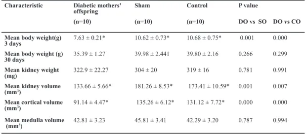
Body weights and kidney dimensions for the offspring of diabetic mothers as well as offspring of sham and control subjects are presented in Table 1.
The mean body weight of newborns of diabetic mothers was significantly lower than that of newborn offspring of control and sham subjects (p=0.000). However, the body weight of 30 day-old offspring of diabetic mothers (DO) was not significantly lower than that in control (CO) and sham (SO) subjects (p=0.218). The kidney weights were similar in all 3 groups (p=0.773).
Table 1: Comparison of the mean body weight, mean kidney weight and volume, and mean cortex and medula volumes P value
Control Sham
Diabetic mothers' offspring Characteristic
DO vs CO DO vs SO
(n=10) (n=10)
(n=10)
0.000 0.001
10.68 ± 0.75* 10.62 ± 0.73*
7.63 ± 0.21*
Mean body weight(g) 3 days
0.299 0.266
39.80 ± 2.16 39.98 ± 2.441
35.39 ± 1.27
Mean body weight (g) 30 days
0.991 0.781
319 ± 16 304 ± 20
322.9 ± 22.27
Mean kidney weight (mg)
0.007 0.001
173.41 ± 10.59* 181.26 ± 8.53*
133.66 ± 5.66*
Mean kidney volume (mm3)
0.000 0.000
131.12 ± 7.72* 135.26 ± 6.12*
91.14 ± 4.47*
Mean cortical volume (mm3)
0.994 0.787
42.29 ± 3.20 45.81 ± 3.41
42.81 ± 3.23
Mean medulla volume (mm3)
Results are Mean ± Standard error; * p<0.05 (ANOVA)
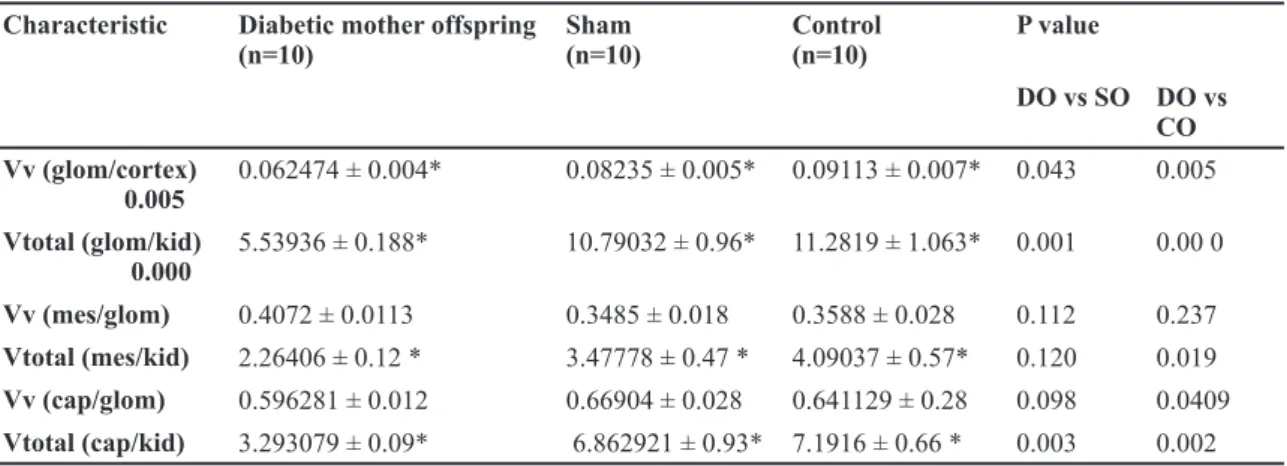
Light-microscopic stereological data are presented in Table 2. The mean offspring kidney and cortical volumes were significantly lower in DO than is CO and SO groups (p<0.005), although medular vol-umes were not changed in the DO group (p>0. 05). The glomerular volume density glomerular volume divided by cortical volume and total glomerular vol-ume were significantly lower in DO than CO and SO groups (p<0.05). Although the numerical glomerular density glomerular number divided by cortical vol-ume (repetition) in DO was greater than in CO and SO groups (p<0.05), the total glomerular number was significantly lower in DO than CO and SO groups (p=0.055). The mesangial volume density was numer-ically but not significantly greater in DO than in CO and SO groups, whereas the total mesangial volume was significantly lower in DO than in CO and SO groups. The offspring capillary volume density in the diabetic group was similar to that in the control and
sham subjects; however, the total capillary volume was lower in diabetic mothers' offspring. The me-sangial cell density (meme-sangial cell numbers divided by glomerular volume) was significantly greater in DO than in CO and SO groups (p=0.000); however the total numbers of mesangial cells were similar in all three groups.
Discussion
In the present experimental study, our results showed that the body weight of diabetic mothers’ offspring was reduced at birth or in neonates. However, at 30 days of life, the body weight was not significantly lower in the DO than in the control and sham groups, suggest-ing that the diabetic fetal environment compromised development. Previous studies also demonstrated that a diabetic pregnancy is associated with suppression in fetal growth, related to reduced protein synthesis (31). Contrary to our results, Wichi et al (32) showed that Table 2: Comparison of volume fractions of renal cortical features between the groups.
P value Control
(n=10) Sham
(n=10) Diabetic mother offspring
(n=10) Characteristic
DO vs CO DO vs SO
0.005 0.043
0.09113 ± 0.007* 0.08235 ± 0.005*
0.062474 ± 0.004*
Vv (glom/cortex) 0.005
0.00 0 0.001
11.2819 ± 1.063* 10.79032 ± 0.96*
5.53936 ± 0.188*
Vtotal (glom/kid) 0.000
0.237 0.112
0.3588 ± 0.028 0.3485 ± 0.018
0.4072 ± 0.0113
Vv (mes/glom)
0.019 0.120
4.09037 ± 0.57* 3.47778 ± 0.47 *
2.26406 ± 0.12 *
Vtotal (mes/kid)
0.0409 0.098
0.641129 ± 0.28 0.66904 ± 0.028
0.596281 ± 0.012
Vv (cap/glom)
0.002 0.003
7.1916 ± 0.66 * 6.862921 ± 0.93*
3.293079 ± 0.09*
Vtotal (cap/kid)
Results are Mean ± Standard error;* p<0.05 (ANOVA)
Vv (glom/cortex): volume density of glomerulus; Vtotal (glom/kid): total glomerular volume; Vv ( mes /glom): volume density of mesangium; Vtotal (mes/kid): total mesangium volume; Vv (cap/glom):volume density of capillary; Vtotal (cap/kid): total capillary volume. DO: offspring of diabetic mothers; SO: offspring of mothers in sham group; CO: offspring of mothers in control group.
Table 3: Comparison of the numerical fractions of renal cortical features between the groups.
P value Control
(n=10) Sham
(n=10) Diabetic mother offspring
(n=10) Characteristic
DO vs CO DO vs SO
0.023 0.041
61.8238 ± 1.78* 63.9635 ± 1.17*
77.9170 ± 5.93*
Nv (glom/cortex) 0.005
0.030 0.046
7639.9 ± 429 * 8349 ± 519 *
6536.72 ± 532*
Ntotal (glom/kid) 0.000
0.000 0.000
596.95 ± 46.4* 592.51 ± 25.23*
984.061 ± 54.71
Nv (mes/glom)
0.330 0.588
6909 ± 1048 6398 ± 645
5438 ± 331
Ntotal (mes/kid)
Results are Mean ± Standard error;* p<0.05 (ANOVA)
Nv (glom/cortex): Numerical glomerular density; Ntotal (glom/kid): total glomerular number per cortex; Nv (mc/glom): Numerical mesangial density;
the body weight of offspring of diabetic mothers was not reduced at birth or in neonates, but was significantly lower 30 days after birth. Both mild gestational diabetes and a reduction in litter size are associated with prena-tal hyperinsulinism, (33). Thus as predicated by Barker, timing of the insult in utero may be critical in the fetal programming of adult disease (34). The experimental induction of diabetes using streptozotocyne (STZ) has dose-dependent effects: low doses of STZ result in mild gestational diabetes associated with fetal macrosomia (33), whereas high doses induce insulin-deficient diabe-tes associated with fetal growth restriction (35).
The present study showed a reduction in kidney volume particularly in cortical zones, but not in medulary por-tions in the DO group. Osmand et al. also demonstrated that slowing of growth in late gestation leads to dispro-portion in the size of organs that develop rapidly in late gestation (e.g. the kidneys). Possibly through the effect of decreased maternal IGF, fetal insulin, insulin like growth factor (IGF), and glucose concentrations fall if a mother decreases her food intak. This leads to a slowing in growth (36).
The numerical glomerular density in DO group was significantly greater than in CO (p=0.041) and SO (p=0.023) groups, and also total glomerular number in DO was significantly lower than in CO and SO groups (p=0.046). This is due to the fact that cortical volume in DO was lower than in CO and SO groups. Our findings were in agreement with the observations of others. Low glomerular numbers in diabetic mothers' offspring have been directly correlated with low birth weights. Hugh-son et al. (22) demonstrated a direct relationship between total glomerular number (Nglom) and birth weight in adult and fully developed postnatal kidneys of infants. A low glomerular number may be the result of imperfect nephrogenesis during kidney development and/or loss of nephrons before nephrogenesis (37). In animal models, in-utero exposure to hyperglycemia was associated with a reduction of number of nephrons in the offspring (38). In this study, the mesangial volume density was numeri-cally, although not significantly (p>0.05), greater in DO than in CO and SO groups. However, total mesangial vol-ume was significantly lower in DO because the cortical volumes were lower than CO and SO groups. The incre-ment in glomerular volume is due to an increase in me-sangial matrix as well as hypertrophy of other glomerular structures (39). Previous studies on the short-term effects of diabetes on kidney morphology have suggested corti-cal hypertrophy accompanied by glomerular mesangial hypertrophy (23, 24), whereas long-term diabetes caused increased glomerular basement membrane thickness (23). In this study, there were significant changes in the mesangial matrix. Further increase in glomerular and me-sangial volumes, as well as capillary length and surface area changes were attributed to compensatory hypertro-phy due to the loss of nephrons (39).
The capillary volume densities in diabetic mothers' offspring were similar to control and sham subjects. However, total volume of capillary was lower in DO
because the cortical volume was lower in this group. Mesangial cell densities however, were significantly greater in diabetic mothers' offspring, suggesting that the increase in cell number is not simply in keeping with the degree of glomerular enlargement. There is a greater proportion of mesangial cells in the DO glomer-ulous. The short-term effects of diabetes have been sug-gested to be increased number of glomerular mesangial cells and interstitial alterations (40-42). These findings are in agreement with the observations of White et.al (43), and are in contrast to those of Steffes et al. (44), who did not find a significant increase in the absolute number of mesangial cell density. However, it must be taken into consideration that the patients in Steffese et al study were at an earlier stage of nephropathy and did not demonstrate any increase in glomerular volume.
Conclusion
This study identifies maternal diabetes as a novel risk factor for inborn nephron deficit in addition to other factors such as fetal growth retardation which alter ne-phrogenesis. It points out the particular risk of a high blood glucose level during the critical period of nephron formation, particularly in early stages, and calls for a strict control of blood glucose in a diabetic pregnancy.
Acknowledgments
We thank Jondishapoor University's director of Medi-cal research for financial support of this work.
The authors wish to thank the staff of Babol Medical Sciences University. There is no conflict of interest in this study.
References
1. Dabelea D, Hanson RL, Lindsay RS, Pettitt DJ, Im-peratore G, Gabir MM, et al. Intrauterine exposure to diabetes conveys risks for type 2 diabetes and obesity: a study of discordant sibships. Diabetes. 2000; 49(12): 2208-2211.
2. Cetin I, Foidart JM, Miozzo M, Raun T, Jansson T, Tsatsaris V, et al. Fetal growth restriction: a workshop report. Placenta. 2004; 25(8-9): 753-757.
3. Becerra JE, Khoury MJ, Cordero JF, Erickson JD. Diabetes mellitus during pregnancy and the risks for specific birth defects: a population-based case-control study. Pediatrics. 1990; 85(1): 1-9.
4. Manderson JG, Mullan B, Patterson CC, Hadden DR, Traub AI, McCance DR. Cardiovascular and metabolic abnormalities in the offspring of diabetic pregnancy. Di-abetologia. 2002; 45(7): 991-996.
5. Holemans K, Gerber R, Meurrens K, De Clerck F, Poston L, Van Assche FA. Maternal food restriction in the second half of pregnancy affects vascular function but not blood pressure of rat female offspring. Br J Nutr. 1999; 81(1): 73-79.
6. Holemans K, Caluwaerts S, Van Assche FA. Unravel-ling the fetal origins hypothesis. Lancet. 2002; 21-28; 360(9350): 2073.
8. Fujisawa Y, Nakagawa Y, Ren-Shan L, Ohzeki T. Streptozotocin-induced diabetes in the pregnant rat re-duces 11 beta-hydroxysteroid dehydrogenase type 2 expression in placenta and fetal kidney. Life Sci. 2004; 75(23): 2797-2805.
9. Martínez-Frías ML. Epidemiological analysis of out-comes of pregnancy in diabetic mothers: identification of the most characteristic and most frequent congenital anomalies. Am J Med Genet. 1994; 51(2): 108-113. 10. Buchanan TA, Denno KM, Sipos GF, Sadler TW. Dia-betic teratogenesis. In vitro evidence for a multifactorial etiology with little contribution from glucose per se. Dia-betes. 1994; 43(5): 656-660.
11.Sadler TW. Mouse embryos in culture: models for un-derstanding diabetes-induced embryopathies and gene function. Int J Dev Biol. 1997;41(2):291-7
12. Mills J, Baker L, Goldman A. Malformations in infants of diabetic mothers occur before the seventh gestational week: implications for treatment. Diabetes 1979; 28; 292-293.
13. Lunch S, Wright C. Sirenomelia, limb reduction de-fects, cardiovascular malformation, renal agenesis in an infant born to a diabetic mother. Clin Dysmorphol. 1997; 6; 75-80.
14. Lowy C, Beard RW, Goldschmidt J. Congenital mal-formations in babies of diabetic mothers. Diabet Med. 1986; 3(5): 458-462.
15. Gomez RA, Norwood VF. Recent advances in renal development. Curr Opin Pediatr. 1999; 11: 135140. 16. Larsson L, Aperia A, and Wilton P. Effect of normal development on compensatory renal growth. Kidney Int. 1980; 18: 29-35.
17. Langley Evans SC, Welham SJ, Jackson AA. Fetal exposure to a maternal protein diet impairs nephrogene-sis and promotes hypertension in the rat. Life Sci. 1999; 64: 965-974.
18. Woods LL, Weeks DA, Rasch R. Programming of adult blood pressure by maternal protein restriction: role of nephrogenesis. Kidney Int. 2004; 65: 1339-1348. 19. Lelievre-Pegorier M, Merlet-Benichou C. The number of nephrons in the mammalian kidney: Environmental influences play a determining role. Clin Exp Nephrol. 2000; 8: 63-65.
20. Bihoreau MT, Ktorza A, Kinebanyan MF, and Picon L. Impaired glucose homeostasis in adult rats from hyperg-lycemic mothers. Diabetes. 1986; 35: 979-984.
21. Thamotharan M, McKnight RA, Thamotharan S, Kao DJ, and Devaskar SU. Aberrant insulin-induced GLUT4 translocation predicts glucose intolerance in the offspring of a diabetic mother. Am J Physiol Endocrinol Metab. 2003; 284: E901–E914.
22. Hugson M, Farris AB 3rd, Douglas-Denton R, Hoy WE, Bertram JF. Glomerular number and size in autopsy kidneys: The relationship to birth weight. Kidney Int. 2003; 63: 2113-2122.
23. Broyer M, Soto B, Gagnadoux MF, Adi M, Rica C, Gubler MC. Oligomeganephronic renal hypoplasia. Adv Nephrol Necker Hosp. 1997; 26: 47-63.
24. Samuel T, Hoy WE, Douglas-Denton R, Hughson MD, Bertram JF. Determinants of glomerular volume in different cortical zones of the human kidney . J Am Soc Nephrol. 2005; 16: 3102-3109.
25. Keller G, Zimmer G, Mall G, Ritz E, Amann K. Ne-phron number in patients with primary hypertension. N Engl J Med. 2003; 348: 101-108.
26. Geoffroy K, Troncy L, Wiernsperger N, Lagarde M, Bawab S: Glomerular proliferation during early stage of
diabetic nephropathy is associated with local increase of sphingosin-1-phosphate levels.FEBS Lett. 2005; 579: 1249-1254.
27. Buzello M. Comparison of two stereological meth-ods for quantitative renal morphology: a modified frac-tionator and modified Weibel-Gomez method. Pathol Res Pract. 2000; 196: 111-117.
28. Heidari Z, Sagheb H, Dezfoulian A, Barbarestani M, Noori H. A stereological analysis of renal glomeruli fol-lowing chronic lead intoxication in rat during a continu-ous period of 8 weeks. Acta Med Iran. 2002; 40: 73-78. 29. Gundersen HJG, Bagger P, Bendtsen TF, Evans S.M, Korbo, L, Marcussen N, et al. The new stereologi-cal tools: Disector, fractionator nucleator and point sam-pled intercepts and their use in pathological research and diagnosis. APMIS. 1988; 96: 857-881.
30. Gilbert JS, Lang AL, Grant AR, Nijland MJ. Mater-nal nutrient restriction in sheep: hypertension and de-creased nephron number in offspring at 9 months of age. J Physiol. 2005; 565: 137-147.
31. Canavan JP, Goldspink DF. Maternal diabetes in rats. II. Effects on fetal growth and protein turnover. Dia-betes. 1988; 37: 1671-1677.
32. Wichi RB, Souza SB, Casarini DE, Morris M, Barre-to-Chaves ML, Irigoyen MC. Increased blood pressure in the offspring of diabetic mothers. Am J Physiol Regul Integr Comp Physiol. 2005; 288(5): R1129-33. Epub 2005 Jan 20. PMID: 15661971.
33. Aerts L and Van Assche F. Intra-uterine transmission of disease. Placenta. 2003; 24: 905–911
34. Alves GM, Barao MA, Odo LN, Nascimento Gomes G, Franco Mdo C, Nigro D, et al. L-Arginine effects on blood pressure and renal function of intrauterine restrict-ed rats. Prestrict-ediatr Nephrol. 2002; 17: 856-862.
35. Holemans K, Aerts L, Van Assche FA. Lifetime con-sequences of abnormal fetal pancreatic development. J Physiol. 2003; 547: 11-20.
36. Holemans K, Aerts L, Van Assche FA. Evidence for an insulin resistance in the adult offspring of pregnant streptozotocin diabetic rats. Diabetologia. 1991; 34: 81-85.
37. Osmand C, Barker D, fetal. infant, and children growth are predictors of coronary heart disease, diabe-tes, and hypertension in adult men and women. Environ Health Perspect. 2000; 108(3): 545-553.
38. Douglas-Dementon RN, Mcnamara BJ, Hoy WE, Hughson MD, Bertram JF. Dose nephron number matter in the development of kidney disease? Ethn Dis. 2006; 16(2): S2-40-5.
39. Amri K, Freund N, Vilar J, Merlet-Benichou C, Le-lievre-Pegorier M. Adverse effects of hyperglycemia on kidney development in rats: in vivo and in vitro studies. Diabetes.1999; 48: 2240-2245.
40. Osterby R. Structural changes in the diabetic kidney. Clin Endocrinol Metab. 1986; 15: 733-751.
41. Bilous R, Mauer S, Sutherland DER, Steffes M. Mean glomerular volume and rate of development of diabetic nephropathy. Diabetes. 1989; 38: 1142-1147. 42. Mauer SM. Structural-functional correlations of dia-betic nephropathy. Kidney Int. 1994; 45: 612-622. 43. White KE, Bilous RW, Marshall SM, Nahas MEI, Re-muzzi G, Piras G, et al. Podocyte number in normoten-sive type 1 diabetic patients with albuminuria.Diabetes. 2002; 51: 3083-3089.