ORIGINAL ARTICLE
Vildagliptin Ameliorates Oxidative Stress and Pancreatic Beta Cell
Destruction in Type 1 Diabetic Rats
Danielle de Lima
Avila,
aGlaucy Rodrigues de Ara
ujo,
aMaisa Silva,
aPedro Henrique de Amorim Miranda,
aMirla Fiuza Diniz,
aMaria L
ucia Pedrosa,
a,bMarcelo Eust
aquio Silva,
a,cWanderson Geraldo de Lima,
a,band Daniela Caldeira Costa
a,b aN
ucleo de Pesquisas em Ci^encias Biologicas-NUPEB,bDepartamento de Ciencias Biol^ ogicas, Instituto de Ci^encias Exatas e Biologicas,cDepartamento de Alimentos, Escola de Nutric¸~ao, Universidade Federal de Ouro Preto, Ouro Preto, Brazil
Received for publication August 21, 2012; accepted March 5, 2013 (ARCMED-D-12-00436).
Background and Aims. It is believed that oxidative stress plays a role in the pathogenesis of diabetes mellitus. Several strategies have been developed with the objective of mini-mizing diabetic complications. Among these, inhibitors of dipeptidyl peptidase-IV (DPP-IV), which act by blocking degradation of incretin hormones, glucagon-like peptide hormone (GLP-1) and glucose-dependent insulinotropic polypeptide (GIP), have been the focus of many studies. It is known that, among the effects of incretins, we highlight its insulinotropic and cytoprotective effects on pancreaticb-cells. The objective of this study was to evaluate the possible protective effects of treatment with vildagliptin, a DPP-IV inhibitor, inb-cells in an experimental model of type 1 diabetes induced by streptozotocin (STZ).
Methods. Rats were treated for 4 weeks with vildagliptin at concentrations of 5 and 10 mg/kg. In order to observe the pancreatic damage and the possible protective effects of vildagliptin treatment, we measured stress markers TBARS and protein carbonyl, antiox-idant enzymes SOD and catalase, and analyzed pancreatic histology.
Results. The treatment was effective in modulating stress in pancreatic tissue, both by reducing levels of stress markers as well as by increasing activity of SOD and catalase. After analyzing the pancreatic histology, we found that vildagliptin was also able to preserve islets and pancreaticb-cells, especially at the concentration of 5 mg/kg.
Conclusion. Thus, our results suggest that vildagliptin ameliorates oxidative stress and pancreatic beta cell destruction in type 1 diabetic rats. However, to evaluate the real potential of this medication in type 1 diabetes, further studies are needed. Ó 2013 IMSS. Published by Elsevier Inc.
Key Words:Inhibitor DPP-IV, Pancreas, Oxidative stress, Antioxidant.
Introduction
Despite substantial advances in our understanding of type 1 diabetes, diagnosis of the condition still requires lifelong daily insulin injections, which are a partially effective therapy at best(1). New therapeutic strategies under inves-tigation include islet transplantation, development of
improved insulin analogues and delivery systems, gene therapy, and the search for novel agents that can protect and/or stimulate the proliferation and regeneration of isletb-cells(1). The importance of the latter strategy is un-derscored by the need for an inexpensive, benign, preven-tive therapy that lacks the considerable side effects of most therapies studied to date (e.g., immunosuppressants)
(2). In this context, glucose-dependent insulinotropic polypeptide (GIP) and glucagon-like peptide-1 (GLP-1) are gastrointestinal hormones that potentiate glucose-stimulated insulin secretion and are classified as incretins. Additionally, these incretins stimulate both insulin
Address reprint requets to: Daniela Caldeira Costa, Universidade Federal de Ouro Preto, Campus Universitario Morro do Cruzeiro, Ouro Preto, Brazil; Phone: (þ55) 31 3559-1761; FAX: (þ55) 31 3559-1680; E-mail:dani.caldeiracosta@gmail.com
0188-4409/$ - see front matter. CopyrightÓ2013 IMSS. Published by Elsevier Inc.
biosynthesis and proliferation of b-cells while inhibiting apoptosis (3e7). Dipeptidyl peptidase-IV (DPP-IV)
termi-nates the actions of GIP and GLP-1 (8e10). DPP-IV is
a multifunctional glycoprotein that contains NH2-terminal serine dipeptidase activity and is present in circulation and on the cell surface (11). Relatively few studies have been conducted on the potential for DPP-IV inhibitors (DPP-IVi) in the treatment of type 1 diabetes. In preclinical studies, the DPP-IVi isoleucine thiazolidine improved glucose toler-ance in streptozotocin (STZ)-induced diabetic rats (2,12), with clear islet protection in the former group(2). The effect of DPP-IVi onb-cells may be small because the destruction of b-cells leads to absolute insulin deficiency by cell-mediated autoimmune attack. Additionally, a recent report showed that DPP-IV may ameliorate an autoimmune attack againstb-cells by restoring or increasing the number of regu-latory T lymphocytes. Altogether, incretin-based therapy may be worth testing in patients with type 1 diabetes(13). It has been reported that the DPP-IVi sitagliptin not only improves glucose metabolism but also protects b-cells, promotes islet neogenesis, and prolongs islet graft survival in rodent diabetic models (2,14,15). Furthermore, DPP-IVi have been reported to delay or suppress the occurrence of hyperglycemia in an animal model of spontaneous diabetes
(16), but the mechanisms have not been completely eluci-dated. Hyperglycemia leads to cellular damage and organ dysfunction (17). In the pancreas, chronic hyperglycemia produces progressive deleterious effects onb-cells (18,19), which are manifested by increased oxidative stress(20,21). Thus, one of the factors that contributes to pancreatic dysfunction is oxidative stress (22). Pancreatic b-cells are vulnerable to oxidative stress due to the low antioxidant enzyme activities(23,24), and enhancing antioxidant defense mechanisms in pancreatic islets helps these cells cope with oxidative stress. Thus, if treatments with DPP-IV modulate oxidative stress in the pancreas, then these treatments can be used to reduce oxidative damage, thus minimizing diabetes-associated complications. In the present study, our objective was to evaluate the parameters of oxidative stress and antioxidant enzymes in the pancreas of type 1 diabetic rats treated with vildagliptin, a DPP-IVi.
Materials and Methods
Reagents
The chemical reagents, including STZ (streptozotocin), 2,4-dinitrophenylhydrazine (DNPH) and thiobarbituric acid (TBA), were purchased from Sigma-Aldrich (St. Louis, MO).
Animals
Eleven-week-old female Fisher rats weighing|180 g were
obtained from the Laboratory of Experimental Nutrition, Escola de Nutric¸~ao, Universidade Federal de Ouro Preto. During the experimental period, the animals were kept in
a well-ventilated environment with controlled temperature, humidity and ventilation. Animals had access to water and commercial rat rationsad libitum. This study was conduct-ed in accordance with the international standards of animal protection and the ethical principles of the Brazilian College of Animal Experimentation(25)and was approved by the Ethics Committee on Animal Use (CEUA) of UFOP (protocol 2011/27).
At the beginning of the experiment, animals showed similar glucose levels. Animals were randomly assigned to a normal control or diabetes group. Age-matched normal control rats were injected with an equal volume of vehicle (sodium citrate buffer).
Forty-eight rats were distributed into six groups accord-ing to the treatment they received: untreated control group (C), control group treated with 5 mg/kg vildagliptin (C5), control group treated with 10 mg/kg vildagliptin (C10), untreated diabetic group (D), diabetic group treated with 5 mg/kg vildagliptin (D5), and diabetic group treated with 10 mg/kg vildagliptin (D10). Three days after diabetes was induced, vildagliptin was administered by gavage for 30 days. Vildagliptin (Novartis), a DPP-IVi, was commer-cially acquired and diluted in phosphate buffer suspended in 0.05% of methylcellulose in a final concentration of 5 or 10 mg/kg body weight/day(26). The vildagliptin dosages used were based on the literature(27e31).
Diabetes Induction
We used streptozotocin (STZ) to induce an experimental model of type 1 diabetes. STZ is the most prominent diabe-togenic chemical in diabetes research and is a cytotoxic glucose analogue. STZ inhibits insulin secretion and causes a state of insulin-dependent diabetes mellitus. Both effects can be attributed to the specific chemical properties of STZ, namely, its alkylating potency.b-cell specificity is primarily the result of selective cellular uptake and accumulation. Streptozotocin enters the b-cell via a glucose transporter (GLUT2) and causes alkylation of DNA(32).
We treated rats i.p. with 35 mg/kg of STZ dissolved in 0.2 mL citrate buffer (0.01 M, pH 4.5). Diabetes was veri-fied 72 h later by evaluating blood glucose levels using Accu-Chek active (Roche Laboratories, San Francisco, CA). After overnight fasting, a blood glucose level of 300 mg/dL was used to establish diabetes.
Insulin
Serum insulin levels were determined using a commercial kit from Crystal Chem Inc (Downers Grove, IL). Using the sandwich ELISA method, this kit is sensitive for determina-tion of insulin in rats.
Preparation of Pancreatic Tissue
concentrations, 200 mg of tissue was homogenized in 50 mmol phosphate buffer (pH 6.7) and 1 mmol EDTA. To determine the concentration of thiobarbituric acid reactive substances (TBARS), superoxide dismutase and catalase activity, 100 mg of pancreatic tissue was homoge-nized in phosphate buffer (pH 7.4). After homogenization, samples were centrifuged at 10,000g for 10 min at 4
C. The supernatant was collected and used as the biological sample.
Determination of Antioxidant Defenses
Catalase (CAT). Catalase activity was determined based on its ability to convert hydrogen peroxide (H2O2) into water and molecular oxygen. Assays were performed as described by Aebi (33). Briefly, 10 mL of sample supernatant was mixed with 50 mL of K2HPO4, 40 mL of milli-Q water (Millipore, Bedford, MA) and 900 mL of 2.5 mmol/L H2O2 before being spectrophotometrically measured at 240 nm at 25
C. Concentrations of H2O2and samples were chosen so that the degradation rate was linearly propor-tional at 30 sec, 1 min, 2 min and 3 min. One unit (U) of catalase is equivalent to the hydrolysis of 1 mmol of H2O2/min.
Superoxide dismutase activity (SOD). Total superoxide dismutase (SOD) activity was measured by the kit from Cayman Chemical Company (Ann Arbor, MI). Briefly, pancreatic cells were homogenized in cold 20 mmol HEPES, pH 7,2, containing 1 mmol EGTA, 210 mmol mannitol and 70 mM sucrose. Ten mL of supernatant was used in the test. The reaction was initiated by adding xanthine oxidase. The plate was incubated on a shaker for 20 min at room temperature, and the absorbance was measured at 450 nm using a plate reader (Biotek ELx808, Green Mountains of Vermont).
Determination of Oxidative Stress Markers
Thiobarbituric acid reactive substances (TBARS). TBARS concentration was determined from thiobarbituric acid (TBA) binding to oxidized lipids. This measurement was performed according to Buege and Aust (34). Briefly, 250 mL of 28% trichloroacetic acid (TCA) was added to 500 mL of sample supernatant, 250 mL of TBA (1% in acetic acid 1:1) and 125 mL of butylated hydroxytoluene (BHT) (5 mmol in ethanol). Samples were then incubated at 95
C for 15 min. The mixture was subsequently centrifuged at 10,000 g for 15 min. Supernatant absorbance was measured at 535 nm using a spectrophotometer. Values were expressed in nmol/mg of protein.
Carbonylated Protein
Protein oxidation by ROS leads to the formation of carbonyl derivatives, which can be measured by sensitive
methods. Methods that use 2,4-dinitrophenylhydrazine (DNPH), which reacts with carbonyl groups to generate the corresponding hydrazone and can then be analyzed spectrophotometrically, are especially useful. Measure-ments of carbonylated protein were performed according to Levine et al. (35). In brief, proteins were precipitated using TCA (10%) and incubated with DNPH and HCl at room temperature for 30 min. TCA (10%) was added to the precipitate and centrifuged at 5,000g for 5 min at 4C. After discarding the supernatant, the precipitate was washed twice with ethanol/ethyl acetate (1:1), dissolved in 6% sodium dodecyl sulfate (SDS) solution and centri-fuged at 10,000g for 10 min at 4C. Supernatant absor-bance was measured at 370 nm using a spectrophotometer. Results were expressed as nmol of carbonyl groups/mg of protein.
Histological Evaluation
Pancreas fragments not exceeding 4 mmol in diameter were fixed in 10% formaldehyde solution and then dehydrated, diaphonized and embedded in paraffin. Paraffin sections of|4mm were obtained by sectioning embedded fragments
on a rotary microtome. Sections were mounted on cleaned and degreased glass slides. The slides were stained with hematoxylin and eosin for visualization of histological damage and Gomori’s trichrome to differentiate phenotypes of pancreatic beta and alpha cells. To determine the average number of inflammatory cells as well as the average size and area of pancreatic islets, digital morphometric analyses were performed using a Leica optical microscope with the Leica Qwin Plus analysis software DM5000 (Leica Micro-systems, Buffalo Grove, IL). The average number of beta and alpha cells was determined using ImageJ software (NIH, Bethesda, MD).
All histological samples from the control and treatment animals underwent blind analysis.
Statistical Analysis
All values are expressed as meanSD. Statistical analysis was performed using unpaired Student t test for between-group comparisons. One-way ANOVA with Tukey post-test was used for analysis between all groups. GraphPad Prism v.5.0 software was used for the analysis;p !0.05 was considered statistically significant.
Results
Evaluation of Body Weight, Blood Glucose and Plasma Insulin Levels
groups (D, D5 and D10) compared to the control groups (C, C5 and C10). Animals in the diabetic groups also showed increased final blood glucose levels when compared to animals in the control groups. Treatment with vildagliptin at 5 and 10 mg/kg failed to improve the glycemic profile of these animals, although treatment reduced blood glucose. Animals with type 1 diabetes showed a significant decrease in serum insulin compared to control animals (Table 1). Treatment with vildagliptin significantly increased insulin levels in the diabetic animals, but these values were still below the values shown in the control animals.
Assessment of Redox Status in Pancreatic Tissue
Oxidative stress markers. To measure oxidative stress markers in pancreatic tissue, we evaluated oxidative damage to proteins and lipids. Specifically, we analyzed carbonylated protein and TBARS. A significant increase in the concentration of carbonylated protein was observed
in the pancreas of diabetic animals compared to controls (Figure 1). Diabetic animals treated with 5 and 10 mg/kg of vildagliptin showed a reduction in the concentration of carbonylated protein compared to untreated diabetic animals. Additionally, a significant increase in the concen-tration of TBARS was observed in diabetic animals compared to control animals (Figure 2). Vildagliptin treat-ment at 5 mg/kg was able to reverse the lipid peroxidation in this tissue.
Antioxidant status. To measure antioxidant status in pancreatic tissue, we evaluated the activity of total super-oxide dismutase (SOD) and catalase (CAT). Diabetic animals exhibited a significant decrease in catalase activity compared to controls (Figure 3). However, vildagliptin was able to significantly increase catalase activity in diabetic animals compared to untreated diabetic animals. Surpris-ingly, diabetic animals exhibited a significant increase in SOD activity compared to controls, and diabetic animals Table 1.Evaluation of body weight, blood glucose and plasma insulin levels
Experimental groups C C5 C10 D D5 D10
Initial weight (g) 182.222.4 183.418.1 182.48.9 178.27.5 180.010.5 180.68.6 Final weight (g) 215.219.5 217.215.6 212.610.2 154.818.9a 168.123.2b 153.819.1c
Initial glycemia (mg/dL) 92.222.3 87.710.6 101.017.7 403.779.5a 375.513 357.176.9
Final glycemia (mg/dL) 88.316.4 87.710.3 97.713.2 389.788.5a 357.8159.6 322.993.1
Insulin (pmol/L) 56.811.2 50.711.7 52.214.9 11.68.7a 34.38.0b,d 32.28.9c,e
C, control (untreated); C5, controlþ5 mg vildagliptin$(kg body mass) 1; C10, control 10 mg vildagliptin$(kg body mass) 1; D, diabetic; D5, diabeticþ5
mg vildagliptin$(kg body mass) 1; D10, diabeticþ10 mg vildagliptin$(kg body mass) 1.
Data are presented as meanstandard deviation (n58).
aIn the same row indicate statistically significant differences between C and D, D and D5, D and D10 (p #0.05).
bRepresents difference between D X D5. cRepresents difference between D X D10. dRepresents difference between C X D5.
eRepresents difference between C and D10 as determined by Student t test.
Figure 2.Data are presented as the meanSEM (n58). C, control (untreated); C5, control þ 5 mg vildagliptin$(kg body mass) 1; C10,
control 10 mg vildagliptin$(kg body mass) 1; D, diabetic; D5,
diabeticþ5 mg vildagliptin$(kg body mass) 1; D10, diabeticþ10 mg
vildagliptin$(kg body mass) 1. Statistically significant differences (p #0.05) are between the following groups: (*) C and D; (**) D and D5.
Figure 1.Data are presented as the meanSEM (n58). C, control (untreated); C5, control þ 5 mg vildagliptin$(kg body mass) 1; C10,
control 10 mg vildagliptin$(kg bodymass) 1; D, diabetic; D5, diabeticþ
5 mg vildagliptin$(kg body mass) 1; D10, diabeticþ 10 mg
vildaglip-tin$(kg body mass) 1. Statistically significant differences (p#0.05) are
treated with 5 and 10 mg/kg vildagliptin showed a dose-dependent increase in SOD activity (Figure 4).
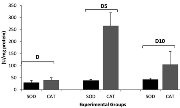
Evaluation of the Relationship Between SOD and CAT Activities
We conducted a joint assessment of SOD and CAT activity. For this experiment, we analyzed the percentage of activa-tion or inhibiactiva-tion of these enzymes in the pancreas from treated or untreated diabetic rats. Increased activity of SOD in the pancreas from diabetic rats was accompanied by decreased CAT activity (Figure 5). However, treatment with vildagliptin increased both SOD and CAT activities, suggesting a balance between SOD and CAT activity.
Histological Evaluation
General histological pictures of the pancreas from all groups did not show macroscopic or microscopic
morphological and staining alterations. Additionally, signals of pancreatitis and other necrotic and degenerative pancreatic diseases were not observed. Morphometrically, the number of pancreatic islets was decreased in the dia-betic group. However, vildagliptin treatment at 5 mg/kg maintained islets in the diabetic groups in numbers similar to their respective controls (Figure 6andFigure 7). Due to the protective effect of treatment on endocrine areas of the pancreas, we evaluated the pancreatic cell populations within the islets. Diabetic animals showed a significant reduction in the number of pancreatic b-cells. Treatment with vildagliptin maintainedb-cells in diabetic animals in numbers similar to those of control animals (Figure 8and
Figure 9). No differences in the populations of alpha cells within the islets were observed (results not shown).
Discussion
DPP-IV is a ubiquitous serine protease that rapidly cleaves and inactivates GIP and GLP-1in vivo. It has been shown to increase circulating active incretin levels, thus increasing the effective concentration of these peptides that reaches the target tissues(9,36,37). Dipeptidyl peptidase-4 inhibi-tors alone or in combination with other therapies are being promoted for the treatment of type 2 diabetes, but little is known about the potential benefits of these inhibitors in type 1 diabetes (38). Pospisilik et al. (2) showed that long-term DPP-IVi treatment of STZ rats improves glucose tolerance, enhances pancreatic insulin content, and stimu-lates survival of pancreaticb-cells. However, mechanisms responsible for preserving b-cells during treatment with DPP-IVi in an experimental model of type 1 diabetes have not been elucidated. Recently, oxidative stress has been re-ported to be responsible, to a certain extent, for b-cell dysfunction caused by glucose toxicity (39). Therefore, the aim of this study was to investigate the possible effects of DPP-IVi as a promoter in modulating the oxidant/anti-oxidant balance and preservation ofb-cells in the pancreas of rats with type 1 diabetes. Accordingly, we conducted Figure 3. Data are presented as the mean SEM (n58). C, control
(untreated); C5, controlþ5 mg vildagliptin$(kg body mass) 1; C10, control
10 mg vildagliptin$(kg body mass) 1; D, diabetic; D5, diabeticþ5 mg
vil-dagliptin$(kg body mass) 1; D10, diabeticþ10 mg vildagliptin$(kg body
mass) 1. Statistically significant differences (p
#0.05) are between the following groups: (*) C and C10; (**) C and D; (**) D and D5.
Figure 4.Data are presented as the meanSEM (n58). C, control (untreated); C5, control þ 5 mg vildagliptin$(kg body mass) 1; C10,
control 10 mg vildagliptin$(kg body mass) 1; D, diabetic; D5,
diabeticþ5 mg vildagliptin$(kg body mass) 1; D10, diabeticþ10 mg
vildagliptin$(kg body mass) 1. Statistically significant differences (p #0.05) are between the following groups: (*) C and D; (**) D and D5; (***) D and D10.
Figure 5.Data are presented as the meanSEM (n58). D, diabetic; D5, diabeticþ5 mg vildagliptin$(kg body mass) 1; D10, diabeticþ10 mg
a comparative study between two concentrations of vilda-gliptin DPP-IVi: 5 mg/kg and 10 mg/kg.
STZ-induced diabetes was characterized by a severe loss in body weight, which has also been reported by other investigators(40). The decrease in body weight in diabetic rats may indicate loss or degradation of structural proteins, which have been reported to contribute to body weight(41). The present study showed that vildagliptin did not prevent weight loss. Moreover, vildagliptin was unable to alter the
plasma glucose levels of diabetic animals (Table 1). Inter-estingly, we found a significant increase in plasma insulin in diabetic rats treated with vildagliptin compared to untreated rats, but the concentration of insulin in these animals was |40% lower when compared to control
ani-mals. Moreover, Jin et al.(27)reported that treatment with vildagliptin partially improves the reduction in insulin levels in STZ-diabetic rats. This outcome occurs in spite of unaffected fasting glucose and glycated hemoglobin (HbA1c) levels but is associated with an increase in GLP-1 plasma levels.
Lipid peroxidation of unsaturated fatty acids is frequently used as an indicator of increased oxidative stress and subsequent oxidative damage(42)and is characteristic of chronic diabetes (43). Lipid peroxidation impairs cell membrane fluidity and alters activity of membrane-bound enzymes and receptors, resulting in membrane malfunction
(44). The high level of the lipid peroxidation marker TBARS in diabetic rats is a reflection of insufficient antiox-idant defenses in combating ROS-mediated damage. Our data show that the pancreas of diabetic animals has in-creased oxidative damage, exemplified by the inin-creased concentration of TBARS. Several studies also showed an increase in the concentration of TBARS in the pancreatic tissue of diabetic rats(43,45,46). However, animals treated with 5 mg/kg vildagliptin showed significantly decreased concentrations of TBARs compared to untreated diabetic animals (Figure 2). Davidson et al. showed that treating Figure 6.Histological analyses of pancreas in nondiabetic and diabetic animals treated with vildagliptin. Histological photomicrography of pancreas. (A) Normal histological aspect of the control group. (B) Controlþ5 mg/kg vildagliptin. (C) Controlþ10 mg/kg vildagliptin. (D) Diabetic, untreated diabetic group. (E) Diabeticþ5 mg/kg vildagliptin. (F) Diabeticþ10 mg/kg vildagliptin. bv, blood vessel, PD, pancreatic duct. Hematoxylin & eosin staining, magnification440.
Figure 7.Data are presented as the meanSEM (n58). C, control (untreated); C5, control þ 5 mg vildagliptin$(kg body mass) 1; C10,
control 10 mg vildagliptin$(kg body mass) 1; D, diabetic; D5,
diabeticþ5 mg vildagliptin$(kg body mass) 1; D10, diabeticþ10 mg
diabetic rats with alogliptin (DPP-4i) at 10e20 mg/kg concentrations per rat per day also lowered serum TBARS, but the difference was not significant compared to untreated diabetic rats. Protein carbonyl contents, reflecting oxidative protein damage, were observed in the pancreas of diabetic animals. Vildagliptin at 5 and 10 mg/kg was effective in reducing the concentration of carbonylated protein in dia-betic animals (Figure 1). Analyzing the results together, we suggest that vildagliptin was able to modulate oxidative
damage, as exemplified by the lower levels of TBARS and carbonylated protein.
Pancreatic b-cells are highly prone to oxidative stress and damage because they have low expression and activity of antioxidant enzymes, which are the first line of defense against oxidative insult (47). SOD and CAT enzymes constitute the first line of cellular antioxidant defense. SOD is one of the most important enzymes in the enzy-matic antioxidant defense system and catalyzes the dis-mutation of superoxide radicals to produce H2O2 and molecular oxygen (48). In this study we found that the activity of SOD was upregulated in the diabetic pancreas. Although SOD is an antioxidant enzyme, some studies have suggested that its overexpression is, in fact, harmful to cells
(49). The toxic effect of ROS observed in many cells with overexpressed SOD has been linked to elevated levels of H2O2 and accompanying oxidative damage following hydroxyl radical formation(50). The implication for SOD upregulation is the high turnover of H2O2. Because CAT, which inactivates H2O2, is an endogenous enzyme and needs to be replenished, the continuous formation of H2O2 may overwhelm this enzyme. Moreover, O2is re-ported to inhibit CAT directly(51); thus, ROS could cause reduced CAT activity in the diabetic rats. The enhanced activity of SOD and reduced CAT activity may generate excessive H2O2, which could increase to other ROS such as hydroxyl radicals (49,50), thus contributing to the increased oxidative stress in the pancreas of diabetic rats. Figure 8.Photomicrographs of histological pancreas sections. Diabetic animals diabetic (D) show a decrease of pancreatic beta cells (arrows) in relation to its control (Figure 7). There were no changes in the number of alpha cells. Gomori trichrome.440. BV, blood vessel.
Figure 9.Data are presented as the meanSEM (n58). C, control (untreated); C5, control þ 5 mg vildagliptin$(kg body mass) 1; C10,
control 10 mg vildagliptin$(kg body mass) 1; D, diabetic; D5,
diabeticþ5 mg vildagliptin$(kg body mass) 1; D10, diabeticþ10 mg
For the first time, our group describes the increase in SOD activity induced by treatment with vildagliptin accom-panied by an increase in catalase activity (Figure 3 and
Figure 4), which could minimize the deleterious effects of H2O2in the pancreas of diabetic rats. Notably, the likely mechanism responsible for the increased catalase activity in animals treated with vildagliptin is independent of the concentration of hydrogen peroxide, as SOD activity is also increased in the pancreas of rats treated with vildagliptin. Moreover, treatment with 5 mg/kg vildagliptin is more effective in maintaining the redox balance of the pancreatic b-cells when compared to treatment with 10 mg/kg (Figure 5). This study also performed tests with 50 mg/kg vildagliptin. However, most animals treated with 50 mg/ kg died during treatment or showed a higher level of stress as measured by protein carbonyl and TBARS (data not shown). Thus, we believe that higher doses of vildagliptin may be toxic to the animal.
Recent studies in rodent models of diabetes suggest that DPP-IVi have the ability to increase islet mass and preserveb-cell function by immediate reactivation ofb-cell glucose competence as well as enhanced beta-cell prolifer-ation and neogenesis and promotion ofb-cell survival(52). b-cell dysfunction progresses to a reduction inb-cell mass, which is caused byb-cell death in diabetic patients(53,54). We observed a reduction in the number of islet cells in the pancreas of diabetic rats accompanied by a decreased number ofb-cells, whereas islets andb-cell numbers were significantly increased in diabetic rats treated with vilda-gliptin compared to untreated rats (Figure 6 and
Figure 8). Notably, treatment with 5 mg/kg vildagliptin was more effective in reducing oxidative stress in pancre-atic tissue, and this reduction of stress can allow for greater preservation ofb-cells.
Cho et al.(55)showed that DA-1229, a novel DPP-IVi, ameliorated established diabetes after STZ treatment by increasing b-cell mass, which could be explained by increasedb-cell regeneration and enhancedb-cell neogen-esis. Small changes inb-cell mass could be critical in the regulation of the release of insulin whenb-cell mass is crit-ically compromised. This phenomenon could be the reason why an increase in b-cell mass by vildagliptin led to an improvement of the plasma insulin level. This study had several limitations. First, the effect of the DPP-IVi on glucose metabolism was not completely excluded. Second, molecular research should be performed to further investi-gate the mechanisms of cell signaling related to the effect of preservingb-cells, such as the evaluation of the activity of caspases in the pancreas. In addition, future studies should consider further evaluation of the regulation of pro-inflammatory cytokines and anti-inflammatory pancre-atic tissue that may lead to a better understanding of the mechanisms responsible for the amelioration of b-cell destruction.
In conclusion, this study demonstrated that 5 mg/kg vil-dagliptin has a remarkable antioxidant effect, leads to an increase in endogenous antioxidant defense capacity and, consequently, reduces formation of ROS as demonstrated indirectly by decreased levels of TBARS and carbonyl protein in pancreatic tissue.
Moreover, by increasing the circulating levels of incretins, vildagliptin led to an increase in serum insulin levels, alth-ough this increase was not accompanied by a decrease in blood glucose. In addition to minimizing the deleterious effects of oxidative stress in diabetes and improving insulin levels, the analysis of pancreatic histology showed that vilda-gliptin was effective in ameliorate the destruction ofb-cells in animals with type 1 diabetes. However, further studies are necessary to elucidate the mechanisms induced by vilda-gliptin, which are responsible for its antioxidant effect.
References
1. Atkinson M, Eisenbarth G. Type 1 diabetes: new perspectives on disease pathogenesis and treatment. Lancet 2001;358:221e229.
2. Pospisilik JA, Martin J, Doty T, et al. Dipeptidyl peptidase IV inhib-itor treatment stimulates b-cell survival and islet neogenesis in streptozotocin-induced diabetic rats. Diabetes 2003;52:741e750.
3. Brubaker PL, Drucker DJ. Minireview: glucagon-like peptides regu-late cell proliferation and apoptosis in the pancreas, gut, and central nervous system. Endocrinology 2004;145:2653e2659.
4. Drucker DJ. The biology of incretin hormones. Cell Metab 2006;3: 153e165.
5. Ehses JA, Casilla VR, Doty T, et al. Glucose-dependent insulino-tropic polypeptide promotes beta-(INS-1) cell survival via cyclic adenosine monophosphate-mediated caspase-3 inhibition and regula-tion of p38 mitogenactivated protein kinase. Endocrinology 2003; 144:4433e4445.
6. Kim SJ, Winter K, Nian C, et al. Glucose dependent insulinotropic poly-peptide (GIP) stimulation of pancreatic beta-cell survival is dependent upon phosphatidylinositol 3-kinase (PI3K)/protein kinase B (PKB) signaling, inactivation of the forkhead transcription factor Foxo1, and downregulation of bax expression. J Biol Chem 2005;280:22297e22307.
7. Kim SJ, Nian C, Widenmaier S, et al. Glucose-dependent insulino-tropic polypeptide (GIP) mediated up-regulation ofb-cell antiapop-totic Bcl-2 gene expression is coordinated by cAMP-response element binding protein (CREB) and cAMP-responsive CREB coac-tivator 2 (TORC2). Mol Cell Biol 2008;28:1644e1656.
8. Mentlein R, Gallwitz B, Schmidt WE. Dipeptidyl-peptidase IV hydro-lyses gastric inhibitory polypeptide, glucagon-like peptide-1(7e36)
amide, peptide histidine methionine and is responsible for their degra-dation in human serum. Eur J Biochem 1993;214:829e835.
9. Kieffer TJ, McIntosh CHS, Pederson RA. Degradation of glucose-dependent insulinotropic polypeptide and truncated glucagon-like peptide 1in vitroandin vivoby dipeptidyl peptidase IV. Endocri-nology 1995;136:3585e3596.
10. Deacon CF, Johnsen AH, Holst JJ. Degradation of glucagon-like peptide-1 by human plasmain vitroyields an N-terminally truncated peptide that is a major endogenous metabolitein vivo. J Clin Endo-crinol Metab 1995;80:952e957.
11. De Meester I, Korom S, Van Damme J, et al. CD26, let it cut or cut it down. Immunol Today 1999;20:367e375.
12. Pospisilik JA, Ehses JA, Doty T, et al. Dipeptidyl peptidase IV inhibition in animal models of diabetes. Adv Exp Med Biol 2003;524:281e291.
14. Mu J, Woods J, Zhou YP, et al. Chronic inhibition of dipeptidyl peptidase-4 with a sitagliptin analog preserves pancreaticb-cell mass and function in a rodent model of type 2 diabetes. Diabetes 2006;55:1695e1704.
15. Kim SJ, Nian C, Doudet DJ, et al. Inhibition of dipeptidyl peptidase IV with sitagliptin (MK0431) prolongs islet graft survival in streptozotocin-induced diabetic mice. Diabetes 2008;57:1331e1339.
16. Sudre B, Broqua P, White RB, et al. Chronic inhibition of circulating dipeptidyl peptidase IV by FE 999011 delays the occurrence of dia-betes in male zucker diabetic fatty rats. Diadia-betes 2002;51:1461e1469.
17. Poungvarin N, Lee JK, Yechoor VK, et al. Carbohydrate response element-binding protein (ChREBP) plays a pivotal role in beta cell glucotoxicity. Diabetologia 2012;55:1783e1796.
18. Unger RH, Grundy S. Hyperglycaemia as an inducer as well as a conse-quence of impaired islet cell function and insulin resistance: implications for the management of diabetes. Diabetologia 1985;28:119e121.
19. Leahy JL, Bonner-Weir S, Weir GC. Beta-cell dysfunction induced by chronic hyperglycemia. Current ideas on mechanism of impaired glucose-induced insulin secretion. Diabetes Care 1992;15:442e455.
20. Kaneto H, Katakami N, Kawamori D, et al. Involvement of oxidative stress in the pathogenesis of diabetes. Antioxid Redox Signal 2007;9: 355e366.
21. Piro S, Anello M, Di PC, et al. Chronic exposure to free fatty acids or high glucose induces apoptosis in rat pancreatic islets: possible role of oxidative stress. Metabolism 2002;51:1340e1347.
22. Cerf ME. High-fat diet modulation of glucose sensing in beta-cell. Med Sci Monit 2007;13:RA12eRA17.
23. Ammon HP, Bumiller G, D€uppenbecker H, et al. Pentose phosphate shunt, pyridine nucleotides, glutathione, and insulin secretion of fetal islets. Am J Physiol 1983;244:E354eE360.
24. Lenzen S, Drinkgern J, Tiedge M. Low antioxidant enzyme gene expression in pancreatic islets compared with various other mouse tissues. Free Radic Biol Med 1996;20:463e466.
25. Colegio Brasileiro de Experimentac¸~ao Animal. Princıpioseticos na experimentac¸~ao animal do Colegio Brasileiro de Experimentac¸~ao Animal. S~ao Paulo: COBEA; 1991.
26. Akarte AS, Srinivasan BP, Gandhi S. Relationships between the islets blood flow, nitric oxide, insulin, and cytosolic calcium in rat pancre-atic islets: Effects of DPP-IV inhibitor vildagliptin. Eur J Pharmaceut Sci 2012;45:546e551.
27. Jin HY, Liu WJ, Park JH, et al. Effect of dipeptidyl peptidase-IV (DPP-IV) inhibitor (Vildagliptin) on peripheral nerves in streptozotocin-induced diabetic rats. Arch Med Res 2010;40:536e544.
28. Akarte AS, Srinivasan BP, Gandhi S, et al. Chronic DPP-IV inhibi-tion with PKF-275-055 attenuates inflammainhibi-tion and improves gene expressions responsible for insulin secretion in streptozotocin induced diabetic rats. Eur J Pharmaceut Sci 2012;47:456e463.
29. Burkey BF, Li X, Bolognese L, et al. Acute and chronic effects of the incretin enhancer vildagliptin in insulin-resistant rats. J Pharmacol Exp Ther 2005;315:688e695.
30. Akarte AS, Srinivasan BP, Gandhi S. Vildagliptin selectively ameliorates GLP-1, GLUT4, SREBP-1c mRNA levels and stimulatesb-cell prolifer-ation resulting in improved glucose homeostasis in rats with streptozotocin-induced diabetes. J Diabetes Complic 2012;26:266e274.
31. Matsui T, Nishino Y, Takeuchi M, et al. Vildagliptin blocks vascular injury in thoracic aorta of diabetic rats by suppressing advanced gly-cation end product-receptor axis. Pharmacol Res 2011;63:383e388.
32. Lenzen S. The mechanisms of alloxan and streptozotocin-induced diabetes. Diabetologia 2008;51:216e226.
33. Aebi H. Catalasein vitro. Methods Enzymol 1984;105:121e126.
34. Beuge JA, Aust SD. Microsomal lipid peroxidation. Methods Enzy-mol 1978;52:302e310.
35. Levine RL, Williams JA, Stadtman ER, et al. Carbonyl assays for determination of oxidatively modified proteins. Methods Enzymol 1994;233:346e357.
36. Pauly RP, Rosche F, Wermann M, et al. Investigation of glucose-dependent insulinotropic polypeptide-(1e42) and glucagon-like
peptide-1-(7e36) degradationin vitroby dipeptidyl peptidase IV using
matrix-assisted laser desorption/ionization-time of flight mass spectrom-etry. A novel kinetic approach. J Biol Chem 1996;271:23222e23229.
37. Deacon CF, Nauck MA, Meier J, et al. Degradation of endogenous and exogenous gastric inhibitory polypeptide in healthy and in type 2 diabetic subjects as revealed using a new assay for the intact peptide. J Clin Endocrinol Metab 2000;85:3575e3581.
38. Davidson JA. Incorporating incretin-based therapies into clinical prac-tice: differences between glucagon-like peptide 1 receptor agonists and dipeptidyl peptidase 4 inhibitors. Mayo Clin Proc 2010;85:S27eS37.
39. Evans JL, Goldfine ID, Maddux BA, et al. Are oxidative stress-activated signaling pathways mediators of insulin resistance andb -cell dysfunction? Diabetes 2003;52:1e8.
40. Chen V, Ianuzzo CD. Dosage effect of streptozotocin on rat tissue enzyme activities and glycogen concentration. Can J Physiol Phar-macol 1982;60:1251e1256.
41. Rajkumar L, Govindarajulu P. Increased degradation of dermal collagen in diabetic rats. Indian J Exp Biol 1991;129:1081e1083.
42. Hauggard N. Cellular mechanism of oxygen toxicity. Physiol Rev 1968;48:311e373.
43. Bhandari U, Jain N, Pillai KK. Further studies on antioxidant poten-tial and protection of pancreatic beta-cells byEmbelia ribesin exper-imental diabetes. Exp Diabetes Res 2007;2007:15803.
44. Halliwell B. Lipid peroxidation, antioxidants and cardiovascular disease: how should we move forward? Cardiovasc Res 2000;47:410e448.
45. Dahech I, Belghith KS, Hamden K, et al. Antidiabetic activity of le-van polysaccharide in alloxan-induced diabetic rats. Int J Biol Mac-romol 2011;49:742e746.
46. Babujanarthanam R, Kavitha P, Mahadeva Rao US, et al. Quercitrin a bioflavonoid improves the antioxidant status in streptozotocin: induced diabetic rat tissues. Mol Cell Biochem 2011;358:121e129.
47. Lenzen S. Oxidative stress: the vulnerable beta-cell. Biochem Soc Trans 2008;36:343e347.
48. Venarucci D, Venarucci V, Vallese A, et al. Free radicals: important cause of pathologies refer to ageing. Panminerva Med 1999;41:335e339.
49. Gardner R, Salvador A, Moradas-Ferreira P. Why does SOD overexpres-sion sometimes enhance, sometimes decrease, hydrogen peroxide produc-tion? A minimalist explanation. Free Radic Biol Med 2002;32:1351e1357.
50. de Haan JB, Cristiano F, Iannello R, et al. Elevation in the ratio of Cu/Zn-superoxide dismutase to glutathione peroxidase activity induces features of cellular senescence and this effect is mediated by hydrogen peroxide. Hum Mol Genet 1996;5:283e292.
51. Kono Y, Fridovich I. Superoxide radical inhibits catalase. J Biol Chem 1982;257:5751e5754.
52. Portha B, Tourrel-Cuzin C, Movassat J. Activation of the GLP-1 receptor signalling pathway: a relevant strategy to repair a deficient beta-cell mass. Exp Diabetes Res 2011;2011:376509.
53. Butler AE, Janson J, Bonner-Weir S, et al. b-Cell deficit and increasedb-cell apoptosis in humans with type 2 diabetes. Diabetes 2003;52:102e110.
54. Mathis D, Vence L, Benoist C.b-Cell death during progression to diabetes. Nature 2001;414:792e798.