Cop
yright
© ABE&M t
odos os dir
eit
os r
eser
vados
.
Reproductive physiology,
and physical and sexual
development of female offspring
born to diabetic dams
Desenvolvimento físico, sexual e isiologia reprodutiva da prole feminina de ratas diabéticas
Raquel Spadotto1, Débora Cristina Damasceno2, Antonio
Francisco Godinho3, Elaine Manoela Porto Amorim4, Juliana
Elaine Perobelli5, Wilma De Grava Kempinas6
ABSTRACT
Objectives: The objective of this study was to evaluate physical and sexual development and reproductive physiology in female rat offspring that developed in hyperglycemia conditions in utero and during lactation. Materials and methods: Maternal diabetes was induced in female rats by a single IV injection of streptozotocin before mating. Female offspring development was evaluated by means of the following parameters: physical development; age of vaginal opening and irst estrus; weight and histological evaluation of uterus and ovaries; duration of the estrous cycle, sexual behavior, and fertility after natural mating. Results: In the female offspring, maternal diabetes caused delays in initial physical development; diminution in ovary weight and number of follicles; and inferior reproductive performance compared with the con-trol group. Conclusions: The exposure to hyperglycemia in uterus and during lactation caused delays in physical and sexual development, and affected the reproductive physiology of female rats negatively. Arq Bras Endocrinol Metab. 2012;56(2):96-103
Keywords
Sexual behavior; physical development; diabetes; female reproductive physiology; female rat; streptozotocin
RESUMO
Objetivos: O objetivo deste estudo foi avaliar o desenvolvimento físico e sexual e a isiologia reprodutiva de ratas que se desenvolveram em condições hiperglicêmicas in utero e lactação.
Materiais e métodos: Para induzir o diabetes nas ratas, foi utilizada estreptozotocina em dose única via intravenosa antes do acasalamento. A prole feminina foi avaliada por meio dos se-guintes parâmetros: o desenvolvimento físico; a idade de abertura vaginal e do primeiro estro, peso e avaliação histológica do útero e ovários; a duração do ciclo estral, o comportamento sexual e a fertilidade após acasalamentos naturais. Resultados: O diabetes materno provocou, na prole feminina, retardo no desenvolvimento físico; diminuição do peso dos ovários e do número de folículos; a performance reprodutiva foi inferior à do grupo controle. Conclusões:
Concluiu-se que a exposição aos meios intrauterino e lactacional hiperglicêmicos provocou retardo no desenvolvimento físico e sexual e prejudicou a isiologia reprodutiva de ratas. Arq Bras Endocrinol Metab. 2012;56(2):96-103
Descritores
Comportamento sexual; desenvolvimento físico; diabetes; isiologia reprodutiva feminina; rata; estreptozotocina
1 Graduate Program in Geral
and Applied Biology, Institute of Biosciences, Universidade Estadual Paulista (Unesp), Botucatu, Sao Paulo, SP, Brazil
2 Department of Gynecology
and Obstetrics, Botucatu Medical School, Unesp, Botucatu, Sao Paulo, SP, Brazil
3 Centro de Assistência
Toxicológica, Institute of Biosciences, Botucatu, Sao Paulo, SP, Brazil
4 Center of Biological and Health
Sciences (CCBS),, Universidade Estadual do Oeste do Paraná (Unioeste), Cascavel, PR, Brazil
5 Graduate Program in Cellular
and Structural Biology, Institute of Biology, Universidade Estadual de Campinas (Unicamp), Campinas, SP, Brazil
6 Department of Morphology,
Institute of Biosciences, Unesp, Botucatu, Sao Paulo, SP, Brazil
Correspondence to:
Wilma De Grava Kempinas Departamento de Morfologia, Instituto de Biociências, Unesp 18618-000 – Botucatu, SP, Brazil Caixa Postal 510
Cop
yright
© ABE&M t
odos os dir
eit
os r
eser
vados
.
INTRODUCTION
D
iabetes is a disease in which the body does not produce or properly uses insulin, causing hyper-glycemia and affecting many organs (1). Diabetes in pregnancy is characterized by numerous disturbances in both development and fetal growth (2), and preg-nant women with diabetes are more susceptible to mis-carriages than non-diabetic women (3). Furthermore, diabetes is the principal cause of birth defects and peri-natal mortality (4).Several epidemiological (5,6) and experimental stu-dies (7-9)have demonstrated that the chances of deve-loping glucose intolerance, obesity, insulin resistance, and type 2 diabetes mellitus in adulthood are higher among offspring of diabetic mothers (10), suggesting that fetal programming occurs during embryo-fetal de-velopment and is involved in diseases that may appear in adulthood (11). Programming is the term used to describe functional alterations through the course of postnatal life that result from a particular event that oc-curred in the initial stages of development (12).
Animal models to study the effects of diabetes du-ring pregnancy have been utilized to characterize pos-sible alterations in the development and function of the offspring organism that result from the effects of exposure to a metabolically abnormal intrauterine en-vironment, independent of heredity (13). The most often used method to produce experimental diabetes is chemical induction by streptozotocin (STZ), a drug that is cytotoxic to pancreatic b-cells in different ex-perimental animals (14) in a dose-dependent manner (15). Streptozotocin provokes, in rodents, a diabeto-genic state that reproduces the clinical picture of type 1
diabetes mellitus thatis decompensate in humans (16). Diabetes is related to functional disturbances of the reproductive system in laboratory animals because it interferes with the hypothalamic-pituitary-gonadal axis (17). Female rats with streptozotocin-induced diabetes present diminution in the secretion of gonadotropins due to inadequate GnRH release, or a reduction in nor-mal pituitary response when subjected to stimulation with this hormone (18).
The precise mechanisms by which diabetes provokes reproductive and fertility problems in women and labo-ratory animals have not been well elucidated. Thus, the present study aimed at evaluating the physical and sexual aspects of development, as well as the reproductive phy-siology during adulthood in female rats that developed in hyperglycemia conditions in utero and during lactation.
MATERIALS AND METHODS
Animals
Thirty male and 72 females Wistar rats both aged 30 days were supplied by the Multidisciplinary Center for Biological Investigation of the Universidade Estadual de Campinas, CEMIB, Unicamp. The animals were adapted and maintained throughout the experimental period in the Laboratory Animal Facility of the Depart-ment of Morphology in the Institute of Biosciences at Botucatu, Unesp, where they were housed in collective polyethylene cages (43x30x15), under controlled con-ditions of temperature, maintained between 22oC and 25oC, relative humidity of about 55% and 12-12 hour photoperiod (light period beginning at 7 am), with free access to water and feed. The experimental period began when the animals were 90 days of age. The ex-perimental protocol followed the Ethical Principles in Animal Experimentation adopted by the Brazilian Col-lege of Animal Experimentation (COBEA), and was approved by the Commission of Ethics in Animal Ex-perimentation (CEEA) of the Institute of Biosciences at Botucatu (protocol 05/05).
Experimental induction of diabetes in rats
After acclimation, diabetes was induced in female rats with streptozotocin (SIGMA Chemical Company, St. Louis, MO, USA). STZ was administered intravenously (IV) into the tail vein, in a single dose of 40 mg/kg dis-solved in citrate buffer (0.1 M, pH 6.5), in female rats aged 90 days, weighting about 220 g. Control rats re-ceived citrate buffer IV. Blood glucose concentrations were measured 7 days after diabetes induction. The threshold of normality was established at 120 mg/dL and glucose levels greater than this value were irmed as hyperglycemia. The diabetic state was con-irmed when blood glucose concentrations exceeded 200 mg/dL. Only rats with glucose plasma levels ≥ 200 mg/dL were included in the diabetic group.
Natural mating
Cop
yright
© ABE&M t
odos os dir
eit
os r
eser
vados
.
considered day 0 of gestation (GD 0). Timed-pregnant Wistar female rats were housed individually and obser-ved daily for delivery. For indirect evaluation of ma-ternal toxicity, rats were weighed on alternating days (from day 0 to 20 of gestation) to assess weight gain. On the mornings of GD 0, 7, 14, and 21, blood sam-ples were collected by puncture of the distal part of the tail to determine maternal glucose levels according to the procedures used during the diabetogenic period.
On day 20 post-conception, pregnant rats were ob-served until delivery. The day of delivery was deinedas day 0 of postnatal life (PND). All rats were born by spon-taneous vaginal delivery. On PND3, to ensure homoge-neity of the study subjects, litters were standardized at six to eight pups per dam, with as many females as possible.
Rats that did not deliver were anesthetized and kil-led by decapitation; their uterus and ovaries were remo-ved and corpora lutea, implantation sites, and resorp-tions were enumerated.
Evaluation of female offspring after birth
Reproductive development was evaluated in control and diabetic rats at different ages, in groups of 5 to 10 rats each, one or two per litter, to assess the following stages of sexual development: pre-puberty (postnatal day 30 – PND 30), puberty (PND 50) and sexual ma-turity (PND 80).
To prevent maternal rejection, pups were not eva-luated at birth (19). Body weight and glucose levels of female offspring were evaluated at PND 3, 10, 20, and on the day they were killed.
To evaluate external physical aspects of female deve-lopment, we utilized previously described methods (19). After the birth of the female offspring, the following pa-rameters were observed daily from PND 3 to 20: body weight, eye-opening day, fur-appearance day, ear-unfol-ding day, and incisor-emergence day. To investigate pu-berty onset, we determined the average day of vaginal opening per litter. Observations started on PND 30.
To determine the age at irst estrus, vaginal smears of all female rats were collected daily, starting on the day of puberty onset. For this purpose, 10 μL of 0.9% saline were instilled into the vagina, and then aspirated (20). The material found in 10 μL of physiological so-lution was analyzed under a phase-contrast microscope. On PND 60, regularity of the estrous cycle was assessed via cells from vaginal smears collected over a period of 15 days, as previously described (20). Every
morning, 10 μL of 0.9% saline was instilled into the vagina, and then aspirated. The material was observed under a light microscope, and the phase of the estrous cycle was determined by cytology (21).
Collection of reproductive organs
After investigating the regularity of the estrous cycle, female rats in estrus were anesthetized and killed by decapitation. The ovaries and uteruses (with luid) were collected from rats (5 female rats per experimen-tal group), weighed and immersed in a ixative mixtu-re (85% alcohol 80o, 10% formaldehyde, and 5% acetic acid) for 24 h. The tissues were embedded in Paraplast. Sections of 5 µm were cut, and stained with hemato-xylin and eosin (HE) for histopathology and morpho-metric analysis.
In the ovary, follicles were enumerated and classi-ied; corpora lutea were also enumerated. Follicles were classiied according to previous studies (22,23). Primor-dial follicles and primary follicles were enumerated to-gether and contained a single layer of either squamous or cuboidal epithelial cells. Follicles were classiied as preantral when containing 2-4 layers of granulosa cells with no antral space. Antral follicles were deined by the presence of three or more layers of granulosa cells and a clearly deined antral space. Characteristics of atretic follicles included pyknotic granulosa cells, disorganized granulosa cells, degenerating oocytes, and detachment from the basement membrane.
Sexual behavior
On the irst estrus after PND80, control (n = 7, one per littler) and treated (n = 7, one per littler) female rats were used for the mating tests. Rats were maintained in an inverted 12-12h light–dark cycle. Animals were placed individually in polycarbonate clear cages measuring 56 x 35 x 31 cm. For the evaluation of female sexual behavior, sexually experienced males were allowed ten mounts on the female, and the presence or absence of lordosis was observed. Results were expressed as the lordosis quo-tient (LQ, number of lordosis/ten mounts x 100).
Reproductive physiology
previou-Cop
yright
© ABE&M t
odos os dir
eit
os r
eser
vados
.
sly proven fertile males in their cages (one female per male), at the beginning of the morning period (in-verted dark-light cycle). At the end of the afternoon, vaginal smears of each female were examined for the presence of sperm. The day when sperm was found in the vaginal smear was considered day 0 of gestation. On GD 20, females were anesthetized and killed by decapitation for collection of the uteruses and ovaries. Afterwards, corpora lutea and implantation sites were enumerated. Based on these results, the following pa-rameters were determined: fertility potential (eficiency of implantation): implantation sites/corpora lutea x 100; gestation rate: number of pregnant females/ number of inseminated females x 100; rate of pre-im-plantation loss: number of corpus luteum – number of implantations/number of corpora lutea x 100; rate of post-implantation loss: number of implantations – number of live fetuses/number of implantations x 100; sex ratio: number of male fetuses/number of female fetuses x 100.
Statistical analysis
To analyze the percentages of pregnant females and those that presented gestation at term, Fisher exact test was utilized and data were expressed as percentages. To compare the rest of the parameters evaluated between the experimental groups, Student’s t test or Mann--Whitney test were utilized, depending on the nature of data distribution. Statistical analyses were carried out in Instat (version 3.0; GraphPad, Inc., San Diego CA, USA). Data were expressed as mean ± standard error of mean (SEM) or median. Statistical signiicance was set at p < 0.05.
RESULTS
Pregnancy results of female rats with induced diabetes
Female rats with diabetes presented lower body-weight gain than those females with normal blood glucose. Mean blood glucose among female rats (Table 1) of the diabetic group, evaluated on DG 0, 7, 14, and 20 was approximately 400 mg/dL (severe diabetes), while blood glucose of control animals was always lower than the maximum threshold of normality (120 mg/dL). Hyperglycemia was conirmed in dams throughout pregnancy (Table 1).
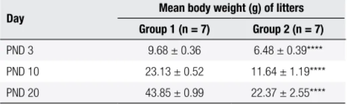
Evaluation of female offspring after birth
Mean body weight (g) of female offspring of diabe-tic female rats was signiicantly lower on PND 3, 10, and 20 than that of control female offspring (Table 2). Mean blood glucose (mg/dL) in offspring of diabetic female rats was statistically lower on PND 3 (Group 1 = 104.78 ± 2.27; Group 2 = 67.93 ± 4.38) and PND 10 (Group 1 = 138.58 ± 3.31; Group 2 = 111.43 ± 9.78), than values presented by control offspring, while on PND 20 blood glucose means were similar in both groups (data not shown).
There was a delay in the mean time (days) for ear unfolding, fur appearance, and eye opening in the offs-pring of diabetic females, compared with the control group. The time of incisor emergence did not differ signiicantly between groups (Table 3).
Table 1. Blood glucose concentrations (mg/dL) in diabetic and
non-diabetic rats during pregnancy
Blood glucose concentrations (mg/dL)
Non-diabetic group (n = 17)
Diabetic group (n = 55)
Day 0 111.16 ± 3.64 476.86 ± 10.06****
Day 7 106.63 ± 3.50 468.71 ± 1.77****
Day 14 96.94 ± 2.75 525.48 ± 24.47****
Day 21 84.76 ± 3.19 561.32 ± 13.43****
Values are expressed as mean ± SEM. **** p < 0.0001, Mann-Whitney test.
Table 2. Mean body weight (g) of litters, assessed on PND 3, 10, and 20
Day Mean body weight (g) of litters Group 1 (n = 7) Group 2 (n = 7)
PND 3 9.68 ± 0.36 6.48 ± 0.39****
PND 10 23.13 ± 0.52 11.64 ± 1.19****
PND 20 43.85 ± 0.99 22.37 ± 2.55****
Group 1: litters of control rats; Group 2: litter of diabetic dams. Values expressed as mean ± SEM. **** p < 0.0001, Student’s t-test.
Table 3. Physical development of female offspring of control and diabetic
rats
Characteristics
Mean (days) of physical development of litters
Group 1 (n = 7) Group 2 (n = 7)
† Ear unfolding 2.92 ± 0.11 3.59 ± 0.18**
† Fur appearance 4.82 ± 0.13 6.03 ± 0.37*
† Incisor emergence 6.16 ± 0.39 6.83 ± 0.27
‡ Eye opening 13.82 ± 0.18 15.12 ± 0.49*
Group 1: litters of control rats; Group 2: litters of diabetic dams. Values expressed as mean ± SEM.
Cop
yright
© ABE&M t
odos os dir
eit
os r
eser
vados
.
Reproductive physiology
Figure 1 shows a signiicant delay in time, in days, to vaginal opening, indicative of the onset of puberty, in the female offspring of diabetic female rats when com-pared with controls.
The evaluation of sexual behavior did not show signiicant differences between the groups (data not shown). In relation to the reproductive performance of female offspring of diabetic rats, it was veriied that exposure to an environment of diabetes in utero and during lactation provoked reductions in inal weight of pregnant females, litter weight, and number of offs-pring, and elevation in placental weight compared with controls (Table 5).
Figure 1. Age of the litters on the day of vaginal opening. Group 1: litters
of control rats (n = 7); Group 2: litters of diabetic dams (n = 7). Values expressed as mean ± SEM. Student’s t-test. **** p < 0.0001.
Similarly, there was a delay in the day of irst estrus among female offspring of diabetic female rats (41.98 ± 0.96) compared with control offspring (36.02 ± 0.45), but there was no difference between the frequency (in days) of pro-estrus, estrus or meta-estrus phases between groups (data not shown).
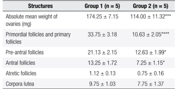
The histological aspects of ovaries and uteruses of female rats in estrus were similar in both experimen-tal groups (data not shown). However, morphometric analysis of the gonads revealed signiicant diminution in the number of primordial/primary, pre-antral, and antral follicles of offspring of diabetic female rats com-pared with controls (Table 4). A reduction was also observed in mean absolute weight of the ovaries (mg) among offspring of diabetic female rats in relation to the control group (Table 4).
Table 4. Absolute mean ovarian weight (mg); classiication and counts of
ovary structures
Structures Group 1 (n = 5) Group 2 (n = 5)
Absolute mean weight of ovaries (mg)
174.25 ± 7.15 114.00 ± 11.32***
Primordial follicles and primary follicles
33.75 ± 3.18 10.63 ± 2.05****
Pre-antral follicles 21.13 ± 2.15 12.63 ± 1.99*
Antral follicles 13.25 ± 1.72 7.25 ± 1.15*
Atretic follicles 1.12 ± 0.13 0.75 ± 0.16
Corpora lutea 9.75 ± 1.03 7.75 ± 1.37
Group 1: female offspring of control rats; Group 2: female offspring of diabetic dams, aged 60 days, in estrus. Values expressed as mean ± SEM. * p < 0.05, *** p < 0.001 and **** p < 0.0001. Student’s t-test.
Table 5. Mean blood glucose and fertility test
Parameter Group 1 (n = 10) Group 2 (n = 10)
b Pregnancy rate (%) 90 80
a Mean blood glucose level (mg/dL) 90.44 ± 3.64 82.75 ± 3.19
a Dam inal body weight (g) 321.80 ± 7.65 286.54 ± 7.49**
a Uterus + fetuses weight (g) 60.99 ± 5.41 42.29 ± 3.43*
a Dam weight – uterus + fetuses (g) 310.05 ± 12.99 264.23 ± 7.25**
a Fetal weight (g) 3.71 ± 0.32 3.05 ± 0.36
a Placenta weight (g) 0.51 ± 0.03 0.70 ± 0.07*
a Number of corpora lutea 14.56 ± 0.87 13.13 ± 0.64
a Number of implantations 11.00 ± 0.80 9.25 ± 1.19
a Number of resorptions 1.11 ± 0.26 1.38 ± 0.38
a Number of fetuses 10.33 ± 0.89 6.00 ± 1.07***
b Sex ratio (%) 50 50
b Fertility potential (%) 80.00 68.76
b Pre-implantation loss (%) 23.99 31.18
b Post-implantation loss (%) 11.24 12.49
Group 1: female offspring of control rats; Group 2: female offspring of diabetic dams. Values expressed as mean ± SEM for Student’s t-testa and as median for Mann-Whitney testb. * p <
0.05, ** p < 0.01, *** p < 0.001.
DISCUSSION
Diverse studies have demonstrated that the program-ming of the function of different body systems and organs occurs during intrauterine development. The results of this programming are manifested throughout life (fetal programming of adult diseases) in respon-se to environmental disturbances in utero and during lactation (10,24). Most of these studies involved the evaluation of the effects of fetal programming on car-diovascular dysfunctions and on the development of diabetes in adults due to gestation in an abnormal ute-rine environment (25). Given this context, the present study aimed at verifying the late effects of fetal/neo-natal programming on female reproductive functions throughout development, and to demonstrate that the exposure of female rats to maternal diabetes during fe-tal development and lactation leads to consequences in
Time (Days)
41
40
39
Group 1 Group 2
38
37
36
35
Cop
yright
© ABE&M t
odos os dir
eit
os r
eser
vados
.
female offspring that are manifested as much prior to puberty as during adulthood.
Among the parameters evaluated, body weight is hi-ghly important, since it provides a general view of the health status of the animal, and indicates possible effects of systemic toxicity. Although body weight gain among diabetic female rats in the present study was similar to that of controls throughout most of pregnancy, there was a diminution in body weight in the diabetic group at the end of this period. A recent study also observed that female rats in which diabetes was chemically in-duced with streptozotocin showed lower body weight gain during the pregnancy than the control group (25).
In the present study, gestation and reproductive success of diabetic female rats was impaired when com-pared with females with normal blood glucose. After the animals were killed, it was observed that the uteru-ses of diabetic female rats that did not deliver offspring revealed the presence of resorptions sites, indicating that chronic hyperglycemia in the intrauterine environ-ment caused by maternal diabetes negatively affected offspring development and survival since the moment of conception. A recent study also reported an eleva-tion in the rates of embryo resorpeleva-tion and fetal death among the offspring of diabetic female rats (26).
In this study of females with severe induced diabetes, it was observed that on PND 3, 10, and 20, mean body weight of female offspring was reduced when compared with control offspring, evidencing that their development was impaired. Epidemiological studies have demonstrated an association between low birth weight and development of chronic diseases in adulthood (27,28). Mean blood glucose among female offspring born to diabetic female rats evaluated on PND 3 and 10 was lower than that of controls, probably due to an exacerbated response of pancreatic b-cells of pups on account of excessive hyperglycemia during intrauterine development (29).
In relation to physical development, delays in ear unfolding, fur appearance, and eye opening was ob-served in the present study among females born to dia-betic dams. This might be related to intrauterine distur-bances during pregnancy caused by maternal diabetes, negatively affecting fetal and neonatal growth and de-velopment, restricting intrauterine growth, characteri-zed by low body weight at birth and at the beginning of the postnatal period (30,31).
In recent decades, several studies have demonstrated a relationship between the restriction of intrauterine/ neonatal growth (RCIU) and pubertal development of
females and males (25,31). The relationship between the mechanisms that control the onset of puberty and the presence of RCIU has not been elucidated yet. Be-cause of that, we performed an analysis of vaginal ing (32). Together with the irst estrus, vaginal open-ing is an external sign of the onset of female puberty (33,34).Our investigation showed that there was a de-lay in the mean age of vaginal opening and irst estrus in offspring of diabetic female rats, suggesting that in-trauterine/neonatal development in an hyperglycemic environment negatively affected initial sexual develop-ment in females. However, in adulthood, estrous cycle became normal.
Uterus weight and morphology underwent normal luctuations due to the alterations in hormone levels that occur during the course of the estrous cycle, with maximum weight observed during pro-estrus in res-ponse to an increase in estrogen secretion (35). In the present study, the absence of changes in the regularity of the estrous cycle, in uterus morphology and weight during the estrus of adult female rats suggests that ex-posure to hyperglycemia in utero and during lactation did not interfere with neuroendocrine secretions during the reproductive cycle.
However, ovary weight was diminished in pubertal female offspring of diabetic dams, probably due to a re-duction, at 60 days of age, in the number of primordial, primary, pre-antral, and antral follicles. These data sug-gest that exposure to hyperglycemic intrauterine and lactation environments may have induced, in this phase of sexual development, a discontinuous evolution of follicular stages, thus diminishing the ovulatory mecha-nism. The impact on reproduction is determined by the stage of follicular development achieved (36,37).
Sexual organization of the female brain requires, during the sensitive period of development, low levels of estrogen of maternal origin to produce characteristic female sexual behaviors, as well as the cyclic release of gonadotropins to maintain the phases of estrus (38). In our studies, the lordosis coeficient of females during the sexual behavior test was similar in both experimental groups, suggesting that maternal hyperglycemia proba-bly did not interfere with the sexual organization of the female brain.
Cop
yright
© ABE&M t
odos os dir
eit
os r
eser
vados
.
follicles provoked by damage in pre-antral and antral follicles, which temporarily interrupted reproductive physiology given the greater recruitment of primordial follicles to replace damaged follicles. Damage in pri-mordial and primary follicles leads to permanent infer-tility, since they are not restored (39).
There was an increase in placental weight in fe-male offspring of diabetic dams, a fact that could be related to a compensatory mechanism, whereas fetuses exposed to hyperglycemic intrauterine environment presented RCIU. However, this increase in placental weight was not suficient to reverse the clinical picture of RCIU. These results corroborate studies of other authors (40).
CONCLUSION
In the present study, chronic hyperglycemia during ges-tation and lacges-tation affected the reproductive functions of female offspring when they reached puberty and se-xual maturity, reinforcing the hypothesis that environ-mental disturbances in utero and during lactation may permanently program the structure and function of the organism. Based only on the parameters evaluated, we cannot determine how the alterations found in the female reproductive system may have directly resulted from the hyperglycemic intrauterine and lactation en-vironment, that is, whether they arose from maternal diabetes or from an indirect effect of a harmful mater-nal-fetal exchange that brought about nutritional dei-ciency and intrauterine restriction of offspring growth, since these two factors are related.
In this context, future studies to verify the mecha-nisms by which maternal diabetes affects reproductive parameters in offspring should be applied to human health to enable better pre- and perinatal care, thus en-suring better quality of life. Diabetes alters psychomo-tor development of female offspring born to diabetic dams; and the occurrence of this disorder has increased in recent decades throughout the globe, becoming an important public health problem. Thus, the utilization of animal models with experimentally-induced mater-nal diabetes (severe or gestatiomater-nal) may help elucidate the mechanisms by which the disease programs the fetal organism to develop chronic diseases and reproductive problems in later phases of life.
Acknowledgements: we would like to thank Fundação de Ampa-ro à Pesquisa do Estado de São Paulo (Fapesp, PAmpa-roc. 04/12948-0 and Proc. 04/12948-04/15664-2) and Fundação para o
Desenvolvimen-to da Universidade Estadual Paulista (Unesp) (Fundunesp, Proc. 01089/05) for their inancial support.
Disclosure: no potential conlict of interest relevant to this article was reported.
REFERENCES
1. ADA. Standards of Medical Care in Diabetes. Care. 2007;30:S4-S41. 2. Takenaka Y, Toyoda N. The effect of alpha 1-blocking vasodilator
on fetal growth and uteroplacental blood low in streptozotocin--induced diabetic rats. Life Sci. 1995;56(13):1127-34.
3. Reece EA, Coustan DR, Gabbe SG. Diabetes in women: adoles-cence, pregnancy and menopause. New York: Lippincott Williams & Wilkins; 2004.
4. Zhao Z, Reece EA. Experimental mechanisms of diabetic embryo-pathy and strategies for developing therapeutic interventions. J Soc Gynecol Investig. 2005;12:549-57.
5. Cho NH, Silverman BL, Rizzo TA, Metzger BE. Correlations between the intrauterine metabolic environment and blood pressure in ado-lescent offspring of diabetic mothers. J Pediatr. 2000;136:587-92. 6. Manderson JG, Mullan B, Patterson CC, Hadden DR, Traub AI,
McCance DR. Cardiovascular and metabolic abnormalities in the offspring of diabetic pregnancy. Diabetologia. 2002;45:991-6. 7. Canavan JP, Goldspink DF. Maternal diabetes in rats II: effects on
fetal growth and protein turnover. Diabetes. 1988;37:1671-7. 8. Thamotharan M, McKnight RA, Thamotharan S, Kao DJ, Devaskar
SU. Aberrant insulin-induced GLUT4 translocation predicts glu-cose intolerance in the offspring of a diabetic mother. Am J Phy-siol Endocrinol Metab. 2003;284:901-14.
9. Fujisaw Y, Nakagawa Y, Ren-shan L, Ohzeki T. Streptozotocin-indu-ced diabetes in the pregnant rat reduces 11 beta-hydroxysteroid dehydrogenase type 2 expression in placenta and fetal kidney. Life Sci. 2004;75:2797-805.
10. Aerts L, Van Assche FA. Intra-uterine transmission of disease. Pla-centa. 2003;24:905-11.
11. Lesage J, Del-Favero F, Leonhardt M, Louvart H, Maccari S, Vieau D, et al. Prenatal stress induces intrauterine growth restriction and programmes glucose intolerance and feeding behaviour dis-turbances in the aged rat. J Endocrinol. 2004;181:291-6.
12. Heindel JJ. Role of exposure to environmental chemicals in the developmental basis of disease and dysfunction. Reprod Toxicol. 2007;23:257-9.
13. Boloker J, Gertz SJ, Simmons RA. Gestational diabetes leads to the development of diabetes in adulthood in the rat. Diabetes. 2002;51:1499-506.
14. Bolzán AD, Bianchi MS. Genotoxicity of streptozotocin. Mutation Research. 2002;512-21.
15. Holemans K, Aerts L, Van Assche FA. Lifetime consequences of abnormal fetal pancreatic development. J Physiol. 2003;574:11-20. 16. Hrabák A, Szabó A, Bajor T, Körner A. Differences in the nitric oxi-de metabolism in streptozotocin-treated rats and children suffe-ring from Type 1 diabetes. Life Sci. 2006;78:1362-70.
17. Baccetti B, La Marca A, Piomboni P, Capitani S, Bruni E, Petraglia F, et al. Insulin-dependent diabetes in men is associated with hy-pothalamo-pituitary derangement and with impairment in semen quality. Hum Reprod. 2002;17(10):2673-7.
18. Jhonson LM, Sidman RL. A reproductive endocrine proile in the diabetes (db) mutant mouse. Biol Reprod. 1979;20:552-9. 19. Smart JL, Dobbing J. Vulnerability of developing brain: II. Effects
Cop
yright
© ABE&M t
odos os dir
eit
os r
eser
vados
.
20. Marcondes FK, Bianchi FJ, Tanno AP. Determination of the estrous cycle phases of rats: some helpful considerations. Braz J Biol. 2002;62(4A):609-14.
21. Mandl AM. The phases of the oestrous cycle in the adult while rat. J Experimental Biology. 1951;28:576-84.
22. Borgeest C, Symonds D, Mayer LP, Hoyer PB, Flaws JA. Metho-xychlor may cause ovarian follicular atresia and proliferation of the ovarian ephitelium in the mouse. Toxicol Sci. 2002;68:473-8. 23. Talsness CE, Shakibaei M, Kuriyama SN, Grande SW,
Sterner--Kock A, Schnitker P, et al. Ultrastructural changes observed in rat ovaries following in utero and lactation exposure to low doses of a polybrominated lame retardant. Toxicol Lett. 2005;157:189-202. 24. Zambrano E, Martínez-Samayoa PM, Bautista CJ, Deás M, Guillén L, Rodríguez-González GL, et al. Sex differences in transgenera-tional alterations of growth and metabolism in progeny (F2) of female offspring (F1) of rats fed a low protein diet during preg-nancy and lactation. J Physiol. 2005;566:225-36.
25. Zambrano E, Bautista CJ, Deás M, Martinez-Samayoa PM, Gon-zalez-Zamorano M, Ledesma H, et al. A maternal low protein diet durin pregnancy and lactation in the rat male reproductive deve-lopment. J Physiol. 2005a;563:275-84.
26. Al Ghali MHM, Padmanabhan R, Kataya HH, Berg B. Effects of alpha-lipoic acid supplementation on maternal diabetes-induced growth retardation and congenital anomalies in rat fetuses. Mol Cell Biochem. 2004;261:123-35.
27. Soto IN, Mericq GV. Fetal growth restriction and insulin resis-tance. New indings and review of the literature. Rev Med Chile. 2005;133(1):97-104.
28. Srinivasan M, Aalinkeel R, Song F, Mitrani P, Pandya JD, Strutt B, et al. Maternal hyperinsulinemia predisposes rat fetuses for hyperinsulinemia, and adult-onset obesity and maternal mild food restriction reverses this phenotype. Am J Physiol Endocri-nol Metab. 2006;290(1):129-34.
29. Van Assche FA, Holemans K, Aerts L. Long-term consequences for offspring of diabetes during gestation. Br Med Bull. 2001;60:173-82.
30. Holemans K, Gerber RT, Meurrens K, Clerck F, Poston L, Van Assche FA. Streptozotocin diabetes in the pregnant rat indu-ces cardiovascular dysfunction in adult offspring. Diabetologia. 1999;42:81-9.
31. Engelbregt MJ, Houdijk ME, Popp-Snijders C, Delemarre-Van de Waal HA. The effects of intra-uterine growth retardation and post-natal undernutrition on onset of puberty in male and female rats. Pediatr Res. 2000;48(6):803-7.
32. Eckstein KL, Oberlander SG, Marx GF. Uterine rupture during ex-tradural blockade. Can Anaesth Soc J. 1973;20(4):566-8. 33. Ojeda SR, Urbanski HF. Puberty in the rat. The physiology of
re-production. New York: Raven Press; 1994. p. 363-409.
34. Beckman DA, Feuston M. Landmarks in the development of the female reproductive system. Birth defects research. Part B. Dev Reprod Toxicol. 2003;68(2):137-43.
35. US EPA. Guidelines for Reproductive Toxicity Risk assessment, U.S. Environmental Protection Agency. Federal Register; 1996. p. 61-212. 36. Hoyer PB, Sipes IG. Assessment of follicle destruction in che-mical-induced ovarian toxicity. Annu Rev Pharmacol Toxicol. 1996;36:307-31.
37. Hoyer PB. Ovotoxic environmental chemicals: indirect endocri-ne disruptors. In: Naz R (ed). Endocriendocri-ne disrupters: effects on male and female reproductive systems. CRC Press. Boca Raton. 1999;57-88.
38. Dohler KD, Cocquelin A, Davies F, Hines M, Shrne JE, Gorski SA. Differentiation of the sexually dimorphic nucleus in the preoptic area of the brain is determined by the perinatal hormone environ-ment. Neurosciences Letters. 1982;33:295-8.
39. Hooser SB, Douds DP, DeMerell DG, Hoyer PB, Sipes IG. Long--term ovarian and gonadotropin changes in mice exposed to 4-vinylcyclohexene. Reprod Toxicol. 1994;8:315-23.