Iranian Journal of Otorhinolaryngology No.4, Vol.24, Serial No.69, Autumn 2012
161 Original Article
Evaluation of Tissue Expression and Salivary Levels of HER2/neu
in Patients with Head and Neck Squamous cell Carcinoma
Soheil Pardis1, Yasaman Sardari1, Mohammad Javad Ashraf2,*Azadeh Andisheh Tadbir1, Hooman Ebrahimi3, Sara Purshahidi4, Bijan Khademi4, Mohammad Javad Fattahi5,
Marzieh Hamzavi2
Abstract
Introduction:
HER2/neu, a member of the epidermal growth factor receptor family, has been shown to be over-expressed in some tumors. The purpose of this study was to determine the salivary levels and tissue expression of HER2/neu in patients with head and neck squamous cell carcinoma (HNSCC) and their correlation with clinicopathologic parameters.
Materials and Methods:
Enzyme-linked immunosorbent assays (ELISA) were used to evaluate the salivary levels of HER2/neu and immunohistochemistry was used to measure tissue expression of HER2/neu in 28 patients with HNSCC and 25 healthy control subjects.
Results:
The salivary levels of HER2/neu in patients with HNSCC were not significantly higher compared to healthy control subjects. There was no apparent correlation between salivary HER2/neu levels and clinicopathological features such as age, sex, tumor grade, tumor size and nodal status. All HNSCC specimens were positive (membranous or/and cytoplasmic) for HER2/neu, except one sample. Only one HNSCC specimen showed staining purely in the tumor-cell cytoplasm. All control specimens were also positive for both membranous and cytoplasmic HER2/neu but there was a significant difference between the level of cytoplasmic staining in the HNSCC specimens and in the control specimens (P<0.05).
Conclusion:
In our study, no overexpression of HER2/neu was observed. Thus, identification of HER2/neu levels plays no role in differentiating between normal and squamous cell carcinoma tissues or detecting the carcinogenesis process. Our findings suggest that the use of HER2/neu as a salivary marker of HNSCC is not recommended, because no significant preoperative elevation and no association with clinicopathological features were found.
Keywords:
Carcinoma, HER2/neu, Salivary Gland, Squamous cell of head and neck, Tissue expression.
Received date: 10 Mar 2012 Accepted date: 17 Jun 2012
1
Department of Oral and MaxillofacialPathology, School of Dentistry, Shiraz University of Medical Sciences, Shiraz, Iran.
2
Department of Pathology, School of Medicine, Shiraz University of Medical Sciences, Shiraz, Iran.
3
Department of Oral Medicine, School of Dentistry, Shiraz University of Medical Sciences, Shiraz, Iran.
4
Department of Otolaryngology, Khalili Hospital, Shiraz Institute for Cancer Research, Shiraz University of Medical Sciences, Shiraz, Iran.
5
Shiraz Institute for Cancer Research, Shiraz University of Medical Sciences, Shiraz, Iran. *
Corresponding Author:
Ghom Abad, Ghasrodasht Avenue, Shiraz, Iran.
162, Iranian Journal of Otorhinolaryngology No.4, Vol.24, Serial No.69, Autumn 2012
Introduction
Squamous cell carcinoma is the most common head and neck cancer (1), it is focally invasive and its behavior depends on the region where it originates. Each anatomic area has its own growth patterns and prognosis (2). Head and neck squamous cell carcinoma (HNSCC) has been a challenge to treat for a long time because of the high rates of recurrence and the advanced disease state usually present at the time of diagnosis. Molecular identification of tissue biomarkers in diagnostic biopsy specimens may not only identify patients at risk for developing HNSCC but may also identify patients who will benefit from more aggressive treatment modalities (3).
The HER2/neu (ErbB) protein or epidermal growth factor receptors (EGFR) are a family of four structurally related receptor tyrosine kinases (4). The c-erbB-2 proto-oncogene (HER/NEU/neu) encodes a 185 transmembrane protein product of the tyrosine kinase family, with an extensive homology to EGFR, which has been mapped to region 21 of chromosome 17 (5), and can be activated by hetero-oligomerization with the other members of the ErbB family (6). Activation of the EGFR family (HER2/neu) by a variety of ligands is necessary for normal growth and differentiation (7). Increased levels of receptor ligands, co-expression of EGFR mutants, and cross-talk with HER2 or other receptors are mechanisms that can enhance EGFR signaling output and potentially alter the response to EGFR inhibitors (8). The dysregulation of these receptors is linked to multiple features of malignant tumors, including a loss of cell cycle control, resistance to apoptotic stimuli, invasiveness, chemo-resistance, and the induction of angiogenesis (9,10). The use of targeted agents against molecular markers belonging to the EGFR family has recently become integrated into the treatment protocols of many malignancies, such as breast cancer (11-14). Despite recent improvements in
the diagnosis and treatment of cancer, there are still many difficulties in evaluating the prognosis of head and neck carcinomas. Thus, recent reports have attempted to find more significant information to predict the biological behavior of this neoplasm, and to look at the possible relationship between tumoral progression and the products of genes that regulate cell proliferation and differentiation, such as the proto-oncogenes, anti-oncogenes and apoptosis-regulating genes. However, there is doubt over the prognostic significance of oncogene EGFR in these tumors, and thus its utility as a target of new therapies is still unclear (15-18).
The aim of this study was primarily to examine the expression of HER2/neu in normal human oral epithelium and patients with HNSCC, to validate the controversial results of various studies and to determine whether or not HER2/neu could be considered as a useful marker for head and neck cancer. Thus, we investigated salivary levels of HER2/neu in healthy subjects and patients with HNSCC and also compared the tissue expression of the protein between the two groups.
Materials and Methods
Iranian Journal of Otorhinolaryngology No.4, Vol.24, Serial No.69, Autumn 2012,163
Clinical data, such as age, gender, location of the tumor, TNM, and tobacco consumption, were obtained from medical records. The Ethical Committee of Shiraz University of Medical Sciences approved the study. All subjects were informed about the research and agreed to participate in the study by signing an informed consent form.
Saliva collection and analysis: Salivary samples were taken from subjects before any surgical procedures or chemotherapy protocols commenced. Prior to the collection of unstimulated whole saliva, subjects were asked to refrain from eating, drinking, smoking or performing oral hygiene procedures for 30 minutes. The lip area was cleaned and each subject rinsed their mouth once with plain water. Typically, patients donated 5 to 10 ml of saliva. Samples were then centrifuged at 2,600×g for 15 minutes at 4ºC. The resulting supernatant was then stored at -80ºC until use (19). Salivary protein levels were measured by sandwich ELISA, in accordance with the procedures recommended by the manufacturer (BMS 207: Bender Med System GmbH, Germany 4).
Immunohistochemical analysis: Sections of tumor tissue (4 µm thick) were mounted on positively charged microscope slides. After dewaxing in xylene, sections were dehydrated in ethanol and rinsed in distilled water. Antigen retrieval was performed using DAKO Target Retrieval Solution (DAKO, Carpinteria, CA). The endogenous peroxidase was quenched with 3% H2O2. Peroxidase-labeled polymer conjugated to goat anti-mouse HER2 was then used to detect the antigen-antibody reaction (DAKO EnVision System; DAKO Corporation, Carpinteria, CA). The HER2 antibodies (ErbB 2 antibody ab2428, DAKO Corporation, Denmark; 1:200 dilution) were incubated with the tissue sections for 1 hour at room temperature. Sections were then visualized
with 3,3-diaminobenzidine as a chromogen for 5 minutes and counterstained with
Harris’s hematoxylin. Slides were washed
in tap water, dehydrated, and mounted with glass coverslips. Positive controls were sections of breast cancer tissue that had previously been found to be positive for HER2/neu and negative controls consisted of duplicated sections of the same specimens in which the primary antibody had been excluded and replaced with PBS.
164, Iranian Journal of Otorhinolaryngology No.4, Vol.24, Serial No.69, Autumn 2012
were evaluated again by both pathologists using a multi-headed microscope.
Statistical analysis: The Mann-Whitney
test, Student’s independent t-test,
Chi-Squared test and Fisher’s exact test were used for statistical analysis. The level of significance was set at 0.05.
Results
The clinical data for the patients with HNSCC included in this study of HER2/neu expression are presented in (Table 1).
Table 1: Clinico-pathological profile of 28
patients with head & neck squamous cell carcinoma
Age (years) 58.0 ± 12.3
Gender
Male 19 (67.8%)
Female 9 (32.2%)
Tumor size
T1 0 (0%)
T2 28 (100%)
T3 0 (0%)
Regional lymph node involvement
N0 24 (85.7%)
N1 4 (14.3%)
N2 0 (0%)
N3 0 (0%)
Distant metastasis
M0 28 (100%)
M1 0 (0%)
TNM stage
I 0 (0%)
II 24 (85.7%)
III 4 (14.3%)
IV 0 (0%)
Histological grade
I (well differentiated) 8 (28.6%) II (moderately differentiated) 17 (60.7%) III (poorly differentiated) 3 (10.7%)
Larynx 22 (78.5%)
Oral cavity 6 (21.5%)
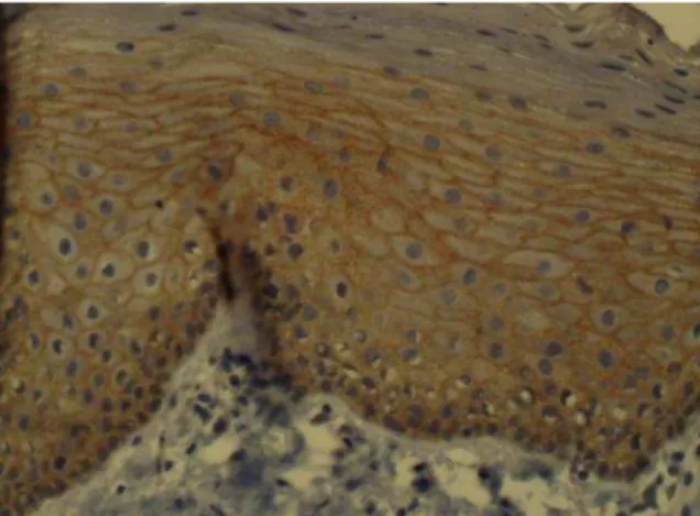
All samples of normal oral epithelium were positive for membranous and cytoplasmic HER2/neu. Cytoplasmic staining was limited to the basal and parabasal layers in normal epithelium (Figure 1).
Fig 1: Cytoplasmic staining in the basal and
parabasal layers of normal epithelium
All HNSCC specimens were positive for membranous or/and cytoplasmic HER2/neu, except one sample (Figure 2,3).
Fig 2: Membranous staining in squamous cell
carcinoma tissue (×400)
Fig3: Cytoplasmic and membranous staining
Iranian Journal of Otorhinolaryngology No.4, Vol.24, Serial No.69, Autumn 2012,165
Only one HNSCC specimen contained only cytoplasmic staining. The mean percentage of membranous staining in HNSCC samples was 63.0 ± 30.8% and in normal epithelium it was 66.4 ± 16.4%. There was no significant difference in the
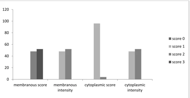
percentage of membranous staining between the patient and control groups. The distribution of membranous immune-staining according to each of the grades is presented in (Figure 4).
Fig 4: Distribution of score and intensity of membranous and cytoplasmic HER-2/neu
immunohistochemical staining in HNSCC specimens
There was no significant difference between the grade of membranous immune-staining in the case and control groups. However, the mean percentage of cytoplasmic staining in samples of HNSCC was 48.6 ± 29.1% and in normal
epithelium it was 13.2 ± 6.7%. There was a significant difference in the percentage of cytoplasmic staining between the case and control groups (P<0.001).The distribution of cytoplasmic immune-staining grades is presented in (Figure 5).
Fig 5: Distribution of score and intensity of membranous and cytoplasmic HER-2/neu
immunohistochemical staining in control specimens
0 20 40 60 80 100 120
membranous score membranous intensity
cytoplasmic score cytoplasmic intensity
score 0
score 1
score 2
166, Iranian Journal of Otorhinolaryngology No.4, Vol.24, Serial No.69, Autumn 2012
There was a significant difference between the grade of cytoplasmic immune-staining in the case and control groups (P<0.001). There was no correlation between the percentage of HER-2 staining and any clinicopathologic factors.There was a high tendency for cytoplasmic staining around keratin pearls in well-differentiated HNSCC samples.
The mean salivary level of HER2/neu in patients with HNSCC was 3.12 ± 4.58 ng/ml and for the control group was 13.2± 6.75 ng/ml. There was no significant difference in salivary concentrations of HER2 between the case and the control groups. There was also no significant correlation of salivary levels with the percentage of overall immune-staining in samples of tumor tissue from patients with HNSCC, and there was no significant correlation between salivary HER-2 levels and the clinic-pathological data of patients. However, salivary HER-2 levels had a reverse relationship with the percentage of cytoplasmic staining in tumor samples from patients with HNSCC, but there was no significant correlation(co.co.= -0.025,
P= 0.90).
Discussion
We studied HER2/neu expression in patients with HNSCC and its correlation with salivary levels of this marker. Although a lack of specificity of tumor markers and a lack of sensitivity of testing systems has been noted, which limits their clinical use, finding biomarkers for cancers would allow physicians to identify individuals who are susceptible to certain types and stages of cancer. It would also allow the development of tailored preventive and therapeutic modalities based on a patient’s genotype and phenotype information. These biomarkers should be cancer specific and detectable at a high level of sensitivity in a wide range of specimens containing cancer-derived materials, including body fluids (plasma, serum, urine, saliva, etc.), tissue samples, and cell lines (5).
The amplification of the HER2 gene has been demonstrated in many carcinomas of glandular origin, and immunohistochemically-identified expression of the gene has been proven to be closely associated with its amplification level (21). The amplification and consequent over-expression of the HER2 gene, as well as its relationship with tumorogenesis was first reported by Schechler et al. (1985) in neuroglioblastomas in rats (22). The dysregulation of these receptors is linked to multiple features of malignant tumors, including a loss of cell cycle control, resistance to apoptotic stimuli, invasiveness, chemo-resistance, and the induction of angiogenesis (10).
Reports on the role of the HER2/neu proto-oncogene product in HNSCC are less conclusive than those detailing the role of EGFR(HER1), as over-expression of HER2/neu has been shown to be present in anywhere from only a few to all HNSCC specimens investigated and correlations with clinical parameters are controversial (23-26). In our study, all samples except one expressed HER2/neu and there was no significant difference between the overall level of staining in the case and control specimens. All non-tumoral and 96.4% of HNSCC samples were positive for cytoplasmic and/or membranous HER2/neu. One of the HNSCC specimens was positive only for cytoplasmic HER2/neu and another one was negative for the marker. Thus, we found no over-expression of HER2/neu in HNSCC tissues. This implies that abnormal expression and over-expression of HER2/neu do not play a role in the carcinogenesis of HNSCC. This result is in accordance with several other studies that found no HER2/neu over-expression in cases of HNSCC (26-29).
Iranian Journal of Otorhinolaryngology No.4, Vol.24, Serial No.69, Autumn 2012,167
are dynamic changes in HER2/neu expression during the process of carcinogenesis in head and neck cancer (31). Wilkman et al.in 1998, reported an increase in HER2/neu expression during the progession from normal mucosa to hyperkeratosis and to dysplasia and HNSCC (32). In our study there was no difference in the percentage of membranous staining of HER2/neu in the case and control groups but there was a significant difference in cytoplasmic staining between the case and control groups. It should be mentioned that cytoplasmic staining in the normal epithelial specimens was specific to the basal and parabasal layers.
In various studies purely membranous (25) or cytoplasmic (25,26), and mixed membranous-cytoplasmic (15,23,25,33) expression have been reported. In squamous cell carcinomas, cytoplasmic staining has been widely reported; however, its interpretation is not clear at present. It has been argued that cytoplasmic staining may be a technical artifact due to cross-reactivity of antibodies, possibly with keratin or antigen retrieval (14). Others, however, propose that it may represent true protein over-expression that is probably due to incomplete receptor degradation (34). Some antibodies, such as CB11, have a tendency towards producing cytoplasmic staining, thus some manufacturers suggest that purely cytoplasmically stained samples should be designated as negative (35). However, the importance of cytoplasmic staining and whether or not it may be evaluated when determining HER-2 expression in OSCC/ HNSCC is controversial.
In our study we evaluated cytoplasmic and membranous staining separately to make it clear and found a significant difference between cytoplasmic immune-reactivity in the case and control groups. In the literature, the percentage of HER-2/neu positivity in patients with HNSCC is
extremely variable. It is possible that these discrepancies in results may be attributed to an initial lack of standardization of the assay methods (14). Another reason for controversial results in different studies might be due to use of different immunohistochemical methods (direct, indirect), type of antibody (clone CerbB2, CB11, ICR1b, polyclonal DAKO, monoclonal zymed), no specific criteria for positive staining of HER2/neu protein (membrane and/ or cytoplasmic) and/or using different techniques (immunosorbent assay, radioimmunoassay, IHC) or the different locations of the lesions and sex of patients with HNSCC.
There was no significant correlation between HER2/neu expression and age, gender, tumor size, lymph node and distant metastasis, tumoral stage and histologic differentiation in our study. In 1997 Xia et al. found a strong correlation between cerbb2 over-expression and overall survival of patients with OSCC (34). In a review study in 2001, Quon et al. indicated that high expression levels of EGFR and HER2/neu are prognostic markers which correlate with poor clinical outcome in patients with HNSCC (36).By contrast, in a case review study in 2009 by Tse et al. HER2 was associated with longer survival in node-positive patients (37). Some studies had evaluated the correlation of clinicopathologic data such as tumor stage with HER2/neu expression. In 2008, Fong et al. found that HER2/neu expression was significantly higher in patients with advanced stage IV tumors than in those with stage I–III tumors (31).
168, Iranian Journal of Otorhinolaryngology No.4, Vol.24, Serial No.69, Autumn 2012
recent years, increasing interest has developed in the use of saliva as an adjunct test medium to help in conventional medical assessment of serious systemic diseases (38). Due to the simplicity of collection, saliva may be collected repeatedly with minimal discomfort to the patient. This noninvasive process thereby renders saliva a very desirable diagnostic medium. More importantly, saliva contains constituents that are frequently altered in the presence of systemic diseases (39). Because of these significant characteristics, finding biomarkers in saliva for the detection of serious systemic illnesses, such as cancer, is of great interest to most salivary researchers.
In our study, there was no significant difference in salivary levels of HER2/neu between the HNSCC and control groups. The salivary level of HER2/neu in patients with HNSCC was lower than in the control group but the difference was not significant. Also, there was an inverse relationship between salivary levels of HER2/neu and the percentage of cytoplasmic staining. One of the reasons for the presence of lower HER2/neu salivary levels in the case group could be the high level of cytoplasmic staining of this marker in HNSCC specimens, which could cause decreased membranous transportation of this marker to saliva. The inverse relationship between salivary levels of HER2/neu and cytoplasmic staining is proof for this claim.
In this study there was no significant correlation between the salivary levels of HER2/neu and the clinicopathological data
of the patients. Briefly, we found that there was no increase in the presence of HER2/neu protein in saliva in patients with more progressive or aggressive lesions. In a similar study in 2010, Vanessa et al. also found no significant association between the salivary levels of the proteins and patient clinicopathological data, such as age, tumor site, histological grading, T status, nodal involvement of the tumor or stage. Salivary levels of HER-2 also showed no difference between patients pre-surgery and healthy control groups; however, both demonstrated an increase after surgical removal of the tumor (19).
In our cases, no overexpression of HER2/neu was observed. Thus, the protein cannot be used to differentiate between normal and squamous cell carcinoma tissues or identify the occurrence of carcinogenesis.
Conclusion
Our findings suggest that the use of HER2 as a salivary marker of HNSCC is not recommended because no significant preoperative elevation of HER2 or association with clinicopathological features was found.
Acknowledgment
The authors would like to thank the Vice-Chancellery for Research of Shiraz University of Medical Sciences for providing financial support for this study (Grant#90-5562).This manuscript is relevant to the post graduate thesis of Dr.Yasaman Sardari.
References
1. Patel SG, Shah JP. TNM staging of cancers of the head and neck: striving for uniformity among diversity.CA Cancer J Clin2005; 55(4):242-58
2. Anthony SF, Euqene B, Dennis LK, Stephen LH, Dan LL, Larry J et al. Harrison's principles of internal medicine. 17th ed. McGraw-Hill; 2008:548-51.
Iranian Journal of Otorhinolaryngology No.4, Vol.24, Serial No.69, Autumn 2012,169
4. Lafky JM, Wilken JA, Baron AT, Maihle NJ. Clinical implications of the ErbB/epidermal growth factor (EGF) receptorfamily and its ligands in ovarian cancer. Biochim Biophys Acta 2008; 1785(2): 232-65.
5. Albuquerque RL Jr, Miguel MC, Costa AL, Souza LB. Correlation of c-erbB-2 and S-100 expression with the malignancy grading and anatomical site in oral squamous cell carcinoma. Int J Exp Pathol 2003; 84(6):259-65.
6. Silva SD, Agostini M, Nishimoto IN, Coletta RD, Alves FA, Lopes MA et al. Expression of fatty acid synthase, ErbB2 and Ki-67 in head and neck squamous cell carcinoma. A clinicopathological study. Oral Oncol 2004; 40(7):688-96.
7. Seifi S, Shafaei SN, Nosrati K, Ariaeifar B. Lack of elevated HER2/neu expression in epithelial dysplasia and oral squamous cell carcinoma in Iran. Asian Pac J Cancer Prev 2009; 10(4):661-4. 8. Arteaga CL. Epidermal growth factor receptor dependence in human tumors: more than just expression? Oncologist 2002; 7(Suppl 4): 31-9.
9. Salem M, Kinoshita Y, Tajiri T, Souzaki R, Tatsuta K, Higashi M et al. Association between the HER2expression and histologicaldifferentiation in Wilms tumor. Pediatr Surg Int 2006; 22(11):891-6. 10. Woodburn JR. The epidermal growth factor receptor and its inhibition in cancer therapy. Pharmacol Ther 1999; 82(2-3): 241-50.
11. Ciardiello F, Tortora G. A novel approach in the treatment of cancer: targeting the epidermal growth factor receptor. Clin Cancer Res 2001; 7:2958-70.
12. Soulieres D, Senzer NN, Vokes EE, Hidalgo M, Agarwala SS, Siu LL. Multicenter phase II study of erlotinib, an oral epidermal growth factor receptor tyrosine kinase inhibitor, in patients with recurrent or metastatic squamous cell cancer of the head and neck. J Clin Oncol 2004; 22(1): 77-85. 13. Crombet T, Osorio M, Cruz T, Roca C, Castillo R d, Mon R, et al. Use of the humanized anti-epidermal growth factor receptor monoclonal antibody h-R3 in combination with radiotherapy in the treatment of locally advanced head and neck cancer patients. J Clin Oncol 2004; 22(9):1646-54. 14. Cavalot A, Martone T, Roggero N, Brondino G, Pagano M, Cortesina G.Prognostic impact of HER-2/neu expression on squamous head and neck carcinomas. Head Neck 2007; 29(7):655-64. 15. Xia W, Lau YK, Zhang HZ, Xiao FY, Johston DA, Liu AR, et al. Combination of EGFR, HER-2/neu, and HER-3 is a stronger predictor for the outcome of oral squamous cell carcinoma than any individual family members. Clin Cancer Res 1999; 5:4164-74.
16. Shiga H, Rasmussen AA, Johnston PG, Langmacher M, Baylor A, Lee M, et al. Prognostic value of c-erbB2 and other markers in patients treated with chemotherapy for recurrent head and neck cancer. Head Neck 2000; 22(6): 599-608.
17. Werkmeister R, Brandt B, Joos U. Clinical relevance of erbB-1 and -2 oncogenes in oral carcinomas. Oral Oncol 2000; 36(1):100-5.
18. Camp ER, Summy J, Bauer TW, Liu W, Gallick GE, Ellis LM. Molecular mechanisms of resistance to therapies targeting the epidermal growth factor receptor. Clin Cancer Res 2005; 11: 397-405.
19. Bernardes V, Gleber-Netto F, Sousa S, Silva T, Aguiar M. Clinical significance of EGFR, Her-2 and EGF in oral squamous cell carcinoma: a case control study. J Exp Clin Cancer Res 2010; 29(1): 40.
20. Albanell J, Codony-Servat J, Rojo F, Del Campo JM, Sauleda S, Anido J, et al. Activated extracellular signal-regulated kinases: association with epidermal growth factor receptor/transforming growth factor alpha expression in head and neck squamous carcinoma and inhibition by anti-epidermal growth factor receptor treatments. Cancer Res 2001; 61(17):6500-10. 21. Sugano S, Mukai K, Tsuda H, Hirohashi S, Furuya S, Shimosato Y. Immunohistochemicalstudy of c-erbB-2 oncoprotein overexpression in human major salivary gland carcinoma: an indicator of aggressiveness. Laryngoscope 1992; 102(8): 923-7.
22. Schechler AL, Hung MC, Vaidyanathan L, Weinberg RA, Yang-Feng TL, Francke U. The neu gene: an erbB-homologous gene distinct from and unlinked to the gene encoding the EGF receptor. Science 1985; 229(4717): 976-78.
170, Iranian Journal of Otorhinolaryngology No.4, Vol.24, Serial No.69, Autumn 2012
24. Reviere A, Becker J, Loning T. Comparative investigation of c-erbB-2/neu expression in head and neck tumors and mammary cancer. Cancer 1991; 67(8): 2142-9.
25. Craven JM, Pavelic ZP, Stambrook PJ, Pavelic L, Gapany M, Kelley DJ, et al. Expression of c-erbB-2 gene in human head and neck carcinoma. Anticancer Res 1992; 12:2273-6.
26. Field JK, Spandidos DA, Yiagnisis M, Gosney JR, Papadimitriou K, Stell PM. C-erbB-2 expression in squamous cell carcinoma of the head and neck. Anticancer Responsibility 1992; 12(3): 613-19.
27. Ekberg T, Nestor M, Engström M, Nordgren H, Wester K, Carlsson J et al. Expression of EGFR, HER2, HER3, and HER4 in metastatic squamous cell carcinomas of the oral cavity and base of tongue. Int J Oncol 2005; 26(5):1177-85.
28. Angiero F, Sordo RD, Dessy E, Rossi E, Berenzi A, Stefani M et al. Comparative analysis of c-erbB-2 (HER-2/neu) in squamous cell carcinoma of the tongue: does over-expression exist? And what is its correlation with traditional diagnostic parameters? J Oral Pathol Med 2008; 37(3):145-50. 29. Mort Tefler MR, Shepherd JP. Psychological distress in patients attending an oncology clinic after definitive treatment for maxillofacial malignant neoplasia. Int J Oral Maxillofac Surg 1993; 22: 347-9.
30. Lebeau A, Delming D, Kaltz C, Sendelhofert A, Iff A, Luthardt B, et al. HER-2/neu Analysis in Archival Tissue Samples of Human Breast Cancer: Comparison of Immunohistochemistry and Fluorescence In Situ Hybridization . J Clin Oncol 2001; 19(2): 354-63.
31. Fong Y, Chou SJ, Hung KF, Wu HT, Kao SYJ. An investigation of the differential expression of Her2/neu gene expression in normal oral mucosa, epithelial dysplasia, and oral squamous cell carcinoma in Taiwan. Chin Med Assoc 2008; 71(3):123-7.
32. Wilkman TS, Hietanen JH, Malmström MJ, Konttinen YT. Immunohistochemical analysis of the oncoprotein c-erbB-2 expression in oral benign and malignant lesions. Int J Oral Maxillofac Surg 1998; 27: 209-12.
33. Rautava J, Jee KJ, Miettinen PJ, Nagy B, Myllykangas S, Odell EW et al. ERBB receptors in developing, dysplastic and malignant oral epithelia. Oral Oncol 2008; 44(3): 227-35.
34. Xia W, Lau YK, Zhang HZ, Liu AR, Li L, Kiyokawa N et al. Strong correlation between c-erbB-2 overexpression and overall survival of patients with oral squamous cell carcinoma. Clin Cancer Res1997; 3(1):3-9.
35. Hsu CY, Ho DM, Yang CF, Lai CR, Yu IT, Chiang H. Interobserver reproducibility of HER-2⁄neu protein overexpression in invasive breast carcinoma using DAKO HercepTest. Am J Clin Pathol 2002; 118: 693-8.
36. Quon, H, Liu, FF,Cummings BJ. Potential molecular prognostic markers in head and neck squamous cell carcinomas. Head Neck 2001; 23(2): 147-59.
37. Tse GM, Yu KH, Chan AW, King AD, Chen GG, Wong KT et al. HER2 expression predicts improved survival in patients with cervical node-positive head and neck squamous cell carcinoma. Otolaryngol Head Neck Surg 2009; 141(4):467-73.
38. Streckfus CF, Bigler LR. Saliva as a diagnostic fluid. Oral Dis 2002; 8:69-76.