Antibody Profiling in Naïve and Semi-immune
Individuals Experimentally Challenged with
Plasmodium vivax
Sporozoites
Myriam Arévalo-Herrera1,2*, Mary Lopez-Perez1,3, Emmanuel Dotsey4, Aarti Jain4, Kelly Rubiano1, Philip L. Felgner4, D. Huw Davies4, Sócrates Herrera1,3
1Malaria Vaccine and Drug Development Center (MVDC), Cali, Colombia,2Faculty of Health, Universidad del Valle, Cali, Colombia,3Caucaseco Scientific Research Center, Cali, Colombia,4Department of Medicine, University of California Irvine, Irvine, California, United States of America
Abstract
Background
Acquisition of malaria immunity in low transmission areas usually occurs after relatively few exposures to the parasite. A recentPlasmodium vivaxexperimental challenge trial in malaria naïve and semi-immune volunteers from Colombia showed that all naïve individuals developed malaria symptoms, whereas semi-immune subjects were asymptomatic or dis-played attenuated symptoms. Sera from these individuals were analyzed by protein micro-array to identify antibodies associated with clinical protection.
Methodology/Principal Findings
Serum samples from naïve (n = 7) and semi-immune (n = 9) volunteers exposed toP.vivax sporozoite-infected mosquito bites were probed against a custom protein microarray dis-playing 515P.vivaxantigens. The array revealed higher serological responses in semi-immune individuals before the challenge, although malaria naïve individuals also had pre-existing antibodies, which were higher in Colombians than US adults (control group). In both experimental groups the response to theP.vivaxchallenge peaked at day 45 and returned to near baseline at day 145. Additional analysis indicated that semi-immune volun-teers without fever displayed a lower response to the challenge, but recognized new anti-gens afterwards.
Conclusion
Clinical protection against experimental challenge in volunteers with previousP.vivax expo-sure was associated with elevated pre-existing antibodies, an attenuated serological response to the challenge and reactivity to new antigens.
OPEN ACCESS
Citation:Arévalo-Herrera M, Lopez-Perez M, Dotsey E, Jain A, Rubiano K, Felgner PL, et al. (2016) Antibody Profiling in Naïve and Semi-immune Individuals Experimentally Challenged with Plasmodium vivaxSporozoites. PLoS Negl Trop Dis 10(3): e0004563. doi:10.1371/journal.pntd.0004563
Editor:Photini Sinnis, Johns Hopkins Bloomberg School of Public Health, UNITED STATES
Received:January 12, 2016
Accepted:February 29, 2016
Published:March 25, 2016
Copyright:© 2016 Arévalo-Herrera et al. This is an open access article distributed under the terms of the Creative Commons Attribution License, which permits unrestricted use, distribution, and reproduction in any medium, provided the original author and source are credited.
Data Availability Statement:All relevant data are within the paper and its Supporting Information files. Microarray information is publicly available on the NCBI Gene Expression Omnibus (http://www.ncbi. nlm.nih.gov/geo/) and is accessible through accession number GPL18316.
Funding:This work was supported by grants from the National Heart, Lung and Blood Institute at the National Institutes of Health (5R01HL086488; SH); Colombian National Research Council,
Author Summary
Malaria remains an important public health problem worldwide, with 13.8 million cases caused byPlasmodium vivax, a parasite species that predominates in South-East Asia and the American continent. Despite the epidemiological importance of this species, studies of the immune response and their potential for vaccine development are limited. Here we use a high-throughput technique (protein microarray) to identify antibodies in serum from malaria naïve and semi-immune Colombian volunteers experimentally infected withP. vivax. We show a higher response in semi-immune individuals before the challenge. Meanwhile, at day 45 after infection, both groups had the highest antibody response to severalP.vivaxproteins. Additional analysis indicated that semi-immune volunteers with-out fever recognized new antigens, which may represent promising targets for vaccine development. Taken together, these findings represent a significant step forward in the understanding of the humoral immune response toP.vivaxmalaria infection, particularly the extent of immune priming upon a first parasite encounter.
Introduction
Malaria remains an important public health problem worldwide, affecting mainly developing countries in Africa, Asia and Latin America. The World Health Organization estimated that 214 million cases of malaria occurred worldwide in 2015 [1]. Of these cases, 13.8 million cases were calculated to be caused byPlasmodium vivax, a parasite species that predominates in South-East Asia and the American continent where it accounts for more than 50% of malaria cases [1].
In areas of high malaria transmission, individuals continuously exposed toPlasmodium develop partial protection against severe symptoms at an early age and a significant number of asymptomatic infections are recorded [2]. This clinical protection is mediated by both innate and acquired mechanisms that are not well understood [2–4]. Under conditions of hypo- or
meso-endemic transmission, both clinical and subclinical infections are seen in all age groups and, despite the lower frequency of malaria exposure, significant protection against the disease is induced [5]. A high prevalence of uncomplicated and asymptomaticP.vivaxandP. falcipa-rummalaria infections are reported in both hyperendemic and unstable malaria transmission regions, indicating that a significant level of clinical immunity is induced by repeated exposure to the parasite [2,6–9].
Specific antibodies againstP.vivaxandP.falciparumproteins have been reported to be associated with clinical immunity [2,4,10]. However, only a few antigens have been made available through traditional cloning methods or peptide synthesis. SequencedP.vivaxandP. falciparummalaria parasite genomes, along with high-throughput proteomic techniques and bioinformatics are powerful tools currently available for systematic analyses of humoral immune responses associated with naturally and experimentally induced malaria. These analy-ses provide a better understanding of malaria parasite-host interaction, disease pathogenesis, host immune response and the identification of potential vaccine candidate antigens [11–13].
Despite the epidemiological importance ofP.vivax, the immune mechanisms and their poten-tial for vaccine development have been studied less than inP.falciparum. Currently, only two parasite antigens,PvCSP andPvs25 have been assessed in early clinical development [14–16]
as vaccine candidates, although several others are in preclinical development [17–19].
In recent years, the Malaria Vaccine and Drug Development Center (MVDC) in Cali (Colombia) has standardized a safe and reproducible method forP.vivaxsporozoite challenge
Antibody Profiling inP.vivax
International Centers of Excellence for Malaria Research at National Institute of Allergy and Infectious Diseases (AI089686; PLF). The funders had no role in study design, data collection, analysis, decision to publish, or preparation of the manuscript.
byAnopheles albimanusmosquito bites [20,21]. This method enables the evaluation of the protective efficacy ofP.vivaxvaccine candidates under controlled conditions, accelerating their clinical development both by facilitating efficacy studies and antigen discovery. In this context, a challenge study was recently conducted in malaria-naïve and semi-immune volun-teers, who were exposed to controlledP.vivaxinfected mosquito bites [22]. Although all study subjects became parasitemic at the same time point afterP.vivaxchallenge, all naïve volunteers developed symptomatic infections while semi-immune volunteers had either only mild symp-toms or no sympsymp-toms. Antibody responses against two immune-dominantP.vivaxantigens, PvCSP andPvMSP1, showed no differences in the frequency of responders, although naïve vol-unteers exhibited significantly higher antibody responses to these antigens [22]. In order to fully characterize the natural protective antibody responses and to better understand the responses induced byP.vivaxinfection in both study groups, a protein microarray displaying 515P.vivaxantigens was probed with serum samples from these volunteers.
Methods
Ethics statement
This trial was conducted according to ICH E-6 Guidelines for Good Clinical Practices [23] and the protocol was approved by Institutional Review Boards (IRB) of the MVDC and Centro Médico Imbanaco in Cali. Written informed consent was obtained from each volunteer at enrollment. The clinical trial was registered on clinicaltrials.gov, registry number
NCT01585077. The protocol for this trial is available as supporting information (S1 Protocol).
Study participants and sample collection
Blood samples were collected from malaria-naïve (n = 7) and semi-immune (n = 9) adult vol-unteers that participated in a clinical trial carried out at the MVDC [22]. Malaria-naïve volun-teers were recruited in Cali (a non-endemic city) and declared not having suffered malaria and lack of previous malaria exposure was ascertained by negative indirect fluorescent antibody test (IFAT). Semi-immune volunteers were recruited in Buenaventura (malaria-endemic area) and previous malaria exposure was confirmed by clinical history as well as by the presence of antibodies againstP.vivaxblood stages and sporozoites detected by IFAT.
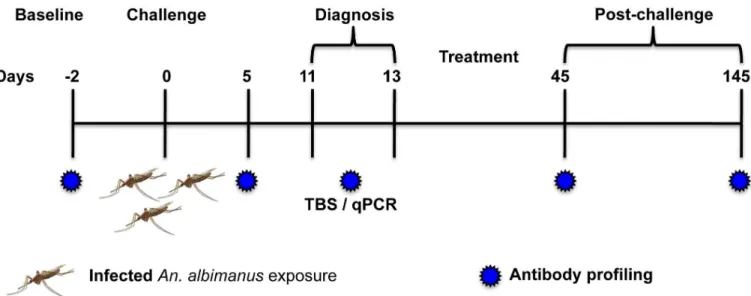
All volunteers were challenged by exposure to bites of two to four mosquitoes previously fed withP.vivax-infected blood obtained from a malaria patient (field strain) as reported before [22]. Volunteers were followed-up for malaria signs and symptoms and were treated orally with curative doses of chloroquine (25 mg/kg) split in three doses and primaquine (0.5 mg/kg daily) for 14 days, as recommended by the official Colombian guidelines, as soon as parasites were detected by microscopy [24]. Serum samples were collected before the challenge (base-line) and five, 11–13 (here day 11), 45 and 145 days after the challenge (Fig 1). Detailed
infor-mation about demographic characteristics of the study participants, challenge infective dose, pre-patent period, parasite density after challenge, and clinical and laboratory evaluations was previously reported [22].
Protein microarray
whole array, data forP.vivaxantigens only are presented in this paper. Microarray information is publicly available on the NCBI Gene Expression Omnibus (http://www.ncbi.nlm.nih.gov/ geo/) and is accessible through accession number GPL18316. Annotation of proteins presented in this study follows gene accession numbers published on PlasmoDB (www.plasmodb.org). Of 515P.vivaxfeatures on the array, 444 mapped to uniqueP.vivaxproteins, of which the major-ity (247; 56%) were classified as hypothetical proteins or hypothetical conserved proteins. Each array contained 24 negative“IVTT-control”reaction spots lacking plasmid template
expres-sion, which provide a donor-specific‘background’signal that was used to normalize data
between individuals.
For probing, serum samples were diluted 1:100 in protein array blocking buffer (Maine Manufacturing, Sanford, ME) supplemented withE.colilysate (GenScript, Piscataway, NJ) to reach a final concentration of 10mg/ml, and pre-incubated at room temperature (RT) for 30 min. Concurrently, arrays were rehydrated in blocking buffer (without lysate) for 30 min. Arrays were probed with pre-incubated serum samples overnight at 4°C with gentle agitation, and then washed at RT five times with TBS-0.05% Tween 20 (T-TBS), followed by incubation with biotin-conjugated goat anti-human IgG (Jackson ImmunoResearch, West Grove, PA) diluted 1:200 in blocking buffer for one hour at RT. After incubation with secondary antibod-ies, arrays were washed three times in T-TBS and bound IgG was visualized using streptavidin-conjugated SureLight P-3 (Columbia Biosciences, Frederick, MD) diluted 1:1000 in blocking buffer for 45 min at RT in the dark. Arrays were washed three times with T-TBS, and once with water. Chips were air-dried by brief centrifugation and scanned in a GenePix 4200AL laser scanner (Molecular Devices, Sunnyvale, CA). All samples in this study were probed at the same time on the same batch of arrays.
Data analysis
Analysis of the protein microarray data was accomplished following our previously published computational methods [3,11]. Briefly, microarray spot intensities (median fluorescence inten-sity, MFI) were quantified using ScanArray Express software (Perkin Elmer, Waltham, MA) Fig 1. Schematic representation of the study.Naïve (n = 7) and semi-immune (n = 9) volunteers were challenged by exposure to the bites of 2–4P.vivax
infected mosquitoes. Patent blood-stage parasitemia was detected by thick blood smear (TBS) and confirmed by real time qPCR on days 11 to 13 post-challenge. All volunteers were treated orally with chloroquine and primaquine and followed-up until day 145 after post-challenge.
doi:10.1371/journal.pntd.0004563.g001
and IVTT spot intensities were normalized by subtraction of the sample-specific median of the IVTT control spots. Antigens were considered seroreactive if the spot intensity of an individual (or the average for a group of individuals) was greater than a cutoff defined as the average plus two standard deviations of the reactivity to allP.vivaxantigens in a US control population. Antibody breadth was used as defined forP.falciparum[26] as the number of seroreactive anti-gens per individual or group. Venn diagrams of group antibody breadths were produced using the BioVenn web application (http://www.cmbi.ru.nl/cdd/biovenn/index.php) [27]. Statistical analyses were performed on data normalized by dividing the IVTT signal by the sample-spe-cific median of the IVTT control spots division (fold-over control, FOC) and taking the base 2 logarithm of the ratio (Log2 FOC). Differentially reactive proteins between both groups were determined using Wilcoxon rank-sum test, and those with Log2 FOC>1 considered
seroposi-tive (Prism v6.0, GraphPad Software Inc., La Jolla CA). A p value<0.05 was considered
statis-tically significant.
Results
Study population characteristics
Volunteers were adults aged between 19 and 38 years. Briefly, all volunteers developed infec-tions, which were confirmed by microscopy and RT-qPCR, with similar median parasitemias between naïve and semi-immune volunteers (36 parasites/μL; IQR 9.0–98.8vs55 parasites/μL;
IQR 29.5–163.5; p = 0.288). All naïve volunteers presented with classical malaria signs and
symptoms, while semi-immune volunteers displayed minor or no symptoms on the day of diagnosis [22].
Characterization of
P.
vivax
reactive targets before challenge
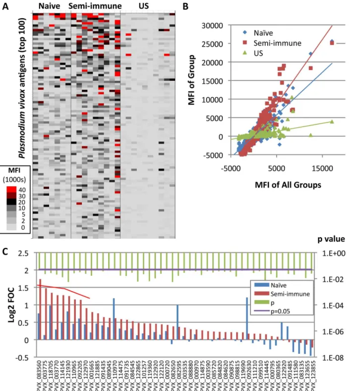
Fig 2Ashows a heat map of‘subtracted’array data (IVTT values minus sample-specific IVTT
controls signals) for each naïve and semi-immune individual, and for US controls. The analysis revealed higher reactive responses in semi-immune than naïve individuals before the challenge, and both groups’responses were higher than those in the US controls. This differential
reactiv-ity is seen more clearly from the slopes of the linear regression lines when average signal inten-sities from each group are plotted against the average of all three groups (Fig 2B). The steeper slope of the semi-immune individuals relative to the naïve individuals confirms an overall higher reactivity in this group. The breadth of the baseline antibody profile, defined as the sum of reactiveP.vivaxantigens per individual, ranged from three to 71 reactive antigens for naïve individuals and three to 89 for semi-immune individuals. While the average group antibody breadth was broader for the semi-immune group (179 antigens) in comparison to naïve volun-teers (113 antigens), both groups shared reactivity for 98 of the antigens. Only a single seropos-itive antigen (PVX_003775, MSP4) was significant when naïve and semi-immune groups were compared (p<0.05). To test whether this small number of differences was influenced by
serum dilution, arrays were probed at 1:200 and 1:400 dilutions. A dilution of 1:200 yielded eight differentially reactive antigens with a Log2 FOC>1 (Fig 2CandTable 1). The majority of
Fig 2. Antibody profiling in Colombian individuals beforeP.vivaxchallenge.Plasmodium vivaxprotein arrays were probed with serum samples collected before challenge (day 0) and at four time-points afterwards, as shown in the schematic inFig 1.A. Heat map showing serological profiles on day 0 prior to challenge for each Colombian naïve and semi-immune individual, and US controls for comparison. Raw signal intensities for each IVTT spot have been subtracted from the sample-specific median of background (IVTT control) spots, and the adjusted signal intensity represented by a color according to the key. Only the top 100 antigens are shown, ranked by average adjusted signals of both Colombian groups.B. Scatter plot of individual antigens, in which the average signal of each group (y-axis) is plotted against the average of all three groups (x-axis); the slope of the regression line is proportional to the overall breadth and intensity of the profile in each group. Each point represents the median fluorescence intensity (MFI) for all individuals examined in the particular group to a particular antigen.C. Bar chart of normalized array data (Log2 FOC) at 1:200 serum dilution. Only antigens with significant reactivity difference (p<0.05) between naïve and semi-immune volunteers are shown (raw p-values; green bars). Of all the significant antigens, nine were considered seropositive (i.e., using Log2 FOC>1 as the cutoff; red bracket); these are shown inTable 1.
doi:10.1371/journal.pntd.0004563.g002
Antibody reactivity induced after
P.
vivax
challenge
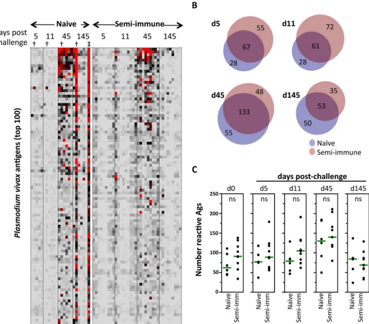
To normalize differences in background reactivity seen between both study groups and to reveal only the signals induced in response to theP.vivaxchallenge, pre-existing background reactivity at baseline (day 0) for each antigen was subtracted from the later time points data. In the semi-immune volunteers, the reactivity after challenge corresponded to a boosting of anti-bodies already present at baseline as well as appearance of new ones. At day five, reactivity of a few proteins was significantly higher in semi-immune than in naïve volunteers: serine-repeat antigen 5 (SERA5; PVX_003830) and three hypothetical proteins with unknown function (PVX_094690, PVX_084120, PVX_113590). However, at diagnosis day (day 11) the antibody response toP.vivaxremained similarly low in both groups (Fig 3A–3C).
Notably, reactivity rose abruptly on day 45 in both groups, followed by a decline to near baseline by day 145 (Fig 3A). The profile in one naïve volunteer (indicated by†inFig 3A) who
presented with a newP.vivaxinfection on day 130 (indicated by‡inFig 3A) did not decline
by the final time point. Indeed, the profile remained strong at a follow-up time point of 145 days. Since the serological dynamic of this individual was different to the others in the group, these data were removed from subsequent analyses. The expansion of the profile as measured by the group antibody breadth (Fig 3B), was marginally more rapid in the semi-immune group, although at response peak (day 45) the breadths were roughly equivalent in both groups (naive = 188; semi-immune = 181; total reactivity = 236). Both group profiles declined thereaf-ter with roughly equivalent breadths at day 145 (naïve = 103; semi-immune = 88; total reactiv-ity = 138). The response dynamics are shown by the dot plots of antibody breadth (Fig 3C). These data were not subtracted from baseline signals to more clearly show the challenge-induced increase in the breadth relative to the pre-challenge baseline.
To determine how long remain the antibodies elicited againstP.vivaxantigens without par-asite re-exposure, the individuals were followed-up for 145 days. On this day several antigens were identified as significant when naïve and semi-immune were compared, although only six were considered seropositive (Log2 FOC>1); two of them were higher in semi-immune
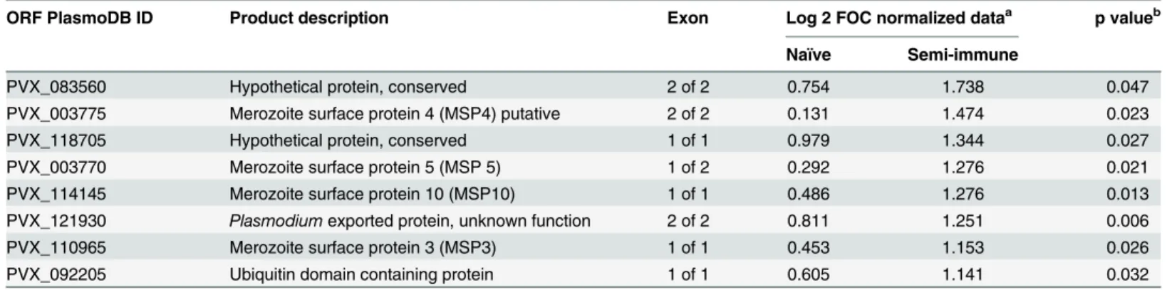
vol-unteers (SERA5 and a hypothetical protein, PVX_094690). In contrast, naïve volvol-unteers had higher response to MSP1, MSP8, ETRAMP and a hypothetical protein with unknown function (PVX_083560;S1 Table).
Table 1. The PlasmoDB gene ID and description of the top antigens that discriminate between naïve and semi-immune individuals at baseline.
ORF PlasmoDB ID Product description Exon Log 2 FOC normalized dataa p valueb
Naïve Semi-immune
PVX_083560 Hypothetical protein, conserved 2 of 2 0.754 1.738 0.047
PVX_003775 Merozoite surface protein 4 (MSP4) putative 2 of 2 0.131 1.474 0.023
PVX_118705 Hypothetical protein, conserved 1 of 1 0.979 1.344 0.027
PVX_003770 Merozoite surface protein 5 (MSP 5) 1 of 2 0.292 1.276 0.021
PVX_114145 Merozoite surface protein 10 (MSP10) 1 of 1 0.486 1.276 0.013
PVX_121930 Plasmodiumexported protein, unknown function 2 of 2 0.811 1.251 0.006
PVX_110965 Merozoite surface protein 3 (MSP3) 1 of 1 0.453 1.153 0.026
PVX_092205 Ubiquitin domain containing protein 1 of 1 0.605 1.141 0.032
aFOC, fold-over control. Values>1 (i.e., two-fold over the IVTT controls spots) were considered seropositive. bp value using Wilcoxon Rank-Sum Test.
Antibody profile associated with
P.
vivax
malaria clinical protection
As described [22], naïve individuals all developed classical malaria symptoms such as head-ache, fever, nausea, chills, and malaise associated withP.vivaxchallenge at the time of parasite patency. In contrast, semi-immune volunteers reported either no symptoms or only minor symptoms associated with theP.vivaxappearance in blood; only 33% presented fever (body temperature38°C). Therefore, semi-immune volunteers were segregated into those that developed symptoms (or“non-protected”) or did not develop symptoms (or“protected”). The
Fig 3. Antibody reactivity after challenge. A. Heat map of array data for all four post-challenge time points. Data were normalized by subtraction of IVTT controls, as described inFig 2, and then subtracted from day 0 values to reveal more clearly the change in the profile due to challenge. The profile in one atypical naïve individual who presented with a newP.vivaxinfections on day 130 (‡), indicated by the dagger (†) in each time point is also shown.B. Venn diagrams of specific and shared antigens at each of the post-challenge time points. An antigen was defined as reactive if the average per group>avg + 2SD of the US controls; data for the atypical naïve individual were removed from this analysis.C. Dot plots showing numbers of reactive antigens for naïve and
semi-immune volunteers at each post-challenge time point; data for the atypical naïve individual were removed for this analysis. Green lines = medians;
ns = not significant (Wilcoxon Rank-Sum test).
doi:10.1371/journal.pntd.0004563.g003
group means of the top 40 individual antigens subtracted from baseline signals showed that those semi-immune individuals that developed fever after challenge had a robust (naïve-like) response that peaked on day 45, while the asymptomatic individuals showed an attenuated response at this time (Fig 4A). Both returned to near baseline by day 145. Segregation using headache as a symptom was also analyzed with similar results (S1 Fig).
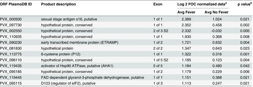
Comparison of the protected and unprotected semi-immune profiles on day 45 identified several antigens as significant when data were segregated by fever (Fig 4BandTable 2), although only 12 were considered seropositive (Log2 FOC>1; indicated by the bracket inFig
4B). Interestingly, all of them were higher in immune volunteers with fever. In semi-immune individuals segregated by headache, several antigens were significant, although only one (PVX_002550; conserved hypothetical) was considered seropositive (Fig 4C).
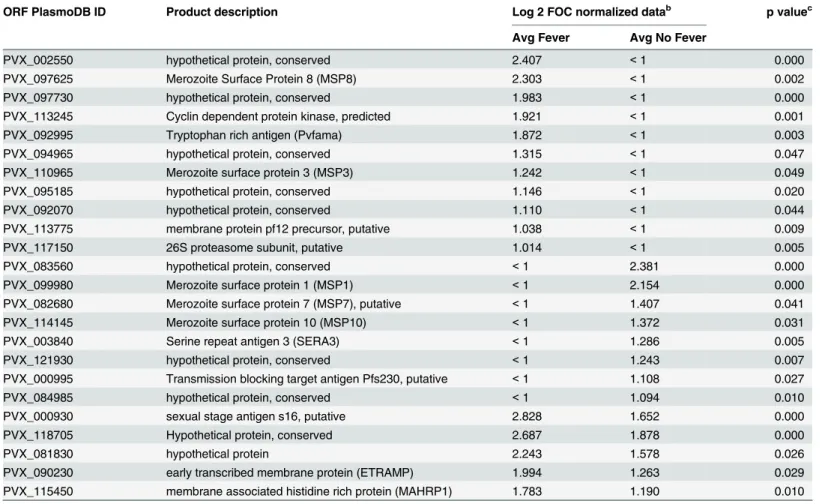
To test the hypothesis that several antigens recognized in semi-immune individuals at the peak of the response after challenge were“new”antibodies absent from the baseline profile, as
opposed to boosted from antibodies present at baseline, statistical comparison between profiles at day 0 (baseline)vs. day 45 (peak) was performed. Those that were protected or non-pro-tected (using fever as the symptom) were analyzed separately. Volunteers without fever devel-oped antibodies to 13 new antigens, including three members of the MSP family (one, seven and 10) and three hypothetical proteins, whereas individuals with fever had reactivity to 16 new antigens. However, antibodies to only five new antigens were shared by both groups, all of them with higher reactivity in volunteers with fever (Table 3). These data suggests that only oneP.vivaxinfection is enough to induce antibody response against new antigens.
Discussion
This study revealed that individuals who were semi-immune toP.vivaxhad pre-existing anti-bodies that although present at low levels were associated with clinical protection toP.vivax sporozoite experimental challenge [22]. As expected, semi-immune volunteers showed higher reactivity than naïve individuals to severalP.vivaxantigens before challenge. Moreover, expo-sure to a presumably low dose of viable sporozoites inoculated by the bites of only 2–4
mosqui-toes was enough to induce a robust antibody response in malaria-naïve volunteers as well as to trigger antibody responses to new antigens in semi-immune volunteers (Table 3). Another valuable observation was that a proportion of the anti-P.vivaxantibodies were short-lived as 138 of the 236 antigens (>40%) recognized by day 45 had disappeared by day 145 after
chal-lenge. The rapid decay of a subset of antibodies indirectly indicated that semi-immune volun-teers had not had recent exposure to the parasites, because several of these antigens were not recognized at pre-challenge time.
Before challenge, the Colombian malaria-naïve individuals had significantly higher serologi-cal reactivity than the US controls, despite being residents of a non-endemic malaria area. They were confirmed as seronegative againstP.vivaxblood stages and sporozoites using IFAT. Although infections or experience with protozoa were not studied here, the reactivity observed in Colombian naïve individuals might be due to other pathogens such asCryptosporidium par-vumor others highly prevalent in Colombia [28];C.parvumshows homology with several Plasmodiumproteins [29]. Nevertheless, this serological reactivity did not appear to have played a role in protection as all naïve volunteers developed malaria-related symptoms and pat-ent parasitemia at the expected time [20–22]. The higher reactivity of the semi-immune
volun-teers to several antigens before challenge as compared to naïve volunvolun-teers indicates that in endemic regions, even with low transmission intensity, they develop and maintainP.vivax spe-cific antibodies to a broad number of antigens even after a few previous malaria episodes (2–5
Fig 4. Antibody profile associated with clinical protection. A. Kinetics of antibody response againstP.vivaxantigens. Semi-immune volunteers were segregated into those that developed fever and those who did not. Average of median fluorescence intensity (MFI) is shown.B-C. Bar graph of normalized array data (Log2 FOC) for top individual antigens in semi-immune volunteers at day 45 segregated by fever (blue bars) or no fever (red bars) (B)and headache (blue bars)vs. no headache (red bars)(C). P values in Log scale (green bars) using the Wilcoxon Rank-Sum Test are shown with the purple line representing the significance threshold (p = 0.05). Red bracket indicates seropositive antigens. Only antigens with significant reactivity (p<0.05) are shown.
doi:10.1371/journal.pntd.0004563.g004
enough to modify the pre-patent period or parasitemia at diagnosis day, although it was highly effective in controlling malaria symptoms.
Interestingly, in the subgroup of semi-immune volunteers that developed fever or headache, as in the naïve, the antibody response to challenge was more vigorous than that in asymptom-atic volunteers who displayed an attenuated antibody response. This is consistent with findings fromP.falciparumvaccination studies in humans where protected individuals did not mount a significant antibody response to challenge, whereas unprotected subjects responded to chal-lenge by elevated signals to many blood stage antigens [11,30]. Although in those studies PfCSP was recognized by both the protected and unprotected subgroups, protected individuals had a significantly higher magnitude of response [11,30].
At day 45 volunteers with fever showed a significantly higher response toP.vivaxantigens such as MSP3, MSP4, MSP5 and MSP10. However, reactivity toPvMSP1 andPvCSP, two established vaccine candidates [14,31], was not different between volunteers with and without fever, as previously seen for the same sera using a recombinantPvMSP1 fragment (r200L) and syntheticPvCSP construct by ELISA [22]. These results partially contrast with those of epide-miological studies onP.vivaxwhere an association between sera reactivity to MSP1, MSP3 and MSP9 proteins and clinical protection has been reported [10,32–34]. The higher reactivity to
the CSP inP.falciparumstudies [30,35] is most likely due to the multiple immunization doses, while here only a few mosquito bites were allowed, with possibly low sporozoite density suffi-cient to induce infection once and a detectable antibody levels against a high number of other P.vivaxantigens in all volunteers.
In summary, the antibody profiles that developed in humans after experimental exposure to P.vivaxsporozoites were defined. It was shown that a single infection was enough to induce detectable specific antibodies in malaria naïve volunteers and to boost the antibodies elicited by natural exposure to malaria in immune individuals. Comparison between semi-immune volunteers segregated by fever showed that those protected had an attenuated serolog-ical response after challenge, but also had reactivity to new antigens, which may represent promising targets for vaccine development. Taken together, these findings represent a Table 2. Top reactive antigens at day 45 after challenge that discriminate between semi-immune individuals with fever or without fever.
ORF PlasmoDB ID Product description Exon Log 2 FOC normalized dataa p valueb
Avg Fever Avg No Fever
PVX_000930 sexual stage antigen s16, putative 1 of 1 2.389 1.024 0.021
PVX_097730 hypothetical protein, conserved 1 of 1 2.352 0.458 0.002
PVX_002550 hypothetical protein, conserved 2 of 3 S2 2.332 -0.032 0.000
PVX_110935 hypothetical protein, conserved 1 of 1 1.930 0.368 0.008
PVX_090230 early transcribed membrane protein (ETRAMP) 1 of 2 1.721 0.632 0.004
PVX_081830 hypothetical protein 2 of 2 1.347 0.643 0.023
PVX_113775 6-cysteine protein (P12) 1 of 1 1.322 0.316 0.001
PVX_096110 hypothetical protein, conserved 1 of 5 S2 1.185 0.123 0.004
PVX_119435 activator of Hsp90 ATPase, putative (AHA1) 5 of 5 1.184 0.480 0.042
PVX_095185 hypothetical protein, conserved 1 of 2 1.179 0.229 0.006
PVX_119445 FAD dependent glycerol-3-phosphate dehydrogenase, putative 1 of 1 1.151 0.368 0.021
PVX_095115 D123 (regulator of eIF2), putative 1 of 3 1.113 0.247 0.021
aFOC, fold-over control. Values>1 (i.e., two-fold over the IVTT controls spots) were considered seropositive. bp value using Wilcoxon Rank-Sum Test.
significant step forward in the understanding of the humoral immune response toP.vivax malaria infection, particularly the extent of priming upon a first parasite encounter.
Supporting Information
S1 Table. Significant reactive antigens at day 145 after challenge.
(DOC)
S1 Fig. Kinetics of antibody response toP.vivaxantigens in semi-immune volunteers seg-regated by headache.The volunteers were segregated into those that reported headache and those who did not. Average of median fluorescence intensity (MFI) of top 40 individual anti-gens is shown.
(TIF)
S1 Protocol. Comparison of the susceptibility of naïve and pre-immune volunteers to the Infectious challenge with viablePlasmodium vivaxsporozoites.
(DOCX)
Table 3. New antigens at day 45 after challenge in semi-immune individuals with fever or without fevera.
ORF PlasmoDB ID Product description Log 2 FOC normalized datab p valuec
Avg Fever Avg No Fever
PVX_002550 hypothetical protein, conserved 2.407 <1 0.000
PVX_097625 Merozoite Surface Protein 8 (MSP8) 2.303 <1 0.002
PVX_097730 hypothetical protein, conserved 1.983 <1 0.000
PVX_113245 Cyclin dependent protein kinase, predicted 1.921 <1 0.001
PVX_092995 Tryptophan rich antigen (Pvfama) 1.872 <1 0.003
PVX_094965 hypothetical protein, conserved 1.315 <1 0.047
PVX_110965 Merozoite surface protein 3 (MSP3) 1.242 <1 0.049
PVX_095185 hypothetical protein, conserved 1.146 <1 0.020
PVX_092070 hypothetical protein, conserved 1.110 <1 0.044
PVX_113775 membrane protein pf12 precursor, putative 1.038 <1 0.009
PVX_117150 26S proteasome subunit, putative 1.014 <1 0.005
PVX_083560 hypothetical protein, conserved <1 2.381 0.000
PVX_099980 Merozoite surface protein 1 (MSP1) <1 2.154 0.000
PVX_082680 Merozoite surface protein 7 (MSP7), putative <1 1.407 0.041
PVX_114145 Merozoite surface protein 10 (MSP10) <1 1.372 0.031
PVX_003840 Serine repeat antigen 3 (SERA3) <1 1.286 0.005
PVX_121930 hypothetical protein, conserved <1 1.243 0.007
PVX_000995 Transmission blocking target antigen Pfs230, putative <1 1.108 0.027
PVX_084985 hypothetical protein, conserved <1 1.094 0.010
PVX_000930 sexual stage antigen s16, putative 2.828 1.652 0.000
PVX_118705 Hypothetical protein, conserved 2.687 1.878 0.000
PVX_081830 hypothetical protein 2.243 1.578 0.026
PVX_090230 early transcribed membrane protein (ETRAMP) 1.994 1.263 0.029
PVX_115450 membrane associated histidine rich protein (MAHRP1) 1.783 1.190 0.010
aNew antigens at day 45 that were absent from the baseline pro
file (day0).
bFOC, fold-over control. Values>1 (i.e., two-fold over the IVTT controls spots) were considered seropositive. cp value using Wilcoxon Rank-Sum Test between day 0 and day 45.
doi:10.1371/journal.pntd.0004563.t003
Acknowledgments
The authors express their sincere gratitude to the volunteers who participated in this study. Special acknowledgment also goes to the MVDC team that provided the logistical support for this study, particularly to all staff involved in the clinical trial, the entomological and data man-agement units and the staff at“Instituto de Salud del Pacífico”(INSALPA) in Buenaventura
(Colombia) for their superb technical support. Ethical Comities (CECIV and CEICMI) greatly contribute to the clinical protocol improvement.
Author Contributions
Conceived and designed the experiments: MAH SH. Performed the experiments: MLP ED AJ KR DHD. Analyzed the data: MLP ED DHD. Contributed reagents/materials/analysis tools: PLF DHD. Wrote the paper: MAH MLP DHD SH. Read and approved the final manuscript: MAH MLP ED AJ KR PLF DHD SH.
References
1. WHO. World Malaria Report 2015. Geneva: WHO, 2015.
2. Doolan DL, Dobano C, Baird JK. Acquired immunity to malaria. Clin Microbiol Rev. 2009; 22(1):13–36. doi:10.1128/CMR.00025-08PMID:19136431
3. Crompton PD, Kayala MA, Traore B, Kayentao K, Ongoiba A, Weiss GE, et al. A prospective analysis of the Ab response to Plasmodium falciparum before and after a malaria season by protein microar-ray. Proc Natl Acad Sci U S A. 2010; 107(15):6958–63. doi:10.1073/pnas.1001323107PMID: 20351286
4. Osier FH, Fegan G, Polley SD, Murungi L, Verra F, Tetteh KK, et al. Breadth and magnitude of antibody responses to multiple Plasmodium falciparum merozoite antigens are associated with protection from clinical malaria. Infect Immun. 2008; 76(5):2240–8. doi:10.1128/IAI.01585-07PMID:18316390 5. Bousema T, Okell L, Felger I, Drakeley C. Asymptomatic malaria infections: detectability,
transmissibil-ity and public health relevance. Nat Rev Microbiol. 2014; 12(12):833–40. doi:10.1038/nrmicro3364 PMID:25329408
6. Arevalo-Herrera M, Lopez-Perez M, Medina L, Moreno A, Gutierrez JB, Herrera S. Clinical profile of
Plasmodium falciparumandPlasmodium vivaxinfections in low and unstable malaria transmission set-tings of Colombia. Malar J. 2015; 14(1):154.
7. Vallejo AF, Chaparro PE, Benavides Y, Alvarez A, Quintero JP, Padilla J, et al. High prevalence of sub-microscopic infections in Colombia. Malar J. 2015; 14:201. doi:10.1186/s12936-015-0711-6PMID: 25971594
8. Laishram DD, Sutton PL, Nanda N, Sharma VL, Sobti RC, Carlton JM, et al. The complexities of malaria disease manifestations with a focus on asymptomatic malaria. Malar J. 2012; 11:29. doi:10.1186/ 1475-2875-11-29PMID:22289302
9. Grobusch MP, Kremsner PG. Uncomplicated Malaria. In: Sullivan DJ, Krishna S, editors. Malaria: Drugs, Disease and Post-genomic Biology. Current Topics in Microbiology and Immunology 295. Ber-lin: Springer Berlin Heidelberg; 2005. p. 81–104.
10. Versiani FG, Almeida ME, Melo GC, Versiani FO, Orlandi PP, Mariuba LA, et al. High levels of IgG3 anti ICB2-5 in Plasmodium vivax-infected individuals who did not develop symptoms. Malar J. 2013; 12:294. doi:10.1186/1475-2875-12-294PMID:23977965
11. Doolan DL, Mu Y, Unal B, Sundaresh S, Hirst S, Valdez C, et al. Profiling humoral immune responses to P. falciparum infection with protein microarrays. Proteomics. 2008; 8(22):4680–94. PMID: 18937256
12. Davies DH, Liang X, Hernandez JE, Randall A, Hirst S, Mu Y, et al. Profiling the humoral immune response to infection by using proteome microarrays: high-throughput vaccine and diagnostic antigen discovery. Proc Natl Acad Sci U S A. 2005; 102(3):547–52. PMID:15647345
13. Ray S, Kamath KS, Srivastava R, Raghu D, Gollapalli K, Jain R, et al. Serum proteome analysis of vivax malaria: An insight into the disease pathogenesis and host immune response. J Proteomics. 2012; 75(10):3063–80. doi:10.1016/j.jprot.2011.10.018PMID:22086083
montanide ISA 720 or montanide ISA 51. Am J Trop Med Hyg. 2011; 84(2 Suppl):12–20. doi:10.4269/ ajtmh.2011.09-0516PMID:21292873
15. Malkin EM, Durbin AP, Diemert DJ, Sattabongkot J, Wu Y, Miura K, et al. Phase 1 vaccine trial of Pvs25H: a transmission blocking vaccine forPlasmodium vivaxmalaria. Vaccine. 2005; 23(24):3131–
8. PMID:15837212
16. Wu Y, Ellis RD, Shaffer D, Fontes E, Malkin EM, Mahanty S, et al. Phase 1 trial of malaria transmission blocking vaccine candidates Pfs25 and Pvs25 formulated with montanide ISA 51. PLoS One. 2008; 3 (7):e2636. doi:10.1371/journal.pone.0002636PMID:18612426
17. Arevalo-Herrera M, Chitnis C, Herrera S. Current status of Plasmodium vivax vaccine. Hum Vaccin. 2010; 6(1):124–32. PMID:20009526
18. Moreno A, Joyner C. Malaria vaccine clinical trials: what's on the horizon. Curr Opin Immunol. 2015; 35:98–106. doi:10.1016/j.coi.2015.06.008PMID:26172291
19. Mueller I, Shakri AR, Chitnis CE. Development of vaccines for Plasmodium vivax malaria. Vaccine. 2015; 33(52):7489–95. doi:10.1016/j.vaccine.2015.09.060PMID:26428453
20. Herrera S, Fernandez O, Manzano MR, Murrain B, Vergara J, Blanco P, et al. Successful sporozoite challenge model in human volunteers withPlasmodium vivaxstrain derived from human donors. Am J Trop Med Hyg. 2009; 81(5):740–6. doi:10.4269/ajtmh.2009.09-0194PMID:19861603
21. Herrera S, Solarte Y, Jordan-Villegas A, Echavarria JF, Rocha L, Palacios R, et al. Consistent safety and infectivity in sporozoite challenge model ofPlasmodium vivaxin malaria-naive human volunteers. Am J Trop Med Hyg. 2011; 84(2 Suppl):4–11. doi:10.4269/ajtmh.2011.09-0498PMID:21292872 22. Arevalo-Herrera M, Forero-Pena DA, Rubiano K, Gomez-Hincapie J, Martinez NL, Lopez-Perez M,
et al. Plasmodium vivax sporozoite challenge in malaria-naive and semi-immune Colombian volun-teers. PLoS One. 2014; 9(6):e99754. doi:10.1371/journal.pone.0099754PMID:24963662
23. International Conference on Harmonisation. ICH Harmonised Tripartite Guideline—Guideline for Good Clinical Practice E6 (R1). 1996.
24. MinSalud. Guía para atención clínica integral del paciente con malaria. Bogotá: Ministerio de la Pro-tección Social. 2010.
25. King CL, Davies DH, Felgner P, Baum E, Jain A, Randall A, et al. Biosignatures of Exposure/Transmis-sion and Immunity. Am J Trop Med Hyg. 2015; 93(3 Suppl):16–27. doi:10.4269/ajtmh.15-0037PMID: 26259938
26. Campo JJ, Aponte JJ, Skinner J, Nakajima R, Molina DM, Liang L, et al. RTS,S vaccination is associ-ated with serologic evidence of decreased exposure to Plasmodium falciparum liver- and blood-stage parasites. Mol Cell Proteomics. 2015; 14(3):519–31. doi:10.1074/mcp.M114.044677PMID: 25547414
27. Hulsen T, de Vlieg J, Alkema W. BioVenn—a web application for the comparison and visualization of biological lists using area-proportional Venn diagrams. BMC Genomics. 2008; 9:488. doi:10.1186/ 1471-2164-9-488PMID:18925949
28. Vergara Castiblanco C, Santos Nunez S, Freire Santos F, Ares Mazas E. [Cryptosporidiosis in the Andean region of Colombia: seroprevalence and recognition of antigens]. Rev Panam Salud Publica. 2000; 8(6):373–9. PMID:11209249
29. Siddiki AZ. Sporozoite proteome analysis of Cryptosporidium parvum by one-dimensional SDS-PAGE and liquid chromatography tandem mass spectrometry. J Vet Sci. 2013; 14(2):107–14. doi:10.4142/ jvs.2013.14.2.107PMID:23814469
30. Trieu A, Kayala MA, Burk C, Molina DM, Freilich DA, Richie TL, et al. Sterile protective immunity to malaria is associated with a panel of novel P. falciparum antigens. Mol Cell Proteomics. 2011; 10(9): M111 007948. doi:10.1074/mcp.M111.007948PMID:21628511
31. Valderrama-Aguirre A, Quintero G, Gomez A, Castellanos A, Perez Y, Mendez F, et al. Antigenicity, immunogenicity, and protective efficacy ofPlasmodium vivaxMSP1 PV200l: a potential malaria vac-cine subunit. Am J Trop Med Hyg. 2005; 73(5 Suppl):16–24. PMID:16291762
32. Cutts JC, Powell R, Agius PA, Beeson JG, Simpson JA, Fowkes FJ. Immunological markers of Plasmo-dium vivax exposure and immunity: a systematic review and meta-analysis. BMC Med. 2014; 12:150. doi:10.1186/s12916-014-0150-1PMID:25199532
33. Stanisic DI, Javati S, Kiniboro B, Lin E, Jiang J, Singh B, et al. Naturally acquired immune responses to P. vivax merozoite surface protein 3alpha and merozoite surface protein 9 are associated with reduced risk of P. vivax malaria in young Papua New Guinean children. PLoS Negl Trop Dis. 2013; 7(11):e2498. doi:10.1371/journal.pntd.0002498PMID:24244763
34. Nogueira PA, Alves FP, Fernandez-Becerra C, Pein O, Santos NR, Pereira da Silva LH, et al. A reduced risk of infection with Plasmodium vivax and clinical protection against malaria are associated
with antibodies against the N terminus but not the C terminus of merozoite surface protein 1. Infect Immun. 2006; 74(5):2726–33. PMID:16622209