INTRODUCTION
W
ith the growing concern and the globalde-bate on the harmful effects of tobacco on the human body, much attention has been direct-ed to the adverse consequences of smoking. The direct and indirect costs related to diseases linked to smoking consume considerable resources for health in our country. Neoplasms, cardiovascular and respiratory diseases associated with smoking are well documented. However, less attention has been paid to surgical complications related to smoking1-4.
Although tobacco products are consumed for hundreds of years, only in the twentieth centu-ry there was a sharp increase in consumption, the cigarette being the most important form of use. Its toxic smoke has more than 5,000 elements, of which nicotine is the primary vasoactive com-ponent, considered to cause the smoker’s
addic-tion, as it strengthens and enhances the desire to smoke3,5,6.
The so-called granulation tissue, essential for a healthy scar development, comes at the begin-ning of the second stage of healing, called prolifera-tive, being mainly composed of newly formed blood vessels (angiogenesis) and modified fibroblasts, called myofibroblasts4. While angiogenesis provides the blood supply suitable for the high level of meta-bolic activity that the healing process requires, myo-fibroblasts, through their contractile force, approach the damaged tissue edges7.
In 1977, Mosely and Finseth8 drew atten-tion in a pioneering way to the undesirable effects of nicotine on tissue healing. Since then, several clinical and experimental studies have tried to explain these effects, demonstrating that this substance causes deficiencies in several factors involved in the healing process, in areas as diverse as Plastic Surgery and Or-thopedics9.
The influence of nicotine in healing of small bowel anastomoses
in rats: angiogenesis and miofibroblasts
A influência da nicotina na cicatrização de anastomoses do intestino delgado em
ratos: angiogênese e miofibroblastos
JaMes skinovsky, tCBC-pr1; osvaldo MalaFaia, eCBC-pr2; MauriCio ChiBata1; Fernanda tsuManuMa3; Flávio panegalli Filho3;
MarCus viníCius dantasde CaMpos Martins,tCBC-rJ4.
A B S T R A C T
Objective: to know the effect of nicotine on angiogenesis and myofibroblast formation in anastomoses of the small bowel of rats. Methods: we randomly divided 60 Wistar rats into the groups Nicotine (N) and control (C), according to the proposed treatment. Each group was subdivided into three subgroups according to the time interval used for the evaluation (7, 14 or 28 days). The N group with 30 animals received nicotine subcutaneously at a dose of 2mg/kg body weight, diluted in 0.3ml of 0.9% saline, twice daily for 28 days prior to the operation, and for more 7, 14 or 28 days, depending on the subgroup. The C group (also 30 animals) received only saline on the same conditions and time intervals. After 28 days we carried out an end-to-end anastomosis 10cm distal to the duodenojejunal flexure in each rat. After 7, 14 or 28 days after surgery, we euthanized ten animals of each group, sent specimens of the anastomosis areas, 1cm proximal to 1cm distal, to counting of blood vessels and myofibroblasts through immunohistochemical staining by the application of monoclonal anti-factor VIII antibodies and anti-smooth muscle alpha-actin. Results: the administration of nicotine led to the decrease in the number of blood vessels measured on the 28th postoperative day and the number of myofibroblasts measured on the seventh day following completion of the anastomoses. Conclusion: administration of nicotine was deleterious on angiogenesis and myofibroblast formation in rats’ small intestine anastomoses.
Keywords: Wound Healing. Nicotine. Intestine, Small. Anastomosis, Surgical. Rats.
Very little is known about the influence of nicotine on healing of the digestive tract, as well as the possible mechanisms involved10-13.
The aim of this study is to analyze the ef-fects of nicotine on the healing process of anasto-moses of the small intestine of rats as for the num-ber of blood vessels and myofibroblasts present in the scar tissue.
METHODS
We performed the experimental procedures in the Surgical Research Center of the Post-Gradu-ation Program in Surgery of the Universidade Fed-eral of Paraná, and the immunohistochemical study at the Pathology Department, Hospital de Clínicas, Universidade Federal do Paraná, and at the Instituto de Pesquisas Médicas (Ipem) of the Hospital Univer-sitário Evangélico de Curitiba, being duly previously reviewed and approved by ethics in research review committees of the aforementioned institutions.
This study used 60 male Wistar rats aged 160-200 days (average 180) and weighting between 270 and 290g, coming from the Instituto de Tec-nologia do Paraná (TECPAR), being held in 12-hour day / night cycles and constant room temperature of 24oC. The animals fed on chow proper for the species and had free access to water throughout the experiment.
We randomly divided the rats into two groups: Group N, with 30 animals submitted to the application of nicotine; and Group C, also with 30
rats, which served as a control. Each group was divid-ed into three subgroups of ten rats each. They were thus named N7, N14, N28, C7, C14 and C28, ac-cording to the postoperative evaluation time.
In group N nicotine was administered (Nic-otine dihydrogen tartrate salt – Sigma, Saint Louis, Missouri, USA) subcutaneously (Figure 1) at the dose of 2 mg per kg bodyweight twice a day (12/12 hours) diluted in 0.3 ml 0.9% saline and adjusted to pH 7.4, for an initial period of 28 days prior to the surgical procedure. In group C we proceeded in an identical manner, though substituting the nicotine for 0.9% saline solution. The rats were weighed weekly, and the dose of nicotine adjusted when necessary. This dosage was established after calculation so that the amount of nicotine were equivalent to two cigarette cards per day in an adult human.
On the 28th day of application we with-drew feeding for 12 hours prior to the operation, maintaining free access to water. The surgical proce-dure took place the next day.
The rats were subjected to inhalation anes-thesia with halothane in a fume cupboard. We asep-tically held a 4 cm long laparotomy and performed an intestinal anastomosis 10 cm distal to the duode-nojejunal flexure, performing a cross section of the isolated bowel loop and an end-to-end anastomosis, with 6-0 polypropylene suture in a single plane with separated full-thickness stitches, in a total of eight stitches (Figure 2). The wall of the abdomen was closed in two planes, with a continuous running su-ture with 3-0 multifilament polyglactin. The animals received free access to water immediately, and to food, 12 hours after the procedure.
The administration of nicotine or saline solution remained even on the day of operation and for more 7, 14 or 28 days according the animal’s sub-group, and on the same already reported conditions, when we held a new inhalational anesthesia and har-vested the anastomosed segments considering 1 cm proximal to 1 cm distal to the anastomotic line, pre-serving the specimen in buffered formalin.
For immunohistochemical staining we ap-plied anti-factor VIII monoclonal antibodies (poly-clonal, Code 0082 - DakoCytomation -
teria, USA) and anti-smooth muscle alpha-actin (Monoclonal Antibody, M0851 Code - DakoCy-tomation - Carpinteria, USA). We identified the myofibroblasts positivity in the areas of brownish pigmentation and we used positive and negative controls.
We identified the number of blood vessels by counting the circular structures positively stained by the anti-factor VIII antibody, which reveals the en-dothelial cells of the vessels’ tunica intima. Count-ing was done in the area of the anastomosis, in a extension 10 mm proximal and 10 mm distal to it (total 20mm included with the anastomosis) with 10x magnification.
As for the quantification of myofibroblasts, we counted the perianastomotic cells positively stained for anti-smooth muscle alpha-actin antibody in a high-power field (40X objective). The scanned images were captured and analyzed by Image Pro Plus (Media Cybernetics, California, USA), through the “Measures” tool.
For each of the quantitative variables we adopted the Mann-Whitney nonparametric test, at a 5% significance level.
RESULTS
The evaluation of the number of blood vessels in the area of the anastomosis in the various phases of the postoperative period can be seen in Table 1. The administration of nicotine lead to a de-crease in the number of blood vessels measured on the 28th postoperative day and in the number of my-ofibroblasts measured on the seventh day after the anastomoses.
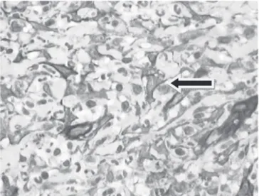
The microscopic appearance of blood ves-sels with positivity for anti-factor VIII in shown in Figure 3.
The quantification of myofibroblasts in the various postoperative stages is shown in Table 2. An image of a myofibroblast stained by anti-alpha-actin antibody is seen in Figure 4.
DISCUSSION
In this experiment we observed that the use of nicotine led to a statistically significant decrease in the number of blood vessels on the 28th postop-erative day and in the number of myofibroblasts on
the seventh day following completion of the anasto-moses. Similarly, many previous studies determined that this drug acts negatively on the healing process in various tissues, leading to tissue vasoconstriction, decreased proliferation of lymphocytes, fibroblasts and collagen, among others14-21. The lymphocytes are an important source of angiogenesis-inducing cy-tokine production. Therefore, reduction of lympho-cytes by nicotine18 can lead to decreased formation of new blood vessels in the scar tissue, as observed in this study. It is important to remember that the oxygen carried through the new vessels is a key fac-tor in the synthesis of collagen, mainly responsible for the resistance in the scar tissue. Thus, decreased angiogenesis, with consequent deficit of oxygen in the scar tissue, may be one explanation for the de-crease in collagen production and rupture strength in the region of intestinal anastomoses identified by previous studies3,7.
Therefore, nicotine may exert inhibiting ef-fect on angiogenesis through several mechanisms: a- inhibition of angiogenic factors, either by direct negative action in their release mechanism or by del-eterious effects on the cells that produce them, such as lymphocytes and fibroblasts5,13,18,19; b- direct deleterious effect on the endothelial cells5,11,19.
Nicotine also reduces the formation of fi-broblasts in the healing process of intestinal anasto-moses, which explains the deficit of collagen, main-ly responsible for local rupture strength5.13, since these cells are the main source of production of that protein. As fibroblasts are the precursor cells of my-ofibroblasts, whose main function is to promote the approximation of the healing wound edges, the very formation of the latter would also be compromised.
Nicotine may therefore have a negative effect on the proliferation of myofibroblasts through different mechanisms: a- inhibition of the fibroblast precursors of
Figure 3. Blood vessels with intima stained in brown, showing positivi-ty for anti-factor VIII (original magnification 100X).
Figure 4. Myofibroblasts stained in brown (anti-smooth muscle al-pha-actin antibody), showing polygonal shape with cy-toplasmic projections and vesicular nuclei (400X original magnification).
Number of
vessels Group Mean ± SD p value
7 days Control 170.89 ± 57.74 1.0000 Nicotine 159.33 ± 72.46
14 days Control 177.56 ± 126.66 0.2973 Nicotine 107.89 ± 59.24
28 days Control 118.67 ± 71.48 0.0027 Nicotine 62.11 ± 57.26
Table 1. Number of blood vessels in the 7th, 14th and 28th postoper-ative days
Table 2. Quantification of myofibroblasts number on the 7th, 14th and 28th postoperative days
Number of
myofibroblasts Group Mean ± SD p value
7 days Control 19.67 ± 6.16 0.0206 Nicotine 13.63 ± 2.62
14 days Control 16.75 ± 6.16 0.6730 Nicotine 17.89 ± 5.99
myofibroblasts, either by direct action or by decreasing the oxygen level at the injury site, hampering multiplica-tion and the funcmultiplica-tioning of these cells13; b- inhibimultiplica-tion of cells producing fibroblast-proliferation stimulating cytokines, such as macrophages and lymphocytes15.
Many can be the mechanisms by which nic-otine can exert harmful effects on the normal healing
process. These should be clarified by further studies, as there are not enough previous works in the litera-ture that have done this analysis for comparison.
In conclusion, according the data presented in this study, administration of nicotine was deleteri-ous to angiogenesis and to myofibroblast formation in anastomoses of the small intestine of rats.
R E S U M O
Objetivo: conhecer o efeito da nicotina sobre a angiogênese e formação de miofibroblastos em anastomoses do intestino delgado de ratos. Métodos: sessenta ratos Wistar foram divididos de maneira aleatória em grupos Nicotina(N) e Controle (C), conforme o tratamento proposto. Cada grupo foi subdividido em três subgrupos, de acordo com o intervalo de tempo utilizado para a avaliação (7, 14 ou 28 dias). O grupo N, com 30 animais, recebeu nicotina por via subcutânea, na dose de 2mg/Kg de peso, diluída em 0,3ml de solução salina a 0,9%, em duas aplicações diárias, durante 28 dias prévios à operação e por mais 7, 14 ou 28 dias, conforme o subgrupo. O grupo C (igualmente com 30 animais) recebeu somente a solução salina nas mesmas condições e intervalos de tempo. Após 28 dias efetuou-se, em cada rato, anastomose término-terminal a 10cm da flexura duodenojejunal. Após 7, 14 ou 28 dias da cirurgia, os dez animais de cada subgrupo foram eutanasiados, sendo que as áreas anastomosadas, 1cm proximal a 1cm distal, foram encaminhadas para contagem de vasos sanguíneos e miofibroblastos, através de coloração imuno-histoquímica por aplicação dos anticorpos monoclonais antifator VIII e anti-alfa-actina muscu-lar lisa. Resultados: a administração de nicotina levou à diminuição do número de vasos sanguíneos aferidos no 28o dia pós-operatório e do
número de miofibroblastos aferidos no sétimo dia após a realização das anastomoses. Conclusão: a administração de nicotina foi deletéria sobre a angiogênese e formação de miofibroblastos em anastomoses do intestino delgado de ratos.
Descritores: Cicatrização. Nicotina. Intestino Delgado. Anastomose Cirúrgica. Ratos.
REFERENCES
1. Bozarth MA, Pudiak CM, Kuolee R. Effect of chronic nicotine on brain stimulation reward. I. Effect of daily injections. Behav Brain Res. 1998;96(1-2):185-8. 2. Coelho ICMM. Estudo comparativo das forças e tensão
entre as cicatrizes das laparotomias paramedianas e das laparotomias transversas em ratos jovens
(Rattus Norvegicus Albonus) [dissertação]. Curitiba:
Universidade Federal do Paraná, Programa de Pós-Graduação em Clínica Cirúrgica; 1999.
3. Frick WG, Seals RR Jr. Smoking and wound healing: a review. Tex Dent J. 1994;111(6):21-3.
4. Silva VLC. Tabagismo: um problema de saúde pública no Brasil. JBM. 1990;59(1):14-24.
5. Giannopoulou C, Geinoz A, Cimasoni G. Effects of nicotine on periodontal ligament fibroblasts in vitro. J Clin Periodontol. 1999;26(1):49-55.
6. Fletcher HG. The history of nicotine. J Chem Educ. 1941;18(7):303-8.
7. Witte MB, Barbul A. General principles of wound healing. Surg Clin North Am. 1997;77(3):509-28. 8. Mosely LH, Finseth F. Cigarette smoking: impairment
of digital blood flow and wound healing in the hand. Hand. 1977;9(2):97-101.
9. Forrest R, Pang Y, Lindsay K. Detrimental effect of nicotine on skin flap viability and blood flow in random skin flap operation on rats and pigs. Surg Forum. 1985;36:611-3.
10. Sørensen LT, Toft BG, Rygaard J, Ladelund S, Paddon M, James T, et al. Effect of smoking, smoking cessation, and nicotine patch on wound dimension, vitamin C, and systemic markers of collagen metabolism. Surgery. 2010;148(5):982-90.
11. Medeiros AC, Carvalho MGF, Medeiros MHO, Uchôa RAC. Efeitos da nicotina na cicatrização intestinal em ratos. Rev Col Bras Cir. 1999;26(6):375-8. 12. Adamsons RJ, Musco F, Enquist IF. The relationschip
of collagen content to wound strength in normal and scorbutic animals. Surg Gynecol Obstet. 1964;119:323-9. 13. Skinovsky J, Malafaia O, Matias JEF, Ioshi SO,
Chibayta M, Campos ACL, et al. Nicotina interfere na cicatrização de anastomoses do intestino delgado em ratos. ABCD, arq bras cir dig. 2001;14(4):151-4. 14. Orgill D, Demling R. Current concepts and approaches
15. Xanthoulea S, Deliaert A, Romano A, Rensen SS, Buurman WA, van der Hulst RR. Nicotine effect on inflammatory and growth factor responses in murine cutaneous wound healing. Int Immunopharmacol. 2013;17(4):1155-64. 16. Watts DT. The effect of nicotine and smoking on
the secretion of epinephrine. Ann N Y Acad Sci. 1960;90(3):74-80.
17. Hesp FL, Hendriks T, Lubbers EJ, deBoer HH. Wound healing in the intestinal wall. A comparison between ileal and colonic anastomoses. Dis Colon Rectum. 1984;27(2):99-104.
18. Neher GH. Nicotine-induced depression of lymphocyte growth. Toxic Appl Pharmacol. 1974;27(2):253-8. 19. Tipton DA, Dabbous MK. Effects of nicotine on
proliferation and extracellular matrix production of human gingival fibroblasts in vitro. J Periodontol.
1995;66(12):1056-64.
20. Benowitz N. Clinical pharmacology of nicotine. Ann Rev Med. 1986;37:21-33.
21. Fawcett A, Shembekar M, Church JS, Vashiht R, Springall RG, Nott DM. Smoking, hypertension, and colonic anastomotic healing; a combined clinical and histopathological study. Gut. 1996;38(5):714-8.
Received in: 23/11/2015
Accepted for publication: 13/03/2016 Conflict of interest: none.
Source of funding: none.
Mailing address: James Skinovsky