* Study carried out in the Laboratory for Thoracic Surgery Research, Post-Graduation Program in Thoracic and Cardiovascular Surgery of the Faculdade de Medicina da Universidade de São Paulo – FMUSP, University of São Paulo School of Medicine – São Paulo, Brazil.
1. Tenured Professor. Faculdade de Medicina da Universidade de São Paulo – FMUSP, University of São Paulo School of Medicine – São Paulo, Brazil. 2. Associate Professor. Pontifícia Universidade Católica de Campinas – PUCCAMP, Pontifical Catholic University of Campinas – Campinas, Brazil.
3. Biologist in the Laboratory for Thoracic Surgery Research. Faculdade de Medicina da Universidade de São Paulo – FMUSP, University of São Paulo School of Medicine – São Paulo, Brazil.
4. Full Professor of Thoracic Surgery. Faculdade de Medicina da Universidade de São Paulo – FMUSP, University of São Paulo School of Medicine – São Paulo, Brazil. Correspondence to: Paulo Manuel Pêgo Fernandes. Av. Dr. Enéas de Carvalho Aguiar, 44, Bloco II, 2º andar, Sala 9, Cerqueira César, CEP 05403-000, São Paulo, SP, Brasil.
Tel 55 11 3069-5248. E-mail: [email protected]
Submitted: 29 January 2007. Accepted, after review: 15 August 2007.
Effects of azathioprine on mucociliary clearance after bronchial
section and anastomosis in a rat experimental model*
Efeitos da azatioprina sobre a depuração mucociliar após secção e anastomose brônquica em um modelo experimental em ratos
Paulo Manuel Pêgo Fernandes1, Marcelo Manzano Said2, Rogerio Pazetti3,
Luis Felipe Pinho Moreira1, Fabio Biscegli Jatene4
Abstract
Objective: To evaluate the effects of azathioprine on the mucociliary system in a model of bronchial section and anastomosis in rats.
Methods: Thirty-six male Wistar-Furth rats were submitted to left bronchial section and anastomosis and divided into two groups to receive either saline solution or azathioprine. After 7, 15 and 30 days of treatment, six animals from each group were killed, after which in situ mucociliary transport velocity, in vitro mucus transportability, and contact angle of mucus in the right (intact) and left (sectioned) bronchi were measured. Results: In situ mucociliary transport velocity was significantly lower in the sectioned bronchi than in the intact bronchi (p < 0.001). In situ mucociliary transport velocity was lower in the intact bronchi of the animals treated with azathioprine for 7 days (p < 0.05), and those bronchi presented full recovery after 30 days of treatment. The contact angle was higher in the mucus samples collected from the sectioned bronchi of the animals treated with saline solution for 30 days (p < 0.001), which is in accordance with the decreased in vitro mucus transportability observed in the same animals (p < 0.001). Conclusions: We conclude that, in the sectioned bronchi of rats, treatment with azathioprine causes only transitory impairment of mucociliary transport, whereas administration of saline solution impairs mucociliary transport for up to 30 days. In addition, azathioprine protects against alterations in mucus surface properties.
Keywords: Mucociliary clearance; Anastomosis, surgical; Immunosuppression; Lung.
Resumo
Objetivo: Avaliar os efeitos da azatioprina sobre o sistema mucociliar em um modelo de secção e anastomose brônquica em ratos.
Métodos: Trinta e seis ratos machos da raça Wistar-Furth foram submetidos à secção e anastomose brônquica esquerda e separados
alea-toriamente em dois grupos para receberem solução salina ou azatioprina. Após 7, 15 e 30 dias de terapia, seis animais de cada grupo foram sacrificados, e foram realizadas as medidas da velocidade de transporte mucociliar in situ, da transportabilidade do muco in vitro e do ângulo de contato do muco nos brônquios direito (intacto) e esquerdo (seccionado). Resultados: A velocidade de transporte mucociliar in situ foi significativamente menor nos brônquios seccionados do que nos brônquios intactos (p < 0,001). Houve redução da velocidade de trans-porte mucociliar in situ nos brônquios intactos dos animais tratados com azatioprina por 7 dias (p < 0,05), havendo completa recuperação após 30 dias de terapia. O ângulo de contato do muco foi maior nos brônquios seccionados dos animais tratados com solução salina por 30 dias (p < 0,001), estando de acordo com a redução da transportabilidade do muco in vitro observada nos mesmos animais (p < 0,001).
Conclusões: Concluímos que, nos brônquios seccionados de ratos, a terapia com azatioprina causa um prejuízo apenas transitório do
trans-porte mucociliar, enquanto a administração de solução salina prejudica o transtrans-porte mucociliar por até 30 dias. Além disso, a azatioprina contribui para prevenir alterações nas propriedades da superfície do muco.
Descritores: Depuração mucociliar; Anastomose cirúrgica; Imunossupressão; Pulmão.
Introduction
Bronchial section during lung transplant interrupts blood flow through the bronchial artery and causes sensory denervation distal to the suture, affecting the cough reflex
as well as to the effects of anesthetics, dehydration,
and mechanical ventilation.(4,6) In addition, since the
drugs used for immunosuppression present a series of side effects, such as nephrotoxicity and hepato-toxicity, we raised the hypothesis that such drugs have a deleterious effect on the airway epithelial tissue as well. Since azathioprine is one of the drugs most commonly used by patients after transplant, and since its direct effect on the mucociliary system is not clearly known, it was chosen as the drug to be tested in this study.
Therefore, the objective of the present study was to evaluate the joint effect of bronchial section/ anastomosis and immunosuppressive therapy with azathioprine, two factors directly involved in lung transplant.
Methods
The present study was approved by the Ethics Committee for the Analysis of Research Projects of the University of São Paulo School of Medicine. We used 36 male Wistar-Furth rats, each weighing approximately 300 g, according to the international norms that regulate laboratory animal
research.(7) All animals were submitted to the same
surgical procedure and then randomly distributed into two groups: the Aza group (n = 18), treated daily with azathioprine; and the Sal group (n = 18), treated daily with saline solution. Six animals from of immunosuppressants, leads to MCT and mucus
alterations, contributing to the greater susceptibility of such patients to respiratory infection in the post-operative period following lung transplant.
As a mechanism to defend the organism against pathogenic agents present in inhaled air, MCT is fundamental. Adequate clearance of the airways occurs through the joint activity of ciliated and mucus-secreting cells present in the tracheobronchial
epithelium.(1) For MCT to be effective, it is essential
that its components be morphologically and
physi-ologically integrated.(2) An adequate mucociliary
clearance is fundamental not only for the normal functioning of the lungs in a healthy state but also, and principally, in the presence of respiratory
infec-tions.(3) Previous studies have shown significant
impairment of MCT after lung transplant.(4) This
alteration, when present, can place graft recipients, who are already immunosuppressed as an effect of the anti-rejection drugs, at an even greater risk of acquiring respiratory tract infections, which consti-tute one of the principal limiting factors for their
survival.(5)
The mechanisms involved in the impairment of mucociliary function after lung transplant are not well established, and could be related to a series of factors present in the peri-operative scenario, such as mechanical injury resulting from bronchial section, which leads to denervation and devascu-larization of the portion distal to the anastomosis,
a b c
CP
MP
CP
PS
DS
PS
DS
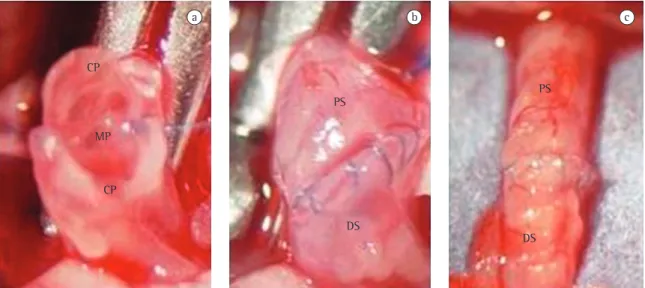
Figure 1 - Bronchial anastomosis technique: stereomicroscope view (×8) of the membrane (a) and cartilage (b)
bronchus, and mucus collection was performed selectively by inserting a small hair paintbrush into the lumen of each bronchus. The mucus adhered to the paintbrush was then placed in an Eppendorf tube containing mineral oil (to prevent dehydration) and stored at −70 °C.
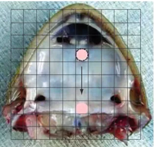
The relative MCT velocity in the mucus samples was measured using an in vitro frog palate model. The mucus samples, previously defrosted at room temperature, were placed on the frog palate ciliated epithelium, and their movement was observed and timed with the aid of a stereomicroscope equipped with a reticulated eyepiece (Figure 2). The MCT velocity in the rat mucus samples was compared to that in the frog mucus itself, and the results were therefore expressed as relative velocity (rat/frog).
Contact angle was measured using an eyepiece (×25) equipped with a goniometer with a scale from 0 to 180 degrees. The samples were placed on slides treated with sulfochromic acid to eliminate electrical currents, after which the angle formed between the air-liquid interface and the slide surface was
regis-tered.(9)
The MCT velocity was monitored through direct observation of the carbon particles deposited on the mucus layer of the distal region of each bronchus using a stereomicroscope with a reticulated eyepiece. each group were killed after 7, 15, and 30 days of
treatment, after which in vitro mucus transportability,
in situ MCT velocity, and contact angle (between a mucus drop and a surface) were analyzed.
The animals were previously anesthetized in a chamber containing isoflurane (Isothane; Baxter Health Care Corporation, Guayama, Puerto Rico) and intubated orotracheally using a polyethylene catheter. Artificial ventilation (model 683; Harvard Apparatus, South Natick, MA, USA) was maintained at a tidal volume of 10 mL/kg of body weight and a respiratory rate of 70 cycles/min. General anesthesia was maintained with 0.5% isoflurane in pure oxygen administered by a nebulizer (model 1223; Takaoka Indústria e Comércio Ltda, São Paulo, SP, Brazil). Access to the thoracic cavity was achieved through a left thoracotomy performed in the fifth inter-costal space. The main left bronchus was carefully dissected, being subsequently clamped near the carina and sectioned in its medial third. Bronchial anastomosis was performed using continuous 8-0 polypropylene sutures (Figure 1). Finally, airflow was restored, and the atelectasis of the left lung was resolved though hyperinflation. Prior to the closure of the incision, a chest tube was inserted. The chest tube was removed, together with the tracheal tube, immediately after spontaneous respiration had been restored. The surgical procedure was performed with the aid of a stereomicroscope, at a magnifica-tion of ×8. All surgical procedures were performed by the same researcher using a clean, albeit non-sterile, technique.
From the day of surgery, azathioprine (Imuran, 50 mg; Merck, Darmstadt, Germany) was administered to the animals via an orogastric tube in a daily dose of 3 mg/kg, diluted in saline solution (1 mg/mL). The animals in the Sal group were submitted to the same therapeutic regimen, receiving only the vehicle in an equivalent volume.
After 7, 15, and 30 days of treatment, six animals from each group were anesthetized intraperitoneally with sodium pentobarbital (30 mg/kg), and killed by exsanguination through section of the abdominal
aorta.(8)
Immediately after the animals were killed, their lungs were removed en bloc from the thoracic cavity and placed in Petri dishes. Only the bronchi that did not present any sign of stenosis, confirmed macroscopically, were included in the study. After dissection, an incision was made in each main
Figure 2 - Photograph of the frog palate with a diagram
the animals treated with saline solution (p < 0,001; Figure 4).
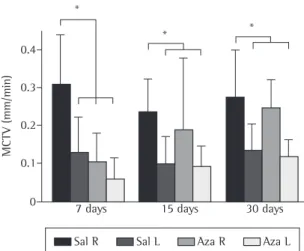
The results show that, in the animals that received saline solution, MCT velocity was lower, at all three time points evaluated, in the left bronchi (submitted to sectioning and anastomosis) than in the right (intact) bronchi (p < 0.05). The analysis of the left (sectioned) bronchi from the animals that received azathioprine showed a decrease in MCT velocity (with a statistical difference) after 7 days of treatment in relation to those that received saline solution, showing an interaction between the factors analyzed. When the right and left bronchi of the animals that received azathioprine were compared, the left (sectioned) bronchi presented an evident decrease in MCT velocity after 7 days of treatment (p < 0.001). The analysis of the right (intact) bronchi of the animals that received azathioprine showed a decrease in MCT velocity after 7 days of treatment (p < 0.05) in relation to the right (intact) bronchi of those that received saline solution, and the bronchi of the animals that received azathioprine presented full recovery after 30 days of treatment (Figure 5).
Discussion
Many advances have been made in the areas of immunosuppression, antibiotic therapy, and post-operative intensive care. Nevertheless, in lung transplants, in which there is manipulation of the bronchi, the most frequent complication is infec-tion resulting from malfuncinfec-tion of the mucociliary
system.(11) The immunosuppressive effect of the
anti-rejection drug is easily identified as a risk factor for complications and constitutes a major hindrance to
the success of the lung transplant.(12)
We observed that various immunosuppressive regimens have been tested in clinical practice, with a retrospective evaluation of the results obtained.
(13) Such testing has become more common since
the introduction of new immunosuppressive drugs aimed at replacing those, such as azathioprine, that have been used for more than 30 years. To date, this ‘competition’ between the new and the old has not produced sufficient scientific data to indicate which immunosuppressive drugs should or should not be used in lung transplant, since not all of their effects have been defined.
We sought to develop, in the present study, the idea of an experimental model that could clarify the The movement of the particles was timed and
regis-tered as the distance covered in one minute. The statistical analysis was performed using the program GraphPad Prism for Windows, version 4.0 (GraphPad Software Inc., San Diego, CA, USA). Analysis of variance was used in order to determine the interference of the factor analyzed, as well as any possible interaction between them. In addition, the Bonferroni test was used in order to determine the differences between the groups for each factor analyzed. The data are presented in the form of
charts and expressed as mean ± standard deviation.
Values of p < 0,05 were considered significant.(10)
Results
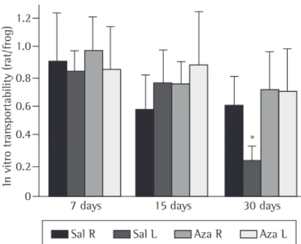
The analysis of the measurements of the contact angle of the mucus samples revealed greater values in the animals evaluated after 30 days of treat-ment, with a significant difference in the sectioned bronchi of the animals treated with saline solution (p < 0,001; Figure 3). This increase in the contact angle demonstrated a lower capacity of the mucus for spreading over a flat surface, characterizing it as more rigid and, therefore, more difficult to transport. This was demonstrated in the overall evaluation of mucus transportability on frog palate, especially in relation to the samples from the sectioned bronchi of
80
60
40
20
0
Contact angle (degrees)
7 days 15 days 30 days *
Sal R Sal L Aza R Aza L
phase, which in turn results in the accumulation of mucus and consequent alterations in its properties. Therefore, sectioning has no direct effect on the formation of mucus or on its properties. However, it has an indirect effect: MCT inefficiency leads to a worsening of mucus quality. Sectioning did not affect MCT or mucus quality on the right (intact) side. This demonstrates that surgery has only a local, rather than a systemic, effect. Indirectly, altered mucus quality promotes the occurrence of infections, and, through this mechanism, systemic complications occur. Therefore, longer-term mucus treatment and the use of mucus-thinning procedures in the post-operative period should be emphasized. Broad-spectrum antibiotics with a protective effect on the mucosa will bring greater benefits than do those without such properties.
Some authors have found a correlation between lung transplant and impairment of
pulmo-nary surfactant activity.(14) Therefore, pulmonary
surfactant might play an important role in normal mucociliary interaction if applied to the bron-chial surface as a therapeutic option in transplant patients. Azathioprine has no direct effect on the
production or quality of pulmonary surfactant.(15)
Other authors have studied, in other animal species, the effects of bronchial denervation on the mucociliary system and observed a decrease in MCT
velocity.(16,17) Bronchial devascularization might
have the additional effect of severely impairing MCT velocity. The principal hypothesis is that the reversibility of MCT impairment is due to vascular neoformation (revascularization of the bronchial tree), since bronchial anastomosis does not lead to
recovery of the nerves.(18) Therefore, our
hypoth-esis was that this effect would add to the effect of azathioprine on the reduction of cell activity and inflammatory activity, thereby prolonging the time to bronchial revascularization.
In the animals that received azathioprine, we found that it reduced MCT velocity in the group killed after 7 days of treatment both on the right (intact) side and on the left (operated) side. Over time, there is evident recovery of MCT velocity on the right and left sides, indicating that MCT velocity is affected by azathioprine only in initial period of up to 7 days of treatment. This mechanism might result from the fact that azathioprine decreases cell activity and reaches the ciliated cells, altering their function. The recovery of ciliated cell function, over time, in effects of immunosuppressive drugs as well as of
bronchial section and anastomosis on the muco-ciliary system, and that could be extrapolated to clinical practice. In this model, we evaluated the effects of bronchial section in animals treated with saline solution or azathioprine. In our model, MCT velocity was determined by direct observation of the India ink solution deposited in the region distal to the anastomosis of the left (sectioned) and right (intact) bronchial epithelium.
Our results demonstrate that there is MCT impairment and altered mucus quality on the left (operated) side but not on the right (intact) side, reinforcing the concept that bronchial denerva-tion is responsible for decreasing MCT velocity. The use of azathioprine minimized these effects by improving mucus quality, contradicting our initial hypothesis that, since azathioprine is a drug that reduces cellular metabolism, it would further impair MCT.
In the left bronchi of all animals studied, there was a reduction in MCT and a worsening of mucus quality. As the scarring process evolved, confirmed through macroscopic analysis, we observed that a tendency toward recovery of MCT after 30 days, although the mucus remained thick, viscous, and very sticky. This results from the fact that sectioning leads to MCT inefficiency in the acute post-operative
1.2
1.0
0.8
0.6
0.4
0.2
0
In vitro tran
sportability (rat/frog)
7 days 15 days 30 days *
Sal R Sal L Aza R Aza L
Figure 4 - Comparison of transportability of mucus
The quality of the mucus worsened over time in the animals submitted to bronchial section and treated with saline solution, and this worsening was greater in the animals killed after 30 days of treat-ment. We found, therefore, that bronchial section in isolation worsens mucus quality over time, and that this worsening is prevented by azathioprine admin-istration. Further scientific evidence regarding drug effects is needed before immunosuppressive proto-cols for use in clinical practice can be defined.
In summary, MCT velocity was impaired in the bronchi submitted to sectioning, and azathioprine had a protective effect on the viscoelastic proper-ties of mucus. The results obtained can add to the understanding of the MCT dysfunction observed after lung transplant, as well as increasing knowl-edge regarding the therapeutic interventions that more rapidly lead to the recovery of this system. Further studies are needed in order to evaluate the joint effect of combining azathioprine with other immunosuppressants.
References
1. Gallagher JT, Richardson PS. Respiratory mucus: structure, metabolism and control of secretion. Adv Exp Med Biol. 1982;144:335-50.
2. Joki S, Toskala E, Saano V, Nuutinen J. Correlation between ciliary beat frequency and the structure of ciliated epithelia in pathologic human nasal mucosa. Laryngoscope. 1998;108(3):426-30.
3. Ceesay SM, Melville GN, Mills JL, Wray SR. Comparative observations of mucus transport velocity in health and disease. Respiration. 1983;44(3):184-8.
4. Brody JS, Klempfner G, Staum MM, Vidyasagar D, Kuhl DE, Waldhausen JA. Mucociliary clearance after lung denervation and bronchial transection. J Appl Physiol. 1972;32(2):160-4.
5. Brooks RG, Hofflin JM, Jamieson SW, Stinson EB, Remington JS. Infectious complications in heart-lung transplant recipients. Am J Med. 1985;79(4):412-22.
6. Rivero DH, Lorenzi-Filho G, Pazetti R, Jatene FB, Saldiva PH. Effects of bronchial transection and reanastomosis on mucociliary system. Chest. 2001;119(5):1510-5.
7. Institute of Laboratory Animal Resources (U.S.). Guide for the Care and Use of Laboratory Animals. Washington, D.C.: National Academy Press, 1996. p. 1-35.
8. AVMA Panel on Euthanasia. American Veterinary Medical Association. 2000 Report of the AVMA Panel on Euthanasia. J Am Vet Med Assoc. 2001;218(5):669-96.
9. Nakagawa NK, Saldiva PHN, Lorenzi-Filho G. Mecanismos de defesa pulmonar. In: Nakagawa N K, Barnabé V. Fisioterapia do sistema respiratório. São Paulo: Sarvier, 2006. p. 207-28. 10. Rosner B. Fundamentals of Biostatistics. 2nd ed. Boston: PWS
Publishers; 1986. p.584.
the group that received azathioprine is probably due to two factors that lead to increased MCT velocity: bronchial scarring and improved mucus formation/ properties. Many immunosuppressive protocols include azathi oprine as a pre-operatively
admin-istered drug.(19) Based on the results of the present
study, we suggest that azathioprine administration, as well as the duration of azathioprine treatment in the post-transplant period, be the object of several other experimental studies.
In the Aza group, there was less worsening of mucus quality over time, which prevented changes in the properties of the mucus. The anti-inflamma-tory effect of azathioprine, as described by various
authors,(20,21) seems to be responsible for this fact.
Acting on the mucus-secreting cells, it presented a regulatory effect on mucus and periciliary fluid production. Our hypothesis was that azathioprine leads to lower MCT velocity and poorer mucus quality. However, these parameters did not worsen after administration of the drug, and the effect of the drug was not diminished after sectioning. The MCT velocity was affected by sectioning, with azathioprine-related worsening, only in the animals killed after 7 days of treatment.
Figure 5 - Comparison of in situ mucociliary transport velocity (MCTV) in the right (R) and left (L) bronchi of the animals treated with saline solution (Sal) or azathioprine (Aza). MCTV was lower in the sectioned bronchi (L) than in the intact bronchi (R) in all animals treated with Sal or Aza at all time points evaluated (*p < 0.05). MCTV in the intact bronchi (R) decreased after 7 days of treatment with Aza (*p < 0.05), and those bronchi presented full recovery after 30 days of treatment.
MCTV (mm/min)
Sal R Sal L Aza R Aza L 0.4
0.3
0.2
0.1
0
7 days 15 days 30 days * *
16. Marelli D, Paul A, Nguyen DM, Shennib H, King M, Wang NS, et al. The reversibility of impaired mucociliary function after lung transplantation. J Thorac Cardiovasc Surg. 1991;102(6):908-12.
17. Pazetti R, Pego-Fernandes PM, Ranzani OT, Parra ER, Lorenzi-Filho G, Jatene FB. Cyclosporin A reduces airway mucus secretion and mucociliary clearance in rats. Clinics. 2007;62(3):345-52.
18. Stone RM, Ginsberg RJ, Colapinto RF, Pearson FG. Bronchial artery regeneration after radical hilar stripping. Surg Forum. 1966;17:109-10.
19. Johnson CA, Porter WA. Compatibility of azathioprine sodium with intravenous fluids. Am J Hosp Pharm. 1981;38(6):871-5.
20. Kaplowitz N. Interaction of azathioprine and glutathione in the liver of the rat. J Pharmacol Exp Ther. 1977;200(3):479-86. 21. Lulin M, Yantang L. Comparison of the immunosuppressive
and toxic effects of TII and azathioprine. Transplant Proc. 1998;30(7):3517-8.
11. Ohori NP, Michaels MG, Jaffe R, Williams P, Yousem SA. Adenovirus pneumonia in lung transplant recipients. Hum Pathol. 1995;26(10):1073-9.
12. Griffith BP, Hardesty RL, Armitage JM, Kormos RL, Marrone GC, Duncan S, et al. Acute rejection of lung allografts with various immunosuppressive protocols. Ann Thorac Surg. 1992;54(5):846-51.
13. Longoria J, Roberts RF, Marboe CC, Stouch BC, Starnes VA, Barr ML. Sirolimus (rapamycin) potentiates cyclosporine in prevention of acute lung rejection. J Thorac Cardiovasc Surg. 1999;117(4):714-8.
14. Erasmus ME, Petersen AH, Oetomo SB, Prop J. The function of surfactant is impaired during the reimplantation response in rat lung transplants. J Heart Lung Transplant. 1994;13(5):791-802.