Major Article
Corresponding author: Prof. Amilcar Sabino Damazo. e-mail: [email protected]
Received 14 October 2016 Accepted 24 March 2017
Analysis of clinical data and T helper 1/T helper 2 responses
in patients with diferent clinical forms of leprosy
Ricardo Wilson de Pinho Rodrigues
[1],[2],
Afonso Bezerra Ribeiro
[2],
Gilcele de Campos Martin Berber
[3],
LeeYun Sheng
[4]and Amilcar Sabino Damazo
[2],[5][1]. Instituto de Ciências da Saúde, Universidade Federal do Mato Grosso, Sinop, MT, Brasil. [2]. Curso de Programa de Pós-Graduação em Ciências da Saúde, Faculdade de Medicina, Universidade Federal do Mato Grosso, Cuiabá, MT, Brasil. [3]. Faculdade de Sinop, Sinop, MT, Brasil. [4]. Instituto de
Ciências Naturais e Humanas, Universidade Federal do Mato Grosso, Sinop, MT, Brasil. [5]. Departamento de Ciências Básicas em Saúde, Faculdade de Medicina, Universidade Federal do Mato Grosso, Cuiabá, MT, Brasil.
Abstract
Introduction:
Currently,
there are no laboratory tests or sensitive and speciic molecular markers for the early diagnosis of leprosy.
The aim of this study was to analyze the clinical characteristics of patients with leprosy and investigate their immunological
proile, comparing this with the type of lesion and the presence or absence of a Bacillus Calmette-Guérin (BCG) vaccination scar.
Methods: Statistical analyzes were performed by employing comparative tests (Pearson´s chi-square) to evaluate the variables in
different clinical forms, considering signiicance at the 5% level.
Results:
The study identiied a predominance of lepromatous
leprosy (26.9%) in patients aged between 34-53 years. Caucasians predominantly had borderline tuberculoid (BT) clinical forms
(42%); a predominance of males with borderline lepromatous (19%) and lepromatous leprosy (26.9%) forms was observed;
and the presence of BCG vaccination scars (27.5%) and lower limb nerves were more affected (38%) predominantly in the BT
clinical form. Signiicant differences were identiied, which included hypochromic lesions predominantly in the BT clinical
form (24%); diffuse-type lesions predominantly in the tuberculoid (TT) clinical form (28%); ill-deined lesion border dominance
in lepromatous leprosy (LL) clinical forms (30%); an irregular lesion limit predominantly in LL clinical forms (32%); and a
predominant Th1 immune response in the BT clinical form (41.7%). Conclusions: The evaluation of the immunological proile
in leprosy patients may contribute to the more detailed diagnosis and possibly better characterization of the prognosis for these
individuals.
Keywords
:
Mycobacterium leprae
. Epidemiology. Th1/Th2 response. Tuberculoid. Lepromatous.
INTRODUCTION
Leprosy has a high rate of infectivity and low pathogenicity;
however, majority of the population do not develop the disease.
This fact indicates that there is an association of genetic and
environmental factors on susceptibility and cause of resistance
1,2.
The correct diagnosis of leprosy requires evolutionary clinical
data of the disease, histopathological analysis and sputum smear
microscopy, enabling determination of the form presented by the
patient, such as: TT (tuberculoid), BT (
borderline
tuberculoid),
BB (
borderline borderline
), BL (
borderline
lepromatous)
and LL (lepromatous leprosy)
3,4,5.
This classiication is necessary
for the appropriate, speciic therapeutic option to be delivered
6,7.
There are major differences in the endemicity of leprosy
between different regions of Brazil. Mato Grosso is the State
with the highest prevalence of leprosy; in 2014, the rate was
10.19 cases/10,000 inhabitants, which exceeds the national
rate of 1.27 cases/10,000 inhabitants
8.
There is a need to
intensify leprosy surveillance, with more effective diagnosis
and treatment of the disease, with an emphasis on the regions
with the highest rates of disease in the country
9.
The immune response is of prime importance to disease
susceptibility or resistance, fundamental to the defense of the
organism against exposure to the bacillus, and is also associated
with the development of the different clinical forms
10.
These
forms range from tuberculoid, with a predominantly cellular
immune response, to lepromatous leprosy dominated by a
humoral response
4,11. These responses are associated with speciic
mechanisms for the recognition of antigens, mediated by receptors
present on the membranes of T and B lymphocytes
12,13.
The
immune response can be categorized into cellular or type 1 and
humoral or type 2. The ability of lymphocytes with the cluster of
(Th), to induce cellular or humoral responses is related to the
types of cytokines secreted at the site of inlammation
14,15,16. The
predominance of a cellular or a humoral immune response to
infection with the bacillus may inluence the evolution of the
disease
17.
TT patients have strong cellular immune response
against
M. leprae
, which could limit the disease to a few
well-deined skin and nerves lesions
18. In patients with the
LL type, there is no speciic cellular immune response against
M. leprae
as bacterial proliferation occurs, with the presence
of many lesions and extensive iniltration of the skin and
nerves
19.
CD4
+T lymphocytes are more abundant in tuberculoid
lesions, whereas CD8
+lymphocytes, which can represent a
suppressor phenotype, predominate in lepromatous lesions
20,21.
In tuberculoid lesions, the distribution of lymphocytes is more
ordered, with CD4
+T lymphocytes in the center of the lesions
and CD8
+T lymphocytes with suppressive function
22.
The aim of this study was to analyze the clinical characteristics
of patients with leprosy and investigating their immunological
proile, comparing this with the type of lesion and the presence
or absence of a scar related to Bacillus Calmette-Guérin (BCG)
vaccination.
METHODS
Patients
This was a cross-sectional study of patients examined in the
Diagnosis and Treatment Leprosy Service section, located at
the
Hospital Universitário Júlio Müller
(HUJM), the Teaching
Hospital of
Universidade Federal de Mato Grosso
(UFMT),
Cuiabá, Mato Grosso State, Brazil, between November 2013
and September 2014. Seventy patients were categorized based
on the clinical forms, according to the established criteria
23.
A standard questionnaire was used to collect information
regarding the age, race, sex, lesion characteristics (color, type,
border), region of the affected nerves, and presence or absence
of a BCG vaccination scar. General physical and dermatological/
neurological examinations were performed on all patients by
the medical doctor responsible for the service. We assessed
the overall condition of the patient's health and the lesion
characteristics such as color (erythematous, hyperchromic,
hypochromic), type (diffuse, plaque or nodular), border (well
deined, ill deined), limit (irregular or regular) and an evaluation
of the affected area of nerves (upper limbs and lower limbs) and
protective sensation of hands and feet, through an esthesiometer
with the use of Semmes-Weinstein (SW)
24.
Patients with comorbities, immunosuppressive diseases,
renal failure and pregnant patients were excluded from our study.
Ethical considerations
This study meets the Resolutions No. 196/96 and No. 347
of 13 January 2005, of the National Health Council and was
approved by the HUJM Ethics Committee, with protocol number
733/CEP-HUJM /09. All patients were asked to voluntarily
participate in the research project. The informed consent form
(ICF) was read to each participant, and the interview process
was done only after the signing of the ICF.
Biopsy for histopathology
Asepsis and local anesthesia with 2% lidocaine without a
vasoconstrictor were performed, which showed that a lesion
has the clinical characteristic of leprosy. Further, a biopsy was
performed using a 4mm
punch
of the lesion. After the biopsy was
collected, the tissue specimen was immersed in 10% buffered
formalin and transported to the histopathology laboratory of
HUJM/UFMT, to examine the lesion. Samples were washed in
the same buffer, dehydrated in solutions with increasing ethanol
concentration, clariied in xylene and embedded in parafin. The
parafin blocks containing the tissue specimens were cut into
sections (5μm) for histology, using the HIRAX M60 microtome
(Carl Zeiss; Germany). Subsequently, the sections were placed on
slides, and after the process of deparafinization and rehydration,
they were stained with hematoxylin-eosin for histopathological
analysis, which allowed identiication of epithelioid and vacuolated
histiocytes, multinucleated giant cells, lymphocytes and plasma
cells, and regions of epithelial damage. In addition, another section
was cut in order to perform the Fite-Faraco staining, which was
used for the identiication of AFB (acid-fast bacilli).
Immunoluorescence for the identiication of Th1 and Th2
cells at the site of inlammation
The identiication of Th1 and Th2 cells was performed
in histological sections (5µm) of skin biopsies from
patients by immunofluorescence staining. Sections for
immunohistochemistry were prepared on slides with biological
adhesive (BIOBOND; British Biocell International, Cardiff,
UK) and subsequently incubated with the following reagents
at room temperature, as described previously
25: a) incubated in
a water bath at 100°C in 0.21% sodium citrate solution for 30
min; b) blocked with 3% hydrogen peroxide in 70% methanol
for 1h; c) permeabilized by incubation with 0.4% Tween 20
in phosphate buffered saline (PBS) for 15 min; d) blocked
with 5% bovine serum albumin (BSA), diluted in PBS for
1h; e) incubated with the primary antibodies: rabbit anti-CD4
(ABCAM, USA) (1:100 in 1% BSA), mouse anti-CC-chemikine
receptor 5 (CCR5) (ABCAM, USA) (1:100 in 1% BSA), for
the detection of membrane markers for Th1, and anti-CCR4
mouse (ABCAM, USA) (1:100 in 1% BSA), for the detection
of membrane marker for Th2. Slides were incubated with the
antibody solution for 18h at 4°C in a moist chamber.
For visualization of the antigen-antibody labeling region, the
following secondary antibodies were used: donkey anti-rabbit IgG
conjugated to ALEXAFLUOR 488 luorochrome (Invitrogen,
USA, 1:50 in 1% BSA) and goat anti-mouse IgG conjugated to
ALEXAFLUOR 546 luorochrome (Invitrogen, USA, 1:50 in
1% BSA). Slides were incubated for 1 h at room temperature and
in a dark chamber. Furthermore, nuclear DAPI
(4',6-diamidino-2-phenylindole) was used for examining nuclear morphology.
Excess luorescent reagent was washed with PBS and the slides
were mounted using Citiluor mounting media (DAKO, USA).
Statistical analysis
Tables were created to describe patient and lesion
Clinical form TT
n (%)
BT n (%)
BB n (%)
BL n (%)
LL
n (%) p-value*
Age
14-33 6 (25.0) 5 (20.8) 6 (25.0) 4 (16.7) 3 (12.5)
34-53 3 (11.4) 7 (26.9) 4 (15.4) 5 (19.2) 7 (26.9)
54 + 4 (21.1) 3 (15.8) 3 (15.8) 6 (31.6) 4 (21.1) 0.74
Race
Caucasians 6 (23.1) 11 (42.3) 3 (11.5) 3 (11.5) 3 (11.5)
Blacks 2 (10.0) 2 (10.0) 5 (25.0) 6 (30.0) 5 (25.0)
mulatto 5 (20.8) 2 (8.3) 5 (20.8) 6 (25.0) 6 (25.0) 0.07
Sex
Female 6 (21.4) 10 (35.7) 5 (17.9) 4 (14.3) 3 (10.7)
Male 7 16.7% 5 11.9% 8 19.1% 11 26.2% 11 26.2% 0.10
TABLE 1
Association of age, race, and sex of patients with the varying clinical forms of leprosy.
TT: Tuberculoid leprosy; BT: Borderline tuberculoid; BB: Borderline borderline; BL: Borderline lepromatous; LL: Lepromatous leprosy. *chi-square test. Leprosy patients examined in Júlio Muller Teaching Hospital ambulatory in Cuiaba, Mato Grosso.
vaccination scar, and Th1/Th2 immunoreactivity, associated
with varying clinical forms of leprosy. Pearson´s chi-square
test was performed to verify the association of each variable to
the clinical form
26,27,28. Data were analyzed using Excel 2010
software and SPSS (version 20), considering a 5% level of
signiicance.
RESULTS
Leprosy clinical form was not statistically associated with
patient age, race or sex (p-value = 0.74, 0.07 and 0.10, respectively)
(Table 1)
. With respect to patient age, 26 patients were aged
between 34-53 years, with a predominance of the BT (n=7;
26.9%) and LL (n=7; 26.92%) clinical forms. Of the reported
cases, 24 were aged between 14-3 years, with a predominance of
TT (n=6; 25.0%) and BB (n=6; 25.0%) clinical forms. Further,
19 patients were older than 54 years, with a predominance of the
TT (n=4; 21.0%) and BL (n=6; 31.6%) clinical forms. Twenty-six
patients were Caucasian with a predominance of the TT (n=6;
2%) and BT (n=11; 42.3%) clinical forms; 24 were Mulatto,
with a predominance of the BL (n=6; 25.0%) and LL (n=6;
25.00%) forms; 20 were Black, with a predominance of the BB
(n=5; 25.0%) and LL (n=6; 30.0%) forms. With respect to sex,
42 were males, predominantly with BL (n=11; 26.2%) and LL
(n=11; 26.2%). The TT (n=6; 21.4%) and BT (n=10; 35.7%)
clinical forms were predominantly found in the female patients.
We found signiicant associations to the varying clinical
forms in relation to the color of the lesion (P = 0.001), type
of injury (P = 0.027), lesion border characteristics (P = 0.001)
and limits (irregular versus regular) of the lesions (P = 0.001)
(Table 2)
. Thirty-three of the cases were hypochromic,
with a predominance of TT (n=12; 36.4%) and BT (n=8;
24.2%) 23 were erythematous, with a predominance of
BL (n=7; 30.4%) and LL (n=7; 30.4%); 14 were hyperchromic,
with a predominance of the BT (n=5; 35.7%) and BB (n=3;
21.4%). As for the type of lesion, 50 cases were diffuse, with
a predominance of the TT form (n=13; 26.0%) and BT (n=11;
22.0%); 12 were plaques with a predominance of the BT (n=4;
33.33%) and BB (n=4; 33.33%) forms; 8 were nodular with a
predominance of the BV (n=3; 37.5%) and LL (n=5; 62.5%)
forms. As for the lesion border characteristics, 43 of the cases
were ill-deined, with a predominance of the BL (n=13; 30.2%)
and LL (n=13; 30.2%) clinical forms; 27 were well-deined,
with a predominance of the TT (n=12; 44.0%) and BT (n=7;
25.9%) forms. With regard to the limits of the lesion, 37 cases
were irregular, predominantly BL (n=10; 27.0%) and LL (n=12;
32.4%); 33 of the cases were regular, with a predominance of
the TT (n=12; 36.4) and BT (n=9; 27.3%) forms. In the analysis
of the region of the affected nerves, no signiicant association
was observed (
P
= 0.639).
Statistical associations were not found between the clinical
form and the presence of a BCG vaccination scar (
P
= 0.359)
(Table 3)
. Forty patients had a BCG scar, with a predominance
of the BT (n=8; 26.7%) and BL (n=10; 25.0%) clinical forms; 30
cases had no scar, with a predominance of the TT (n=8; 26.7%)
and LL (n=7; 23.3%) forms
.
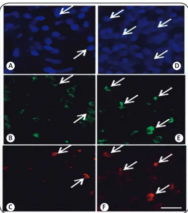
Immunoreactivity, in terms of Th1/Th2 responses, was
observed by immunostaining with the cell markers CD4/CCR5
and CD4/CCR4 (
Figure 1
). The staining proile was associated
with the differing clinical forms (
P
= 0.001); 36 cases were Th1,
with a predominance of the TT (n=11; 30. 6%) and BT (n=15;
41. 7%) forms; 34 were Th2, with a predominance of the BL
(n=12; 35,3%) and LL (n=13; 38,2%) forms
(Table 4).
There was a signiicant association (
P
= 0.001) between
A
B
C
D
E
F
FIGURE 1- Immunoluorescence analysis for CCR5 and CCR4. Figure 1 A, B and C: Tuberculoid leprosy patients with lymphocytes immunostained for DAPI (Panel A), CD4 (Panel B), and CCR5 (Panel C). Figure 1 D, E and F: Lepromatous leprosy patients with lymphocytes immunostained for DAPI (Panel D), CD4 (Panel E) and CCR4 (Panel F). Bar = 50µm.
CCR: CC-Chemokine receptor; DAPI:4',6-diamidino-2-phenylindole;
CD4: cluster of differentiation 4.
Clinical form
TT n (%)
BT n (%)
BB n (%)
BL n (%)
LL
n (%) p-value*
Color of Lesion
Erythematous 1 (4.4) 2 (8.7) 6 (26.1) 7 (30.4) 7 (30.4)
Hyperchromic 0 (0.0) 5 (35.7) 3 (21.4) 3 (21.4) 3 (21.4)
Hypochromic 12 (36.4) 8 (24.2) 4 (12.1) 5 (15.6) 4 (12.1) 0.001
Type of lesion
Diffuse 13 (26.0) 11 (22.0) 9 (18.0) 10 (20.0) 7 (14.0)
Nodular 0 (0.0) 0 (0.0) 0 (0.0) 3 (37.5) 5 (62.5)
Plaques 0 (0.0) 4 (33.3) 4 (33.3) 2 (16.7) 2 (16.7) 0.027
Border of the lesion
well deined 12 (44.4) 7 (25.9) 5 (18.5) 2 (7.4) 1 (3.7)
ill deined 1 (2.3) 8 (18.6) 8 (18.6) 13 (30.2) 13 (30.2) 0.001
Limit of lesion
Irregular 1 (2.7) 6 (16.2) 8 (21.6) 10 (27.0) 12 (32.4)
Regular 12 (36.4) 9 (27.3) 5 (15.2) 5 (15.2) 2 (6.1) 0.001
Affected nerves
Lower limbs 5 (16.7) 9 (30.0) 6 (20.0) 5 (16.7) 5 (16.7)
Upper limbs 4 (20.0) 4 (20.0) 2 (10.0) 4 (20.0) 6 (30.0)
Not affected 4 (20.0) 2 (10.0) 5 (25.0) 6 (30.0) 3 (15.0) 0.639
TABLE 2
Association of color of the lesion, type of lesion, lesion border and limits, and region of nerves affected for patients with varying clinical forms of leprosy.
TT: Tuberculoid leprosy; BT: Borderline tuberculoid; BB: Borderline borderline; BL: Borderline lepromatous; LL: Lepromatous leprosy. *chi-square test. Leprosy patients examined in Júlio Muller Teaching Hospital ambulatory in Cuiaba, Mato Grosso.
scar, and Th1/Th2 immunoreactivity
(Table 5)
. This analysis
showed that in 19 of the cases that had a BCG scar and Th1
immunoreactivity, there was a predominance of the TT clinical
form (n=5; 26.3%), and 17 of the cases with no BCG scar and
the presence of Th1 immune response had a prevalence of the
TT (n= 6; 35.3%) form. Twenty-one cases also had no BCG
scar and a Th2 immune response, with a predominance of the
BL (n=9; 42.7%) form. Thirteen of the cases had no BCG scar
and the presence of a Th2 immune response, with a prevalence
of LL (n= 6; 46.0%).
DISCUSSION
To date, there is no laboratory test or sensitive and speciic
molecular markers for the effective and early diagnosis of
leprosy
29,
with a diagnosis mainly based on clinical examination.
Thus, this study examined the inluence of Th1/Th2 immune
response on the clinical and histopathological variables, with
the objective of identifying possible patterns that may aid in
the prognosis of the different clinical forms of leprosy. Various
researchers have reported techniques for leprosy diagnosis
that may increase our understanding of the epidemiology and
transmission of the disease, with the ultimate goal of developing
new intervention strategies to prevent leprosy
30.
Although this study focused on the patients treated at the
Júlio Müller Teaching Hospital in the city of Cuiabá, many of
Clinical form
TT n (%)
BT n (%)
BB n (%)
BL n (%)
LL
n (%) p-value*
BCG
No 8 (26.7) 4 (13.3) 6 (20.0) 5 (16.7) 7 (23. 3)
Yes 5 (12.5) 11 (7.5) 7 (17.5) 10 (25.0) 7 (17.5) 0.36
TABLE 3
Association of the presence of a BCG vaccination scar with varying clinical forms of leprosy.
BCG: Bacillus Calmette-Guírin; TT: Tuberculoid leprosy; BT: Borderline tuberculoid; BB: Borderline borderline; BL: Borderline lepromatous; LL: Lepromatous leprosy. *chi-square test. Leprosy patients examined in Júlio Muller Hospital Teaching ambulatory in Cuiaba, Mato Grosso.
Clinical form
TT n (%)
BT n (%)
BB n (%)
BL n (%)
LL
n (%) p-value*
Th1
BCG
No 6 (35.3) 4 (23.5) 4 (23.5) 2 (11.8) 1 (5.9)
Yes 5 (26.0) 11 (57. 9) 2 (10.5) 1 (5.3) 0 (0.0)
Th2
BCG
No 2 (15.4) 0 (0.0) 2 (15.4) 3 (23.1) 6 (46.2)
Yes 0 (0.0) 0 (0.0) 5 (23.8) 9 (42.7) 7 (33.3) 0.001
TABLE 5
Association of BCG scar and immunoreactivity with varying clinical forms of leprosy.
TT: Tuberculoid leprosy; BT: Borderline tuberculoid; BB: Borderline borderline; BL: Borderline lepromatous; LL: Lepromatous leprosy. *chi-square test. Leprosy patients examined in Júlio Muller Teaching Hospital ambulatory in Cuiaba, Mato Grosso.
Clinical form
TT n (%)
TB n (%)
BB n (%)
BL n (%)
LL
n (%) p-value*
Immunoreactivity
Th1 11 (30. 6) 15 (41.7) 6 (16.7) 3 (8.3) 1 (2.8)
Th2 2 (5.9) 0 (0.0) 7 (20.6) 12 (35.3) 13 (38.2) 0.001
TABLE 4
Association of the immunoreactivity in patients with varying clinical forms of leprosy.
TT: Tuberculoid leprosy; BT: Borderline tuberculoid; BB: Borderline borderline; BL: Borderline lepromatous; LL: Lepromatous leprosy. *chi-square test. Leprosy patients examined at Júlio Muller Teaching Hospital ambulatory in Cuiaba, Mato Grosso.
individuals in the economically productive population, which
causes high economic impacts due to their having to abstaining
from work, caused by the development of permanent and
physically disabling lesions
31,32. Importantly, as these patients
are not able to work, they have the right to seek sickness beneits
and even disability retirement
33. Associated with this, the most
common clinical forms of leprosy in these patients are the BB,
BL, and LL forms, which have higher transmission capability
and cause greater incapacitation
34.
Regarding the ethnicity of patients, in our study, Caucasians
were the predominant race, which differs from previous indings
in the literature
32,35,36where Mulatos were the most prevalent
affected population. It should be noted that many patients
who are from different regions of the state and who are users
of the HUJM in Cuiaba are predominantly Caucasians. The
population of Mato Grosso is composed of 50.0% mixed race,
38.9% Caucasians and 9.8% Blacks (IBGE, 2010)
37.
Magalhães
38considers that the migration process in Mato Grosso territory
contributed to the spread and evolution of leprosy in the 1970s
and 1980s, with large lows of people of Caucasian origin into
the state. Few studies have reported the inluence of ethnic
variation as a factor of exposure to the bacillus.
Men were more affected in this study, and were associated
with the BL and LL clinical forms (26% each). These data are
consistent with the indings in the literature
39,40. According to
due to lifestyle factors. The higher incidence of physical
disabilities in men may be related to less concern with
self-image, particularly in relation to the body and aesthetics; thus,
men are less likely to access health services and this may
contribute to late diagnosis and subsequently predispose the
patient to become a propagator of the disease
42,43,44,45.
The dermato-histopathological characteristics of skin
lesions found in our study are consistent with indings in the
literature
45,46,47,48.
Assessment of the patients by dermatologic
and histopathological data are fundamental to arrive at a more
accurate diagnosis
49,50.
Regarding the region of the affected nerves, in this study
BT and BB forms were more frequent in the lower limbs
compared with other clinical forms. The involvement of the
peripheral nerves is present in all clinical forms of leprosy,
and in tuberculoid forms, the nerve damage is usually earlier,
while in lepromatous leprosy it appears later
48,51.
This variable
is an important epidemiological indicator responsible for
causing irreversible consequences, resulting in deformities
and disabilities
52,53.
Early diagnosis of neural integrity and
the degree of disability is key to determining follow-up
strategies, in order to guide appropriate treatment, prevent the
advancement of neural disability and contribute to the physical
rehabilitation
54,55,56.
Finally, the association of clinical forms with the
immunoreactivity in terms of Th1 and Th2 responses associated
with the type of leprosy patient was established. The literature
shows that the CCR5 is a good cell marker for Th1 cells, as is
CCR4 for Th2 cells
57. It is important to note that, in this study,
we evaluated the skin lymphocytes, while in most studies,
determination of phenotype of circulating leukocyte populations
in the blood is assessed. In some studies that have compared
the immunoreactivity of cells from the blood and the skin, the
existence of dissimilar responses has been reported
58,59.
This
demonstrates that, at the inlammatory site where bacteria are
concentrated, the immune response may vary according to what
is
classically
reported in the literature
10.
In this study, the majority of patients showed Th1
immunoreactivity, with the BT clinical form present at the
highest prevalence (41%), while the remainder of the patients
had Th2 characteristics, with a high prevalence of LL clinical
forms (38%). These data corroborate findings from other
studies that have shown similar results
58,59,60,61.
However, other
cell components may interfere with local immune responses
and lymphocyte differentiation, such as the interaction with
antigenic cells, like the Langerhans cells
62,extracellular matrix
components such as collagen ibers, ibronectin, and laminin
63,
and other factors such as hormones
64.
Thus, any changes in
Th1 and Th2 response should be considered with caution. In
addition, some studies have reported the tendency for variation
in the immune response, especially in patients with borderline
clinical proiles
58.
The Th1/Th2 responses may have inconsistencies. Some
patients may have different proile, including the presence
of regulatory T cells or Th17 cells along with a classic Th1
or Th2 proile
54,58,65,66,67.
This differential response could lead
to vulnerability to infection with the bacteria, or a change in
the clinical proile of patients from the tuberculoid pole to
lepromatous
59. Future studies may indicate the presence of each
cell type as a way to understand these changes in the immune
proile of patients, and may thus enable further improvements
in the prognosis of these patients.
Finally, the presence of a BCG vaccination scar in leprosy
patients was assessed. Most of the patients had a vaccination
scar, these being predominantly BT and BL clinical types.
Several studies have shown that the BCG vaccine protects
against leprosy, being one of the priority interventions
established by the World Health Organization (WHO) to control
the disease
50. Conversely, some studies have suggested varied
protection by the BCG vaccine and this may be related to
genetic factors
68,69., In this study most patients with leprosy who
had a BCG vaccination scar and a Th1 immune response were
tuberculoid while most patients without a BCG scar and with
a Th2 response were lepromatous. Some studies demonstrate
that BCG vaccination makes the individual most likely to
develop a proile of M1 macrophages, inducing them to produce
pro-inlammatory cytokines such as IL-1 and TNF-α, thereby
developing a greater resistance to the bacteria
70.
In conclusion, immunological evaluation of patients
with leprosy can contribute to the more detailed diagnosis
and possibly better characterization of prognosis in these
individuals. Further, public health policies should encouraged
BCG vaccination for individuals without vaccine scar, in order
to provide greater protection against this disease.
Financial Support
This study was supported by the Research Support Foundation of Mato Grosso (Fundação de Amparo a Pesquisa de Mato Grosso), Brazil, Grant no. 841967/2009 (PRONEX/FAPEMAT/CNPq) and Grant no. 754477/2011 (/FAPEMAT No. 009/2011). ASD was supported by the Brazilian National Council for Scientiic and Technological Development (Conselho Nacional de Desenvolvimento Cientíico e Tecnológico; Grant no. 311986/2014-5). Conlict of Interest
The authors declare that there is no conlict of interest.
REFERENCES
1. Costa RD. Estudo do peril de citocinas inlamatórias, moléculas anti-inlamatórias e BDNF em pacientes com hanseníase. Dissertação de Mestrado em Clínica Médica e Biomedicina. Belo Horizonte: Santa Casa de Misericórdia de Belo Horizonte; 2008. 147 p.
2. Alter A, Grant A, Abel L, Alcais A, Schurr E. Leprosy as a genetic disease. Mamm Genome. 2011;22(1-2):19-31.
3. Foss NT. Aspectos imunológicos da hanseníase. Medicina, Ribeirão Preto. 1997;30:335-9.
4. Mendonça VA, Costa RD, Melo GEBA, Antunes CM, Teixeira AL. Imunologia da hanseníase. An Bras Dermatol. 2008;83(4):343-50. 5. Jopling WH, McDougall AC. Manual de Hanseníase. 4ª edição.
São Paulo: Atheneu, 1991. 183 p.
7. Orsini M, de Freitas MRG, Antonioli RS, Mello MP, Reis JPB, Reis CHM, et al. Estudos clínicos, imunopatológicos e eletroisiológicos dos nervos periféricos na hanseníase. Rev Neurocienc. 2008;16(3):220-30.
8. Ministério da Saúde. Secretaria de Gestão do Trabalho e da Educação na Saúde. Mobilização reforça combate à hanseníase em Mato Grosso. Cuiabá: Universidade Aberta do SUS (UNA-SUS). 2015. Disponível em: http://unasus.gov.br/. Acesso em 20 abril de 2016.
9. World Healthy Organization (WHO). Global leprosy situation, 2012. Wkly Epidemiol Rec. 2012;87(34):317-28.
10. Goulart IMB, Penna GO, Cunha G. Imunopatologia da Hanseníase: a complexidade dos mecanismos da resposta imune do hospedeiro ao Mycobacterium leprae. Rev Soc Bras Med Trop. 2002;35(4): 365-75.
11. Araújo MG. Hanseníase no Brasil. Rev Soc Bras Med Trop. 2003;36(3):373-82.
12. Charo IF, Ransohoff RM. The roles of chemokines and chemokine receptors in inlammation. N Engl J Med. 2006;354(6):610-21. 13. Scapini P, Lapinet-Vera JA, Gasperini S, Calzetti F, Bazzoni F,
Cassatella MA. The neutrophil as a cellular source of chemokines. Immunol Rev. 2000;177:195-203.
14. Lew W, Chang S, Tada Y, Nakmura K, Tamaki K. Serum monocyte chemoattractant protein-1 is elevated in lepromatous leprosy patients with high bacterial indices. Int J Lepr Other Mycobact Dis. 2002;70(2):129-31.
15. Hasan Z, Jamil B, Zaidi I, Zafar S, Khan AA, Hussain R. Elevated serum CCL2 concomitant with a reduced Mycobacterium-induced response leads to disease dissemination in leprosy. Scand J Immunol 2006;63(3):241-7.
16. Mendonça VA, Malaquias LC, Brito-Melo GE, Castelo-Branco A, Antunes CM, Ribeiro AL, et al. Short report: differentiation of patients with leprosy from non-infected individuals by the chemokine eotaxin/CCL11. Am J Trop Med Hyg. 2007:77(3):547-50. 17. Moraes MO, Cardoso CC, Vanderborght PR, Pacheco AG. Genetic
of host response in leprosy. Lepr Rev. 2006;77(3):189-202.
18. Britton WJ. Leprosy. In: Cohen J, Powerly WG, editors. Infectious diseases. 2nd edition. London: Mosby; 2004. p. 1507-13.
19. Britton WJ, Lockwood DN. Leprosy. Lancet. 2004;363(9416): 1209-19.
20. Van Voorhis WC, Kaplan G, Sarno EN, Horwitz MA, Steinman RM, Levis WR, et al. The cutaneous iniltrates of leprosy: cellular characteristics and the predominant T-cell phenotypes. N Engl J Med. 1982;307(26):1593-7.
21. Narayanan RB, Bhutani LK, Sharma AK, Nath I. T cell subsets in leprosy lesions: in situ characterization using monoclonal antibodies. Clin Exp Immunol. 1983;51(3):421-9.
22. Hastings RC, Gillis TP, Krahenbuhl JL, Franzblau SG. Leprosy. Clin Microbiol Rev. 1988;1(3):330-48.
23. Ridely DS, Jopling WH. Classiication of leprosy according to immunity: a ive group system. Int J Lepr Other Mycobact Dis. 1966;34(3):255-73.
24. Lehman LF, Orsini MB, Nicholl AR. The development and adaptation of the Semmes Weinstein monoilaments in Brazil. J Hand Ther. 1993;6(4):290-7.
25. Damazo AS, Yona S, Flower RJ, Perretti M, Oliani SM. Spatial and temporal proiles for anti-inlammatory gene expression in leukocytes during a resolving model of peritonitis. J Immunol. 2006;176(7):4410-8.
26. Pozzebon M, Freitas HMR. Modelagem de casos: uma nova abordagem em análise qualitativa de dados? In: Anais do Encontro Anual da Associação Nacional de Pós-Graduação e Pesquisa em Administração. Foz do Iguaçu: ANPAD; 1998. p. 1-37.
27. Duarte T. A possibilidade da investigação a 3: relexões sobre triangulação (metodológica). CIES - Centro de Investigação e Estudos de Sociologia. CIES e-Working Paper. 2009;60:1-24. 28. Braga LPV. Introdução à Mineração de Dados. 2ª edição. Rio de
Janeiro: E-papers Serviços Editoriais; 2005. 212 p.
29. Stefani MMA. Desaios na era pós genômica para o desenvolvimento de testes laboratoriais para o diagnóstico da hanseníase. Rev Soc Bras Med Trop. 2008;41(Suppl 2):89-94.
30. Pinheiro RO, Salles JS, Sarno EN, Sampaio EP. Mycobacterium leprae-host cell interactions and genetic determinants in leprosy: an overview. Future Microbiol. 2011;6(2):217-30.
31. Lustosa AA, Nogueira LT, Pedrosa JIS, Teles JBM, Campelo V.The impact of leprosy on health-related quality of life. Rev Soc Bras Med Trop. 2011;44(5):621-6.
32. Miranzi SSC, Pereira LHM, Nunes AA. Peril epidemiológico da hanseníase em um município brasileiro, no período de 2000 a 2006. Rev Soc Bras Med Trop. 2010;43(1):62-7.
33. Ministério da Saúde. Secretaria de Vigilância em Saúde. Departamento de Vigilância Epidemiológica. Hanseníase e direitos humanos: direitos e deveres dos usuários do SUS (Série F. Comunicação e Educação em Saúde). Brasília: Ministério da Saúde; 2008. p. 72.
34. Flach DMAM, Andrade M, Valle CLP, Pimentel MIF, Mello KT. Análise da série histórica do período de 2001 a 2009 dos casos de hanseníase em menores de15 anos, no estado do RJ. Hansen Int. 2010;35(1):13 20.
35. Lana FCF, Lanza MF, Melendez VG, Branco CA, Teixeira S, Malaquias LCC. Distribuição da hanseníase segundo o sexo no município de Governador Valadares, Minas Gerais, Brasil. Hansen Int. 2003;28(2):131-7.
36. Ferreira SMB, Ignotti E, Gamba MA. Fatores associados à recidiva em hanseníase em Mato Grosso, Centro-oeste de Brasil. Rev Saúde Pública 2011;45(4):756-64.
37. Instituto Brasileiro de Geograia e Estatística (IBGE). Censo Demográico 2010. Brasília: IBGE, 2011. Disponível em: http:// www.ibge.gov.br/home/estatistica/populacao/censo2010/. (acessado em 20 de abril de 2016).
38. Magalhaes MCC, dos Santos ES, de Queiroz ML, de Lima ML, Borges RCM, Souza MS, et al. Migração e hanseníase em Mato Grosso. Rev Bras Epidemiol. 2011;14(3):386-97.
39. Souza CS. Hanseníase: formas clínicas e diagnóstico diferencial. Simpósio Hanseníase. Medicina, Ribeirão Preto. 1997;30(3):325-34. 40. Lana FCF, Amaral EP, Lanza FM, Lima PL, Carvalho ACN, Diniz
LG. Hanseníase em menores de 15 anos no Vale do Jequitinhonha, Minas Gerais, Brasil. Rev Bras Enferm. 2007;60(6):696-700. 41. Moreira FL, Nascimento AC, Martins ELB, Moreira HL, Lyon AC,
Lyon S, et al. Hanseníase em Alfenas: aspectos epidemiológicos e clínicos na região sul do estado de Minas Gerais. Cad Saude Colet 2009;17(1):131-43.
42. Hinrichsen SL, Pinheiro MRS, Jucá MB, Rolim H, Danda GJN, Danda DMR. Epidemiologic aspects of leprosy in the city of Recife, Pernambuco state, 2002 An Bras Dermatol. 2004;79(4):413-21. 43. Opromolla PA, Dalben I, Cardim M. Análise da distribuição
44. Lana FCF, Amaral EP, Lanza FM, Saldanha ANSL. Desenvolvimento de incapacidades físicas decorrentes da hanseníase no Vale do Jequitinhonha, MG. Rev Latino-Am Enferm. 2008;16(6):993-7. 45. Ministério da Saúde. Secretaria de Vigilância em Saúde. Guia
de Vigilância Epidemiológica. 6ª edição. Brasília: Ministério da Saúde; 2005. 806 p.
46. Coura JR. Síntese das Doenças Infecciosas e Parasitárias. 1ª edição. Rio de Janeiro: Guanabara Koogan; 2008. 322 p.
47. Rotta O. Guia de Dermatologia: Clínica, Cirúrgica e Cosmiátrica. Barueri: Manole; 2008. 725 p.
48. Lockwood DNJ, Suneetha L, Sagili KD, Chaduvula MV, Mohammed I, Van Brakel W, et al. Cytokine and protein markers of leprosy reactions in skin and nerves: baseline results for the North Indian INFIR cohort. PLoS Negl Trop Dis. 2011;5(12):e1327. 49. Barbieri CLA, Marques HHS. Hanseníase em crianças e
adolescentes revisão bibliográica e situação atual no Brasil. Pediatria 2009;31(4):281-90.
50. Lima LS, Jadão FRS, Fonseca RNM, Silva Junior GF, Barros Neto RC. Caracterização clínica- epidemiológica dos pacientes diagnosticados com hanseníase no município de Caxias, MA. Rev Bras Clin Med 2009;7:74-83.
51. Garbino JA, Marques Junior W, Barreto JA, Heise CO, Rodrigues MMJ, Antunes SL, et al. Primary Neural Leprosy: Systematic Review. Arq Neuro-Psiquiatr 2013;71(6):397-404.
52. Ministério da Saúde. Secretaria de Vigilância em Saúde. Manual de prevenção de incapacidades. 3ª edição. Brasília: Ministério da Saúde; 2008. 135 p.
53. Ministério da Saúde. Secretaria de Vigilância em Saúde. Portaria n° 3.125, de 07 de outubro de 2010. Aprova as Diretrizes para Vigilância, Atenção e Controle da Hanseníase. Diário Oicial da União. 2010 (15/10/ 2010); Sessão 1:p. 55.
54. Alves ED, Ferreira TL, Ferreira IN, organizadores. Hanseníase: avanços e desaios. Brasília: NESPROM/UnB; 2014. 492 p. 55. Baialardi KS. O estigma da hanseníase: relato de experiência em
grupo com pessoas portadoras. Hansen Int. 2007;32(1):27-36. 56. Raposo PM, Raposo AVC, Sanchez-González MA, Medeiros JLA,
Nemes MIB. Avaliação de incapacidades em pessoas vivendo com hanseníase: análise do grau de incapacidade em Campina Grande. Cad Saude Colet 2009;17(1):221-3.
57. Andrew DP, Rufing N, Kim CH, Miao W, Heath H, Li Y, et al. C-C chemokine receptor 4 expression deines a major subset of circulating nonintestinal memory T cells of both Th1 and Th2 potential. J Immunol. 2001;166(1):103-11.
58. Howe RC, Wondimu A, Demissee A, Frommel D. Functional heterogeneity among CD4+ T-cell clones from blood and skin lesions of leprosy patients. Identiication of T-cell clones distinct from Th0, Th1 and Th2. Immunology. 1995;84(4):585-94.
59. Saini C, Ramesh V, Nath I. CD4+ Th17 cells discriminate clinical types and constitute a third subset of non Th1, non Th2 T cells in human leprosy. PLoS Negl Trop Dis. 2013;7(7):e2338.
60. Berrington WR, Kunwar CB, Neupane K, van den Eeden SJF, Vary Junior JC, Peterson GJ, et al. Differential dermal expression of CCL17 and CCL18 in tuberculoid and lepromatous leprosy. PLoS Negl Trop Dis. 2014;8(11)e3263.
61. Venturini J, Soares CT, Belone AF, Barreto JA, Ura S, Lauris JR, et al. In vitro and skin lesion cytokine proile in Brazilian patients with borderline tuberculoid and borderline lepromatous leprosy. Lepr Rev. 2011;82(1):25-35.
62. Steinmann RM. The dendritic cell system and its role in immunogenicity. Annu Rev Immunol. 1991;9:271-96.
63. Matusyana T, Yamada A, Kay J, Yamada KM, Akiyama SK, Scholossman SF, et al. Activation of CD4 cells by ibronectin and anti-CD3 antibody. A synergistic effect mediated by the VLA-5 ibronectin complex. J Exp Med. 1989;170(4):1133-48.
64. Daynes RA, Araneo BA, Dowell TA, Huang K, Dudley D. Regulation of murine lymphokine production in vivo. III. The lymphoid tissue microenvironment exerts regulatory inluences over T helper cell functions. J Exp Med 1990;171(4):979-96. 65. Fink S, Finiasz MR, Valdez R, de la Barrera S, Sasiain MC.
Evaluacion de la produccion de citoquinas en enfermos de lepra. Medicina (B Aires). 1996;56(6):705-8.
66. Nath I, Vemuri N, Reddi AL, Jain S, Brooks P, Colston MJ, et al. The effect of antigen presenting cells on the cytokine proiles of stable and reaction all lepromatous leprosy patients. Immunol Lett. 2000;75(1):69-76.
67. Zhu J, Yamane H, Paul WE. Differentiation of effector CD4 T cell populations. Annu Rev Immunol. 2010;28(1):445-89.
68. Merle CS, Cunha SS, Rodrigues LC. BCG vaccination and leprosy protection: review of current evidence and status of BCG in leprosy control. Expert Rev Vaccines. 2010;9(2):209-22.
69. Setia MS, Steinmaus C, Ho CS, Rutherford GW. The role of BCG in prevention of leprosy: a meta-analysis. Lancet Infect Dis 2006;6(3):162-9.