Recebido em 17.08.2001. / Received in August, 17thof 2001.
Aprovado pelo Conselho Consultivo e aceito para publicação em 28.10.2002. / Approved by the Consultive Council and accepted for publication in October, 28thof 2002. * Trabalho realizado no Hospital de Clínicas da Universidade Federal do Paraná. / Work done at "Hospital de Clínicas da Universidade Federal do Paraná".
1Médica dermatologista. / M.D. Dermatologist.
2Médica residente do 3º ano de Dermatologia. / Resident M.D., 3rdyear of Dermatology. 3Médica dermatologista. / M.D. Dermatologist.
4Professor adjunto de Patologia - Universidade Federal do Paraná. / Adjunct professor of Pathology - Federal University of Parana. 5Professor assistente de Dermatologia - Universidade Federal do Paraná. / Assistant professor of Dermatology - Federal University of Parana.
©2004by Anais Brasileiros de Dermatologia
Fenômeno de Lúcio (eritema necrosante) na gestação
*The Lucio's phenomenon (necrotizing erythema)
in pregnancy
*Karin Adriane Helmer
1Isabela Fleischfresser
2Luciana D. Kucharski-Esmanhoto
3José Fillus Neto
4Jesus Rodriguez Santamaria
5Resumo:O fenômeno de Lúcio, variante do estado reacional hansênico tipo 2, provavelmen-te mediado por imunocomplexos, caracprovavelmen-teriza-se por reação cutânea necrosanprovavelmen-te grave que ocorre principalmente em doentes portadores de hanseníase virchowiana não nodular. Os autores relatam o caso de uma paciente de 27 anos, do sexo feminino, gestante de 32 sema-nas, com quadro de lesões eritêmato-purpúricas nos membros, bem delimitadas, confluentes, encimadas por bolhas, algumas necróticas e ulceradas, com uma semana de evolução, acom-panhadas de febre e intensa dor local. A baciloscopia da linfa evidenciou bacilos álcool-aci-dorresistentes formando globias, e a histopatologia da pele confirmou hanseníase virchowia-na, compatível com fenômeno de Lúcio. Foi iniciada prednisona e poliquimioterapia multi-bacilar com rifampicina, clofazimina e sulfona, com boa evolução. A gravidez tem sido asso-ciada à elevada incidência de aparecimento dos primeiros sinais ou ao agravamento da han-seníase por alterações hormonais que levam ao desequilíbrio imunológico, sendo considera-do crítico o períoconsidera-do compreendiconsidera-do entre o último trimestre de gestação e os primeiros três meses da lactação, quando a imunossupressão atinge seu ápice. Apesar da recomendação de se restringir a ingestão de drogas na gestação, o tratamento da hanseníase deve ser realizado, visto que seus benefícios superam os riscos.
Palavras-chave: eritema; gravidez; hanseníase.
Summary: The Lucio's phenomenon, a type 2 reactional condition in leprosy probably
mediated by immune complexes, is a severe necrotizing skin reaction that occurs mainly in patients with non-nodular lepromatous leprosy. This report presents a 27-year-old woman, in her 32nd week of pregnancy, with a one-week history of painful skin lesions in extremities, reddish-purple, sharply delineated, confluent, with bullae and occasional necrosis and ulceration. The patient also referred fever. Baciloscopy showed acid-fast bacilli and globi, and the histopathologic findings of a skin biopsy were consistent with lepromatous leprosy and Lucio's phenomenon. Prednisone and multidrug therapy with rifampin, clofazimine and dapsone were given, with remission. Pregnancy has been asso-ciated with a high incidence of first diagnosis of leprosy or with an exacerbation of symptoms in patients with the established disease because hormonal alterations cause immunological imbalance, particularly between the last three months of pregnancy and the first three months of lactation, when immunosuppression is higher. Despite the recom-mendation not to take drugs during pregnancy, the multidrug therapy regimen must be used, since the benefits achieved with the treatment surpass the risks.
Keywords: erythema; pregnancy; leprosy.
INTRODUÇÃO
A hanseníase é doença infecciosa crônica causada pelo bacilo álcool-acidorresistente Mycobacterium leprae, trans-mitido por via inalatória após exposição a doente bacilífero não tratado, geralmente por contato íntimo e prolongado. Dependendo da imunidade do paciente, a infecção pode evoluir para cura ou para as formas clínicas da doença: inde-terminada, tuberculóide, dimorfa ou virchowiana, as quais podem cursar com episódios inflamatórios agudos denomina-dos "reações ou estadenomina-dos reacionais". As reações do tipo 1 são mediadas pela imunidade celular e se apresentam clinica-mente por neurite ou reação reversa; as do tipo 2 envolvem imunocomplexos e produção de citocinas, como o fator de necrose tumoral alfa,1
e incluem o eritema nodoso hansênico, o eritema multiforme, o fenômeno de Lúcio e neurites.2
O fenômeno de Lúcio foi descrito por Lúcio e Alvarado em 1852, no México, e recebeu essa denominação em 1948 por Latapi e Zamora.3
Representa uma variante da reação hansênica tipo 2 e histopatologicamente caracteriza-se como vasculite aguda necrosante, tendo como sinonímia a expressão "eritema necrosante".2
Pode ocorrer na hanseníase de Lúcio (forma lepromatosa pura e primitiva) e em outras formas de hanseníase virchowiana.4,5
A exacerbação da hanseníase no final da gestação e no puerpério foi pela primeira vez descrita por Ryrie, em 1938, que declarou: "A hanseníase não apresenta qualquer efeito sobre o curso da gravidez, que, entretanto, exerce importante efeito no curso da hanseníase".6
Desde então a gestação tem sido associada à elevada incidência de aparec-imento dos primeiros sinais ou ao agravamento da hanseníase.
Os autores apresentam uma gestante de 27 anos, na trigésima segunda semana de gestação, com lesões ulceronecróticas nas extremidades, diagnosticadas como hanseníase difusa não nodular com vasculite do tipo fenô-meno de Lúcio.
RELATO DO CASO
Paciente de 27 anos, do sexo feminino, gestante de 32 semanas, referia queimadura com água escaldante na perna esquerda há sete meses, revelando-se de difícil cica-trização. Há duas semanas apresentou infecção secundária no local, tendo feito uso de cefazolina, heparina e medi-cação sintomática, sem melhora. Evoluiu com surgimento de lesões purpúricas nos membros inferiores e superiores e no abdômen, acompanhadas de dor e febre.
De antecedentes pessoais, estava na segunda ges-tação, não tendo apresentado intercorrências na primeira; negava tabagismo, etilismo e drogadição. De antece-dentes familiares, negava conhecimento de quadro seme-lhante.
Na admissão encontrava-se em regular estado geral, com dados vitais estáveis e afebril. Ao exame der-matológico apresentava fácies infiltrada com telangec-tasias e madarose (Figura 1), edema e eritema
impor-INTRODUCTION
Leprosy is a chronic infectious disease caused by the acid-fast bacillus Mycobacterium leprae, transmitted by inhalation following exposure of a person with an untreat-ed bacilliform infection, usually during intimate and pro-longed contact. Depending on the patient's immunity, the infection may develop toward recovery or the clinical forms of the disease: indeterminate, tuberculoid, dimorphic or lepromatous, each of which may course with episodes of acute inflammation called "reaction or reactional states". Type 1 reactions are mediated by cellular immunity and clinically present neuritis or a reverse reaction. Those of type 2 involve the immunocomplex and cytokine production, such as tumoral necrosis factor alpha,1
and include erythe-ma nodosum leprosum, multiform erytheerythe-ma, Lucio's leprosy phenomenon and neuritis.2
Lucio's leprosy phenomenon was described by Lucio and Alvarado in 1852 in Mexico, and was so named in 1948 by Latapi and Zamora.3
It represents a variant of leproma-tous reaction type 2 and histopathologically is characte-rized by acute necrotic vasculitis, being synonymous with the expression "necrotizing erythema".2
This can occur in Lucio's leprosy (where it forms a pure and primitive lepro-matosis) and in other forms of lepromatous leprosy.4,5
The exacerbation of leprosy at the end of gestation and in the puerperium was described for the first time by Ryrie, in 1938, who declared: "Leprosy does not present any effects on the course of the pregnancy, however, the pregnancy exercis-es an important effect on the course of the leprosy".6
Since then, gestation has been associated with the high incidence of onset of the first signs or aggravation of leprosy.
This work presents the case of a 27-year-old preg-nant woman, in the thirty-second week of gestation, with ulcerous necrotic lesions in the extremities, diagnosed as diffused non-nodular leprosy with vasculitis of Lucio's lep-rosy phenomenon.
CASE REPORT
The authors herein present a 27 year old female patient, 32 weeks pregnant, who had reported a burn with scalding water on the left leg seven months previously, resulting in a lesion of difficult cicatrization. Two weeks earlier, a secondary infection had presented itself in that area, there had been no improvement despite having used cefazolin, heparin and symptomatic medication. It deve-loped with the appearance of purpuric lesions in the inferi-or and superiinferi-or members and in the abdomen, accompa-nied by pain and fever.
Regarding personal history, this was her second ges-tation and she did not refer any problems during the first. She denied smoking, alcoholism or drug addiction. When questioned about family antecedents, she affirmed that she was not aware of similar occurrences.
Figura 1: Face infiltra-da com telangec-tasias e madarose /
Figure 1: Face infil-trated with telangiecta-sia and madarosis
Figura 3: Lesão eritê-mato-purpúrica enci-mada por bolha
Figure 3: Erythematous pur-puric lesion, well delineated and topped by a blister
showed infiltrated facies with telangiectasia and madarosis (Figure 1), edema and extensive erythema in the members, with disseminated and confluent erythematous-purpuric lesions, topped by blisters with well-defined borders, some with ulcerated and necrotic areas (Figures 2 and 3) and bilateral inguinal enlarged lymph nodes.
Laboratory exams showed hypochromic microcytic anemia, leukocytosis with deviation to the left, increase in the erythrocyte sedimentation rate, anti-HIV, VDRL and non-reagent anticardiolipin antibodies.
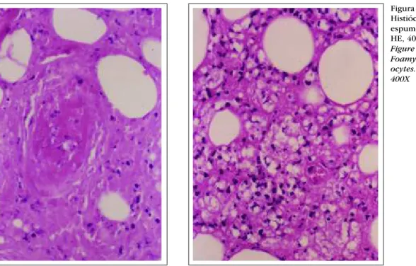
Bacilloscopy of the lymph revealed acid-fast bacilli with the formation of globi, bacilloscobic index = 1, with 5% complete bacilli, and 95% granular bacilli. Examination of the biopsy of apparently healthy skin but with lesions using hematoxylin and eosin stain showed leukocytoclas-tic vasculitis with fibrinoid necrosis (Figure 4), foamy histiocytes occupy-ing the lobular portion of the hypo-dermis (Figure 5) and presence of numerous granular bacilli determined by Fite-Faraco (Figure 6). This pat-tern is compatible with Virchow's lep-rosy and Lucio's leplep-rosy phenomenon. Multibacillary polychemother-apy was initiated with rifampicin 600mg/month, clofazimine 300mg/month and 50mg/day and dap-sone 100mg/day administered with
tantes nos membros, com lesões eritêmato-purpúricas disseminadas e confluentes, encimadas por bolhas, de bordas bem delimitadas, com algumas áreas ulceradas e necróticas (Figuras 2 e 3) e linfonodomegalia inguinal bilateral.
Os exames laboratoriais mostraram anemia hipo-crômica microcítica; leucocitose com desvio à esquer-da; aumento da velocidade de hemossedimentação; anti-HIV, VDRL e anticorpo anticardiolipina não reagentes.
A baciloscopia da linfa mostrou bacilos álcool-aci-dorresistentes com formação de globias, índice baciloscópi-co = 1, baciloscópi-com 5% de bacilos íntegros, e 95% de bacilos gran-ulosos. O exame da biópsia de pele
aparentemente sã e com lesão mostrou, pela hematoxilina-eosina, vasculite leucocitoclásica com necrose fibrinóide (Figura 4), histióc-itos espumosos com ocupação da porção lobular na hipoderme (Figura 5) e presença de numerosos bacilos granulosos pelo Fite-Faraco (Figura 6), compatível com hanseníase vir-chowiana e fenômeno de Lucio.
Foi iniciada poliquimioterapia multibacilar com rifampicina 600mg/mês, clofazimina 300mg/mês e 50mg/dia e dapsona 100mg/dia associadas à prednisona 60mg/dia, a
Figura 2: Lesões eritê- mato-pur-púricas bem delimitadas encimadas por bolhas, algumas com necrose e ulceração, no membro inferior /
Figura 4: Vasculite leucocito-clásica. HE, 400X
Figure 4: Leukocytocl astic vas-culitis. HE, 400X
Figura 5: Histiócitos espumosos. HE, 400X
Figure 5: Foamy histi-ocytes. HE, 400X
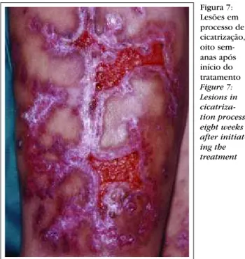
qual foi retirada progressivamente ao longo de oito sem-anas, com boa evolução (Figura 7).
A gestação evoluiu para parto normal a termo, sem intercorrências, com recém-nato de peso adequado à idade gestacional e sem malformações.
DISCUSSÃO
O fenômeno de Lúcio representa reação cutânea necrosante grave que pode ocorrer na hanseníase de Lúcio e em outras formas de hanseníase virchowiana.4,5
A hanseníase de Lúcio, mais comum no México e na América Central e pouco encontrada em outros grupos étni-cos,2,7
caracteriza-se por infiltração difusa da pele, madarose e perda dos cílios, dando aspecto "suculento, mixede-matoso" à pele.4
Em geral entre três e quatro anos após o início da doença,3
usualmente em pacientes não tratados,7
instala-se o fenômeno de Lucio, com aparecimento de máculas eritematosas dolorosas, que evoluem com necrose e ulceração,3,8
de forma ascendente (membros inferiores, superiores, glúteos, tronco e, com menor freqüência, face)9
e se resolvem em período que varia de duas a quatro sem-anas, deixando cicatriz atrófica.
Em outras formas de hanseníase virchowiana, o fenômeno de Lúcio caracteriza-se por necrose em lesões de eritema nodoso ou polimorfo surgidas na evolução de reação hansênica, em pacientes com ou sem hansenomas (hanseníase virchowiana não nodular).4,5
Embora o Brasil ocupe o segundo lugar do mundo em número de casos de hanseníase, relatos de fenômeno de Lúcio são raros.10
Laboratorialmente sempre estão presentes anemia, leucocitose e desvio à esquerda, e o exame da linfa em geral apresenta alto índice baciloscópico4,8
(não encontrado no caso descrito, provavelmente por falha técnica na realização
prednisone 60mg/day, which was reduced progressively over an eight-week period, with good clinical results (Figure 7). The gestation developed to term for a normal child-birth, without concurrent disease. The newborn was of appro-priate weight for gestational age and without malformations.
DISCUSSION
Lucio's leprosy phenomenon represents a serious cutaneous necrotizing reaction that can occur in Lucio's leprosy and also in other forms of lepromatous leprosy.4,5
Lucio's leprosy is most common in Mexico and Central America and little found in other ethnic groups,2,7
it is characterized by diffuse infiltration of the skin, madaro-sis and loss of eyelashes, giving "a swollen, edematous look" to the skin.4
Generally, three to four years after onset of the disease3
and usually in untreated patients,7 Lucio's leprosy phenomenon establishes itself with the emergence of painful erythematous discoloration that courses into necrosis and ulceration.3,8
This development is ascendant (inferior members, superiors, buttocks, trunk and, with less-er frequency, the face)9
and resolves in a period that varies from two to four weeks, leaving atrophic scars.
In other forms of lepromatous leprosy, Lucio's lep-rosy phenomenon is characterized by necrosis in lesions of nodular or polymorphic erythema that appear in the course of a lepromatous reaction in patients with nodular or non-nodular lepromatous leprosy.4,5
Although Brazil occupies second place in the world according to the number of cases of leprosy, reports of Lucio's leprosy phenomenon are rare.10 Laboratory findings always present anemia, leuko-cytosis and deviation to the left. Examination of the lymph generally presents a high bacilloscobic index4,8
performing the tests). In the histology, infiltrated inflamma-tion with foamy histiocytes are observed together with vas-cular damage due to direct invasion by M. leprae into endothelial cells7
causing vasculitis and thrombosis of the superficial and deeper vessels with consequent hemorrhage and infarction of the skin. Acid-fast bacilli can be detected with Ziehl or Fite-Faraco stains.3,4,10
These findings are observed as much in the clinically altered skin as in the apparently healthy skin.2
Treatment consists of polychemotherapy for multi-bacilli and sometimes systemic corticoids for control of the reactions. Unlike erythematous nodular leprosy, Lucio's leprosy phenomenon when it occurs in Lucio's leprosy does not present a good response to thalidomide.9,11
Pregnancy has been associated with a high incidence of onset of the first signs or aggravation of leprosy. The crit-ical period is considered to be between the last quarter of gestation and the first three months of nursing due to hor-monal, metabolic and immune system alterations. In gesta-tion cellular immunity is relatively suppressed, mainly in the third quarter, leading to the unchaining or worsening of type 2 reactions, as in the described case. There is also the risk of recurrence of the disease.12
In the puerperium there is a rel-ative suppression of the humoral immunity, with a greater risk for the development of type 1 reactions beginning between the third and sixteenth week of postpartum.6
Newborn whose mothers presented leprosy during pregnancy tend to weigh less than the mean for the general population12
and present a higher incidence of respiratory problems,13
due to placental inadequacy and consequent intra-uterine growth retardation. Nevertheless there is no conclusive controlled study that provides evidence of com-plications in a gestation as a result of having been treated
do exame). Na histologia observam-se infiltrado infla-matório com histiócitos espumosos, dano vascular por invasão direta do M. lepraeàs células endoteliais,7
determi-nando vasculite e trombose dos vasos superficiais e profun-dos com conseqüente hemorragia e infarto da pele, e baci-los álcool-acidorresistentes na coloração de Ziehl ou de Fite-Faraco.3,4,10
Esses achados são observados tanto na pele clinicamente alterada como na aparentemente sadia.2
O tratamento consiste no emprego da poliquimiote-rapia para multibacilares e, algumas vezes, de corticóides sistêmicos para controle das reações. Ao contrário do eritema nodoso hansênico, o fenômeno de Lúcio quando ocorre na hanseníase de Lúcio não apresenta boa resposta à talidomida.9,11
A gravidez tem sido associada à elevada incidência de aparecimento dos primeiros sinais ou ao agravamento da hanseníase, sendo considerado crítico o período compreen-dido entre o último trimestre da gestação e os três primeiros meses da lactação, por alterações hormonais, metabólicas e do sistema imune. Na gestação, ocorre supressão relativa da imunidade celular, principalmente no terceiro trimestre, levando ao desencadeamento ou à piora das reações tipo 2, como no caso descrito, havendo também risco de recidiva da doença.12
No puerpério há supressão relativa da imu-nidade humoral, com maior risco de desenvolvimento de reações tipo 1 iniciando-se entre a terceira e a décima sexta semana do pós-parto.6
Os recém-natos cujas mães apresentaram hanseníase na gravidez parecem pesar menos do que a média para pop-ulação geral12
e apresentam maior incidência de problemas respiratórios13
por insuficiência placentária e retardo de crescimento intra-uterino, porém não há estudo prospectivo controlado que evidencie complicações da gestação de
mu-Figura 6: Numerosos bacilos gran-ulosos na hipoderme. Fite-Faraco, 1000X
Figure 6: Numerous granular bacilli in the hypoder-mis. Fite-Faraco, 1000X
Figura 7: Lesões em processo de cicatrização, oito sem-anas após início do tratamento
lheres tratadas com poliquimioterapia.6
Podem ocorrer como intercorrências no recém-nato dermatite esfoliativa nas primeiras horas de vida pela sulfona e impregnação da clofazimina na pele, que apresenta coloração pardacenta.12
Mulheres devem ser encorajadas a tratar a hanse-níase com poliquimioterapia antes de considerar uma futu-ra gfutu-ravidez.13
Em recente publicação do Ministério da Saúde (Portaria 1073-26 set 2000),14
preconiza-se o trata-mento de gestantes com os hansenostáticos (rifampicina, clofazimina e dapsona) no esquema padrão, a despeito da recomendação de se restringir a ingestão de drogas no primeiro trimestre da gravidez, pois os benefícios do trata-mento superam os riscos.
O quadro clínico, os exames de laboratório e a histopatologia no caso descrito selam o diagnóstico de hanseníase virchowiana não nodular com fenômeno de Lucio, tendo sido a gestação associada ao diagnóstico da infecção e ao estado reacional tipo 2 exuberante, provavel-mente relacionado à imunossupressão fisiológica desse
período. q
with polychemotherapy.6
Although the newborn may present intercurrent disease, such as foleate dermatitis in the first hours of life due to the sulfone and impregnation of clofazi-mine in the skin, which presents a brownish coloration.12
Women should be encouraged to treat leprosy with polychemotherapy before considering a future pregnancy.13 In a recent publication, the Ministry of Health (Ordinance 1073-26 Sept. 2000),14
recommended the treatment of preg-nant women with leprostatics (rifampicin, clofazimine and dapsone) in the standard regimen, despite the usual recom-mendation of restricting the ingestion of drugs in the first quarter of pregnancy. It was considered that the benefits of the treatment outweigh the risks.
The clinical picture, laboratory exams and histopathology in the described case confirm the diagnosis of non-nodular lepromatous leprosy with Lucio's leprosy phenomenon. The gestation being associated with the diag-nosis of infection and to the virulent reactional state type 2, which was probably related to the physiologic
immunosup-pression of this period. q
10. Souza CS, Roselino AMF, Figeiredo F, Foss NT. Lucio's Phenomenon: Clinical and Therapeutic Aspects. Int J Lepr Other Mycobact Dis 2000 Dec; 68(4): 417-425.
11. Rea TH, Levan NE. Lucio's Phenomenon and Diffuse Nonnodular Lepromatous Leprosy. Arch Dermatol 1978(Jul); 114: 1023-1028.
12. Lopes VGS, Sarno EN. Hanseníase e Gravidez. Rev Ass Med Brasil 1994; 40(3): 195-201.
13. Morrison A. A woman with leprosy is in double jeopardy. Lepr Ver 2000; 71(2): 128-143.
14. Portaria n º 1073/GM de 26 de setembro de 2000. Publicada no D.O.U. - 100-E - página 18 - Seção 1 de 28 de setembro de 2000.
REFERÊNCIAS / REFERENCES
1. Foss NT. Hanseníase: aspectos clínicos, imunológicos e tera-pêuticos. An Bras Dermatol 1999 (Mar-Abr); 74(2): 113-119. 2. Gilbert E, Cubria JL, Gratacos R, et al. Lepra de Lucio. Med Cut ILA 1982; 10: 41-46.
3. Pursley TV, Jacobson RR, Apisarnthanarax P. Lucio's Phenomenon. Arch Dermatol 1980(Feb); 116: 201-04.
4. Pereira Jr AC. Hanseníase de Lucio. An Bras Dermatol 1993(Jan-Fev); 68(1): 33-40.
5. Buffon LP, Leal R, Vidigal MR, Gatti TSR, Pires MC, Reis VMS. Fenômeno de Lucio (eritema necrosante) na gestação: rela-to de caso e revisão da literatura. An Bras Dermarela-tol 2001 (Jul-Ago); 76(4): 441-448.
6. Lockwood DNJ, Sinha HH. Pregnancy and leprosy: A Comprehensive Literature Review. International Journal of Leprosy 1999; 67(1): 06-12.
7. Rea TH, Ridley DS. Lucio's Phenomenon: A Comparative Histological Study. International Journal of Leprosy 1979; 47(2): 161-66.
8. Saúl A, Novales J. La Lèpre de Lucio-Latapi et le Phénomène de Lucio. Acta leprol 1983 (Jul-Sep);1(3) : 115-32
9. Bernadat JP, Faucher JF, Huerre M. Lèpre lépromateuse diffuse révélée par une vasculite cutanée. Ann Dermatol Venereol 1996; 123: 21-23.
ENDEREÇO PARA CORRESPONDÊNCIA: / MAILINGADDRESS:
Karin Adriane Helmer
R. Padre Anchieta, 1721 - apto. 102 80730-000 Curitiba PR
Tel.: (41) 336-0161