Cop
yright
© ABE&M t
odos os dir
eit
os r
eser
vados
.
851
case report
Arq Bras Endocrinol Metab. 2014;58/8
Papillary thyroid cancer case
masked by subacute thyroiditis
Caso de carcinoma papilar de tiroide mascarado por tireoidite subaguda
Bekir Ucan1, Tuncay Delibasi1, Erman Cakal1, Muyesser Sayki Arslan1, Nujen
Colak Bozkurt1, Taner Demirci1, Mustafa Ozbek1, Mustafa Sahin2
SUMMARY
Subacute thyroiditis (SAT) association with thyroid carcinoma has been rarely reported in the literature. We present a patient with SAT and papillary thyroid cancer that was suspected by ultrasonographic evaluation (US) following SAT treatment. A ifty-four-year old female patient referred to our department due to tachycardia, jitteriness and pain in cervical region for the past one month. SAT diagnosis was established by physical examination, laboratory and ultrasono-graphic indings. After treatment, control thyroid US revealed regression of the hypoechogenic regions seen in both lobes, and a previously unreported hypoechogenic lesion with microcal-ciication focus that had irregular borders and was not clearly separated from the surrounding parenchyma located in the posterior aspect of the lobe (Elasto score: 4, Strain index: 7.08). Fine needle aspiration biopsy was taken from this nodule; cytology was assessed to be compatible with papillary thyroid carcinoma. Postsurgical pathology evaluation showed a papillary mi-crocarcinoma. SAT may produce ultrasound changes that obscure the coexistence of papillary carcinoma. We recommend that patients with SAT have ultrasonography after they recover. Hy-poechogenic regions bigger than 1 cm that are present in the follow-up post-therapy US should be assessed by biopsy. Arq Bras Endocrinol Metab. 2014;58(8):851-4
SUMÁRIO
A associação da tireoidite subaguda (TSA) com o carcinoma de tiroide foi raramente relatada na literatura. Apresentamos uma paciente com TSA e tumor papilar de tiroide suspeito na ultrasso-nograia (US) após o tratamento para a TSA. Uma mulher de 54 anos de idade foi encaminhada ao nosso departamento com taquicardia, agitação e dor na região cervical, com duração de 1 mês. O diagnóstico de TSA foi estabelecido pelo exame físico, e pelos achados laboratoriais e ultrassonográicos. Depois do tratamento, o US para controle da tiroide mostrou regressão das regiões hipoecoicas vistas em ambos os lobos e uma lesão hipoecoica anteriormente não observada com focos de microcalciicação, bordas irregulares, não claramente separada do parênquima circundante e localizada na região posterior do lobo. (Escore elástico: 4, índice de deformação: 7,08). Foi feita uma biópsia do nódulo por meio de aspiração por agulha ina. A ci-tologia mostrou-se compatível com um carcinoma papilar de tiroide. A avaliação pós-cirúrgica mostrou um microcarcinoma papilar. A TSA pode produzir alterações ultrassonográicas que obscurecem a coexistência de carcinoma papilar. Recomendamos que pacientes com TSA pas-sem por exame ultrassonográico após a recuperação. Regiões hipoecoicas maiores que 1 cm encontradas no US para acompanhamento pós-tratamento devem ser avaliadas por biópsia.
Arq Bras Endocrinol Metab. 2014;58(8):851-4
1 Diskapi Yildirim Beyazit
Training and Research Hospital, Department of Endocrinology and Metabolism, Ankara, Turkey
2 Ankara University, School
of Medicine, Department of Endocrinology and Metabolism, Ankara, Turkey
Correspondence to:
Bekir Ucan
Diskapi Yildirim Beyazit Training and Research Hospital, Department of Endocrinology and Metabolism, Ankara, Turkey [email protected]
Received on Jan/12/2014 Accepted on Mar/11/2014
DOI: 10.1590/0004-2730000003222
INTRODUCTION
S
ubacute thyroiditis (SAT) is one of the inlamma-tory thyroid diseases known as de Quervain’s dise-ase. This disease affects women three to ive times more often than men. In addition to that, viral infection isCop
yright
© ABE&M t
odos os dir
eit
os r
eser
vados
.
852 Arq Bras Endocrinol Metab. 2014;58/8
Ultrasonography (US) is a useful tool for diagnosing and monitoring thyroid pathology, including subacute thyroiditis. Characteristic ultrasonographic features of thyroiditis are enlargement of thyroid gland, focal hy-poechoic zones with indeinite borders or diffuse hypo-echoecogenicity, and lack or low low on color Doppler in these areas (5). These indings usually accompany hard and tender thyroid gland (3). However these ul-trasonographic indings are not pathognomic for SAT. Diffuse hypoechogenity can be seen in Graves’ di-sease and Hashimoto thyroiditis, whereas benign no-dules and thyroid carcinomas may present as focal hy-poechoic regions (4,5). In pathological examinations, thyroid gland is seen as engorged and edematous in SAT. There is follicular cell destruction and the iniltra-tion of inlammatory cells. These inlammatory cells are neutrophils, lymphocytes, histiocytes and multinuclear giant cells and are seen in the hypoechoic regions de-termined in the biopsy taken under US guidance. With the regression of the inlammatory process, there are different stages of ibrosis and granuloma formation in the thyroid gland (6).
SAT’s association with thyroid carcinoma has been rarely reported in the literature. In this case report, we presented a 54 years old female patient who was diag-nosed subacute thyroiditis and papillary thyroid cancer by US evaluation following treatment.
CASE REPORT
Fifty-four years old female referred to our department due to tachycardia, jitteriness and pain in servical region for the past 1 month. The pain in the neck that radiated to the ear and jaw and was worsening with swallowing. Asthma, gastritis and carpal tunnel syndrome was pres-ent in her history. There were no thyroid carcinoma in her family history. In the physical examination, thyroid gland was bilaterally enlarged and hard. Also tender-ness was found in the thyroid region by palpation. Thy-roid gland was not ixated to the surrounding tissue. No cervical lymph node was apparent.
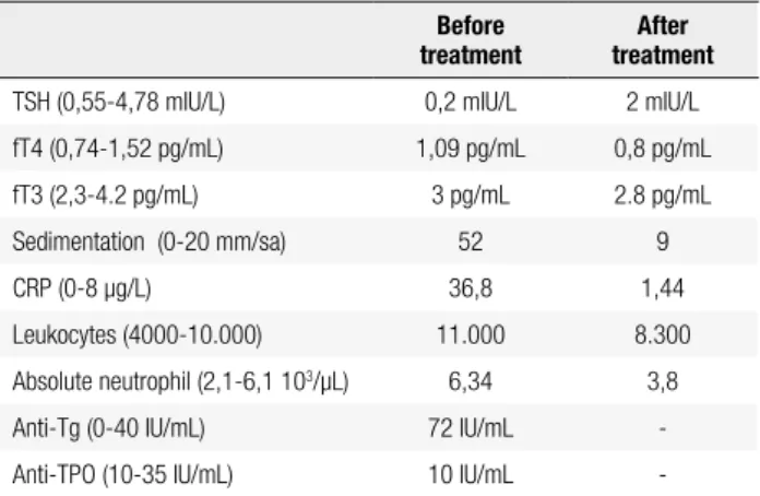
Her laboratory examinations were as follows; TSH: 0.2 (0,55-4,78 mIU/L), sT4: 1.09 (0.74-1.52), sT3: 3 (2.3-4.2 pg/mL), anti-TPO: 10 (10-35 IU/mL), anti-tg: 72 40 IU/mL), sedimentation rate: 52 (0-20 mm/h), C-reactive protein (CRP): 36.8 (0-8 μg/L), neutrophils 11 (4-10 103/μL), absolute neutrophil 6.34
(2,1-6.1 103/μL), haemoglobin: 12.6 (11.4-16.4 g/dL),
and thrombocyte count was 269.000 (150-372 103/μL).
In the thyroid ultrasonography, bilaterally enlar-ged, diffuse and tenderness with contact of the probe in the thyroid gland was detected. Moreover, hypoe-chogenic heterogeneous regions of irregular borders in both thyroid lobes. SAT diagnosis was established by physical examination, laboratory and ultrasonographic indings. Medical therapy was initiated. On the second day of the steroid treatment, there were signiicant re-gression in the symptoms and signs of the patient. Ste-roid treatment was continued for a month with gradual decreases. The patient’s control levels were as follows; sedimentation rate 9 mm/h, CRP:1.441 mg/L, TSH: 2 (0.55-4.78 mIU/L), sT4: 0.8 (0.74-1.52 pg/mL), sT3:2.8 (2.3-4.2 pg/mL), neutrophil: 8.3 (1.69-8.3 103/μL). Her control thyroid US revealed regression
of the hypoechogenic regions seen in both lobes and a previously unreported hypoechogenic region or nodule with microcalciication focus that had irregular borders and was not clearly seperated from the surrounding parenchyma localized in the posterior aspect of the ri-ght thyroid lobe (elasto score: 4, strain index: 7,08). The ine needle aspiration biopsy taken from this no-dule revealed a malign cytology and was compatible with papillary thyroid carcinoma. Total thyroidectomy and central neck lymph node dissection were planned. Postsurgical pathology evaluation was reported to be papillary microcarcinoma (9 mm).
DISCUSSION
Subacute granulomatous thyroiditis (SAT) is a self-limiting, painful, non-infectious inlammatory disease of the thyroid gland. Although the etiology of subacute thyroiditis is not completely known, it is assumed to be related to viral infections and genetic factors. Infectious etiology is not clearly proven that no proliferation was found in viral cultures, however viral antibodies were found to be high (7).
Thyroid carcinoma rarely co-exists with subacute thyroiditis. Fatourechi and cols. found no thyroid can-cer in 160 SAT patients (8). But there are several case reports about this co-existence worldwide in the litera-ture (9-11).
Ultrasonography provides important clues for diag-nosis of SAT however hypoechogenic regions that are typical ultrasonographic indings of subacute thyroiditis are not speciic to this disease (12). Hypoechogenity, nodule shape, tumor magrin, hypoechoic rims are im-portant factors in evaluating nodule for thyroid
Cop
yright
© ABE&M t
odos os dir
eit
os r
eser
vados
.
853 Arq Bras Endocrinol Metab. 2014;58/8
noma (13). Also ultrasonographic features such as mi-crocalciication and blurred borders are very important for deciding biopsy even there is no obvious nodule. Without any other suspicious clue than diffuse hypo-echogenic regions, it is very dificult to determine the coocurrence of SAT and papillary carcinoma. In our case, the presence of microcalciication focus and blur-red borders accompanying the hypoechogenic regions in the ultrasonography was a red lag.
In the subacute thyroiditis, initial sonographic exa-mination is important, unlike typical indings on the presence of a suspicious lesion should be considered. In other words, at the irst ultrasonography may be missed the suspicious lesion. Because, it was dificult to point out the coexistence of papillary carcinoma with SAT, based on no clues other than diffuse hypoechoic area at initial ultrasonographic examination. Nishihara and cols. were also not determined a suspicious lesion at the irst ultrasound examination in their study (11). Thyroid US is useful for the follow-up of patients with subacute granulomatous thyroiditis. The follow--up US indings of SAT were a reduction of an enlarged thyroid volume and a decreased or absent hypoechoic areas. A follow-up US exam is recommended rather than immediately performing ine needle aspiration biopsy (FNAB). However, FNAB is indispensable in cases of lesions that are seen as a focal mass mimicking thyroid malignancy (11). Also, as in our case, control ultrasonography is very important in SAT for undetec-table suspicious lesions in irst ultrasound.
In a study 15 patients with subacute thyroiditis, Tokuda and cols. have reported that sonographic in-dings changed during the course of the disease. They have reported the presence of hypoechogenic regions during the active phase, and their regression and
di-sapperance when the clinical symptoms ameliorated. They have shown in 3 patients that hypoechogenic regions reoccured with the recurrence of the disease (12). In our case, there was also signiicant decrease in hypoechogenic regions following therapy. This enabled the detection of the suspicious region.
Lymphadenopathy (LAP) might accompany to thyroid carcinoma and SAT. Nishihara and cols. detec-ted that two patients had enlarged and rounded cervi-cal lymph nodes and diffusely hypoechoic areas in their thyroid (11). In our case, there was no LAP accom-panying suspicious lesions. Elastography may assist in the diagnosis and monitoring of SAT. Also it may help in deciding biopsy requirement in SAT coexistent with suspicious lesions or nodules.
It is also uncertain whether a coexisting thyroid inlammation might inluence the result of elastogra-phic evaluation of thyroid focal lesions (14). As cases of coexistence of SAT with nodular goiter or thyroid cancer have been described, it seems best to postpone the assessment of the nodule stiffness until a complete recovery from SAT (11).
In our case, we found a previously unreported sus-picious hypoechogenic lesion in control thyroid US. This nodule has an increased elastography score and strain index according to uninvolved and other hypoe-choic regions. US examination made by an experinced doctor and elastography indings help us to take biopsy decision.
We have reported a case with presentation of diffuse hypoechogenic regions in ultrasonography compatible
Table 1. Laboratory indings of the patient
Before
treatment treatmentAfter
TSH (0,55-4,78 mIU/L) 0,2 mIU/L 2 mIU/L
fT4 (0,74-1,52 pg/mL) 1,09 pg/mL 0,8 pg/mL
fT3 (2,3-4.2 pg/mL) 3 pg/mL 2.8 pg/mL
Sedimentation (0-20 mm/sa) 52 9
CRP (0-8 μg/L) 36,8 1,44
Leukocytes (4000-10.000) 11.000 8.300
Absolute neutrophil (2,1-6,1 103/μL) 6,34 3,8
Anti-Tg (0-40 IU/mL) 72 IU/mL
-Anti-TPO (10-35 IU/mL) 10 IU/mL
-Figure 1. Longitudinal thyroid ultrasonography and elastography taken when the patient symptoms disappeared with prednisolone treatment, showing a heterogeneous thyroid nodule with microcalciications and persistent hypoechoic area.
Cop
yright
© ABE&M t
odos os dir
eit
os r
eser
vados
.
854 Arq Bras Endocrinol Metab. 2014;58/8
with SAT that masked the papillary carcinoma focus. In conclusion, SAT may accompany to the papilla-ry thyroid carcinoma. Therefore we propose a careful thyroid examination due to the the possibility of SAT and thyroid carcinoma co-existence. Also patients with SAT diagnosis should be followed up by USG after the therapy and hypoechogenic regions greater than 1 cm should be assessed by biopsy.
REFERENCES
1. Zacharia TT, Perumpallichira JJ, Sindhwani V, Chavhan G. Gray-scale and color Doppler sonographic indings in a case of sub-acute granulomatous thyroiditis mimicking thyroid carcinoma. J Clin Ultrasound. 2002;30:442-4.
2. Slatosky J, Shipton B, Wahba H. Thyroiditis: differential diagnosis and management. Am Fam Physician. 2000;61:1047-52. 3. Benker G, Olbricht T, Windeck R, Wagner R, Albers H, Lederbogen
S, et al. The sonographical and functional sequelae of de Quer-vain’s subacute thyroiditis: long-term follow-up. Acta Endocrinol. 1988;117:435-41.
4. Jhaveri K, Shroff MM, Fatterpekar GM, Som PM. CT and MR im-aging indings associated with subacute thyroiditis. Am J Neuro-radiol. 2003;24:143-6.
5. Tokuda Y, Kasagi K, Iida Y, Yamamoto K, Hatabu H, Hidaka A, et al. Sonography of subacute thyroiditis: changes in the indings during the course of the disease. J Clin Ultrasound. 1990;18:21-6.
6. Meachim G, Young MH. De Quervain’s subacute granulomatous thyroiditis: histological identiication and incidence. J Clin Pathol. 1963;16(3):189-99.
7. Luotola K, Hyöty H, Salmi J, Miettinen A, Helin H, Pasternack A. Evaluation of infectious etiology in subacute thyroiditis lack of association with coxsackievirus infec-tion. APMIS. 1998;106(4):500-4.
8. Fatourechi V, Aniszewski JP, Fatourechi GZ, Atkinson EJ, Jacobsen SJ. Clinical features and outcome of subacute thyroiditis in an incidence cohort: Olmsted County, Minnesota, study. J Clin En-docrinol Metab. 2003;88(5):2100-5.
9. Choia YS, Kima BK, Kwon HJ, Lee JS, Heo JJ, Jung SB, et al. Sub-acute thyroiditis with coexisting papillary carcinoma diagnosed by immediately repeat ine needle aspiration: a case report. J Med Case Rep. 2013;7(1):3.
10. Lam KY, Lo CY. Papillary carcinoma with subacute thyroiditis. En-docr Pathol. 2002 Fall;13(3):263-5.
11. Nishihara E, Hirokawa M, Ohye H, Ito M, Kubota S, Fukata S, et al. Papillary carcinoma obscured by complication with subacute thyroiditis: sequential ultrasonographic and histopathological indings in ive cases. Thyroid. 2008;18(11):1221-5.
12. Lu CP, Chang TC, Wang CY, Hsiao YL. Serial changes in ultrasound-guided fine needle aspiration cytology in subacute thyroiditis. Acta Cytol. 1997;41:238-43.
13. Jeh SK, Jung SL, Kim BS, Lee YS. Evaluating the degree of con-formity of papillary carcinoma and follicular carci-noma to the reported ultrasonographic findings of malignant thyroid tumor. Korean J Radiol. 2007;8:192-7.
14. Xie P, Xiao Y, Liu F. Real-time ultrasound elastography in the di-agnosis and differential didi-agnosis of subacute thyroiditis. J Clin Ultrasound. 2011;39(8):435-40.