ABSTRACT
Cushing’s syndrome (CS) results from sustained pathologic hypercortisolism. The clinical features are variable and the most specific features for CS include abnormal fat distribution, particularly in the supraclavicular and temporal fos-sae, proximal muscle weakness, wide purple striae, and decreased linear growth with continued weight gain in a child. Clinical presentation of CS can be florid and in this case the diagnosis is usually straightforward. However, the diagnosis can be difficult particularly in states of mild or cyclical or periodical hypercortisolism. Several tests based on the understanding of the physiologic characteristics of the hypothalamic–pituitary–adrenal axis have been used extensively to confirm the diagnosis of Cushing’s syndrome, but none has proven fully capable of distinguishing all cases of CS from normal and/or pseu-do-Cushing individuals. Three first-line diagnostic tests are currently used to screen for CS: measurement of free cortisol in 24-hour urine (UFC), cortisol suppressibility by low doses of dexamethasone (DST), and assessment of cor-tisol circadian rhythm using late-night serum and/or salivary corcor-tisol. This paper discusses the effectiveness regarding best cut-off values, the sensitivity and the specificity of these tests to screen for CS. Late-night salivary cortisol appears to be the most useful screening test. UFC and DST should be per-formed to provide further confirmation of the diagnosis. (Arq Bras Endocrinol Metab 2007;51/8:1191-1198)
Keywords:Cushing’s syndrome; Hypercortisolism; Salivary cortisol; Urinary free cortisol; Dexamethasone suppression; Circadian rhythm
RESUMO
Rastreamento e Diagnóstico da Síndrome de Cushing.
A síndrome de Cushing (SC) resulta de um hipercortisolismo patológico man-tido. As manifestações clínicas são variáveis e os achados mais específicos para a SC incluem distribuição anormal de gordura, particularmente nas fossas supraclaviculares e temporais, fraqueza muscular proximal, estrias purpúreas largas e interrupção do crescimento linear com ganho contínuo de peso na cri-ança. A apresentação clínica da SC pode ser florida e, neste caso, o diagnósti-co é usualmente direto. Entretanto, o diagnóstidiagnósti-co pode ser dificultado particu-larmente em estados de hipercortisolismo leve ou cíclico/periódico. Vários testes baseados na compreensão das características fisiológicas do eixo hipotálamo–hipófise–adrenal têm sido usados extensivamente para confirmar o diagnóstico da SC, mas nenhum deles mostrou-se totalmente capaz de dis-tinguir todos os casos de SC dos indivíduos normais e/ou portadores de pseu-do-Cushing. Três testes diagnósticos de primeira linha são atualmente empre-gados para rastrear SC: a medida do cortisol livre em urina de 24-horas (CLU), a supressão do cortisol por doses baixas de dexametasona (TSD) e a avaliação do ritmo circadiano do cortisol usando a dosagem do cortisol sérico ou salivar às 23–24 hs. Este artigo discute a efetividade com relação aos melhores valores de corte e a sensibilidade e especificidade destes testes no rastreamento da SC. O cortisol salivar às 23–24 hs parece ser o teste mais útil de rastreamento. O CLU e o TSD devem ser realizados na tentativa de fornecer confirmação adi-cional ao diagnóstico. (Arq Bras Endocrinol Metab 2007;51/8:1191-1198)
Descritores:Síndrome de Cushing; Hipercortisolismo; Cortisol salivar; Corti-sol livre urinário; Supressão com dexametasona; Ritmo circadiano
revisão
MARGARET DECASTRO AYRTONC. MOREIRA
Division of Endocrinology & Metabolism, Department of Internal Medicine, School of Medicine of Ribeirão Preto — University of São Paulo, Ribeirão Preto, SP.
C
USHING’S SYNDROME (CS) results from lengthyand inappropriate exposure to excessive concen-trations of circulating free glucocorticoids. CS may be caused by excess ACTH production (80–85%), usual-ly by a pituitary corticotroph adenoma — Cushing’s disease (CD), less frequently by an extrapituitary tumor (ectopic ACTH syndrome), or very rarely by a tumor secreting CRH (ectopic CRH syndrome). CS can also be ACTH-independent (15–20%) when it results from excess secretion of cortisol by unilateral adrenocortical tumors, either benign or malignant, or by bilateral adrenal hyperplasia or dysplasia (1,2). However, it is important to point out that the most common cause of Cushing’s syndrome is use of supra-physiological amounts of exogenous glucocorticoids, including topical or inhaled corticosteroids (iatrogenic Cushing’s syndrome). Therefore, the diagnosis of endogenous CS should begin with a careful case his-tory and a thorough physical examination, looking for the characteristic features while excluding the use of oral, parenteral, inhaled, or topical corticosteroids.
The suspicion of CS in a patient arises in the presence of central obesity with supraclavicular fat accu-mulation, a cervical fat pad, thinned skin, purple striae, proximal muscle weakness, fatigue, high blood pressure, glucose intolerance, acne, hirsutism, and menstrual irre-gularity. Neuropsychological disturbances including de-pression, emotional irritability, sleep disturbances, and cognitive deficits are also frequently observed. Muscular atrophy and purple striae are particularly helpful stigma-ta in adults, whereas in children growth restigma-tardation is frequently present (1,2).
Clinical presentation of CS can be florid and in this case the diagnosis is usually straightforward. How-ever, the diagnosis can be difficult particularly in states of mild or cyclical or periodical hypercortisolism (3-5). CS diagnosis suspicion should also arise with a less complete picture, particularly if concomitant recent weight gain, impaired glucose tolerance, and high blood pressure are present (6). The epidemic of obesi-ty and metabolic syndrome increased the number of patients with the Cushing’s phenotype, which might also require screening for CS, especially if young and resistant to conventional treatment. In addition, two studies found that 2% to 3% of patients with poorly controlled type 2 diabetes mellitus may have unrecog-nized Cushing’s syndrome (7,8). Many of adrenal incidentalomas demonstrate subtle autonomous secre-tion of cortisol (9,10). Subclinical Cushing’s syn-drome caused by adrenal incidentalomas is frequently associated with overweight and insulin resistance. A recent study to evaluate the prevalence of occult CS in
overweight, type-2 diabetic patients demonstrated a relatively high number of patients with occult CS (11). However, the authors suggested that further studies are needed to evaluate the impact of the cure of occult CS on obesity and diabetes mellitus in these patients. In addition to these conditions, screening might also be warranted in certain groups of patients without classic clinical features, such as hypertensive patients and men with unexplained osteoporosis (12). There-fore, the diagnosis of CS is often a challenge for clini-cians due to the variable pattern and the nonspecifici-ty of clinical manifestations.
Primary care physicians may suspect CS and proceed to the initial biochemical screening tests. However, due to the potential complexity of investi-gation, further evaluation and treatment of this syn-drome should be conducted in specialized endocrinol-ogy referral centers. The choice of optimal laboratory screening procedures for patients in whom the suspi-cion of CS has arisen is not firmly established. In addi-tion, in cases where the diagnosis of CS is suspected clinically but initial screening tests are normal, the patient should be reevaluated at a later date, and inva-sive procedures should be postponed.
BIOCHEMICAL DIAGNOSIS OF HYPERCORTISOLEMIA
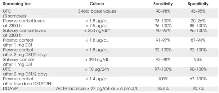
Several tests based on the understanding of the physio-logic characteristics of the hypothalamic– pituitary –adrenal axis have been used extensively to confirm the diagnosis of the Cushing syndrome, but none has proven fully capable of distinguishing all cases of CS from normal and/or pseudo-Cushing individuals. Among these, four diagnostic tests are currently used to screen for CS: measurement of free cortisol in 24-hour urine (UFC), cortisol suppressibility by low doses of dex-amethasone, and assessment of cortisol circadian rhythm using late-night serum and salivary cortisol levels (6) and the dexamethasone-CRH test. Table 1 shows the diag-nostic criteria and sensitivities and specificities for these tests.
Measurement of free cortisol in 24-hour urine
mL/min, UFC can be normal despite the presence of excessive cortisol production. In children, the urinary cortisol excretion should be corrected for body surface
area/1.72 m2. Due to the possibility of intermittent
hypercortisolism, up to three 24-h urine collections should be performed and if they are normal, CS is highly unlikely. On the other hand, values four-fold greater than the upper limit of normal strongly suggest Cushing’s syndrome (12). Milder elevations of urinary cortisol can be found in conditions such as chronic anxiety, depression, and alcoholism, all of which are also known as pseudo-Cushing states (14), and in normal pregnant women.
The reference range for urine free cortisol levels depends on the type of assay used (15). Measurement of urinary cortisol by immunoassays (RIA or
immuno-metric assays; normal range: < 80–120 µg/24h or <
220–330 nmol/24h) is influenced by various metabo-lites of cortisol and some synthetic glucocorticoids, whereas measurements using high-performance liquid chromatography (HPLC) allow the separation of vari-ous urinary glucocorticoids and metabolites (normal
range: < 50 µg/24h or < 138 nmol/24h). Thus,
HPLC or gas chromatography coupled with mass spectrometry provides the best specificity for measur-ing UFC (16,17) and is replacmeasur-ing older immunoassay methods because of its higher specificity. Differences in assay methodology may make interpretation of the data very challenging. Although HPLC has a high sen-sitivity and specificity, occasionally interfering
sub-stances, such as carbamazepine and digoxin, can also coelute with cortisol and produce false elevations of the UFC (18). The sensitivity of UFC for Cushing’s syndrome may be only 45–71% at 100% specificity (19,20). Furthermore, pseudo-Cushing’s states may have false-positive UFC testing (21,22). Therefore, although UFC may be useful to confirm Cushing’s syndrome, its sensitivity and specificity are not optimal as an initial screening test and it cannot be considered as a universal single screening test for the detection of CS (6,23).
Low-dose dexamethasone suppression tests (DST)
Two dexamethasone-suppression tests (DST) are use for screen CS, the overnight and the 48-h DST. These tests depend on the concept that dexamethasone will suppress ACTH and cortisol release in normal sub-jects, while patients with corticotroph adenomas will not suppress below a specified cut-off. The overnight low-dose (1 mg) DST consists of the oral intake of 1 mg dexamethasone between 2300 and 2400 h, fol-lowed by measurement of fasting plasma cortisol between 0800 and 0900 h in the following morning. In the 48-h test, dexamethasone is given at the dose of 0.5 mg every 6 h for 2 days at 0900 h, 1500 h, 2100 h, and 0300 h with measurements of cortisol in serum at 0900 h at the start and end of the test. The original criterion for normal level of suppression was a plasma cortisol level below 5 µg/dL (138 nmol/liter) (24).
Table 1.Screening tests for Cushing’s syndrome: diagnostic criteria, sensitivities and specificities for each test.
Screening test Criteria Sensitivity Specificity
UFC 3-fold basal values 90–98% 45–95%
(3 samples)
Plasma cortisol levels < 1.8 µg/dL 93–100% 20–26%
at 2300 h < 7.5 µg/dL 96–100% 88–100%
Salivary cortisol levels < 250 ng/dL* 90–96% 96–100%
at 2300 h
Plasma cortisol < 1.8 µg/dL 91–97% 87–94%
after 1 mg DST
Plasma cortisol < 1.8 µg/dL 92–100% 92–100%
after 2 mg DST/2 days
Salivary cortisol < 390 ng/dL 93–98% 94%
after 1 mg DST
UFC < 10 µg/24h 97–100% 90–100%
after 2 mg DST/2 days
Plasma cortisol < 1.4 µg/dL 100% 67–100%
after low dose DST/CRH
DDAVP ACTH increase > 27 pg/mL or > 6 pmol/L 86.8% 90.7%
UFC (Urinary free cortisol): normal range: <80–120 µg/24 h or <220–330 nmol/ 24h (RIA); <50 µg/24 h or <138 nmol/24h (HPLC).
* Cut-off value (mean) based on several available studies (95%CI 153–346 ng/dL).
DST: dexamethasone suppression test, CRH: corticotrophin releasing hormone, DDAVP: Desmopressin. To convert plasma cortisol from µg/dl to nmol/L, multiply by 27.6.
More recently, this cut-off level has been reduced to less than 1.8 µg/dL (50 nmol/liter) (2), greatly enhancing the sensitivity of the overnight DST, espe-cially in patients with mild hypercortisolism. However, the cortisol assay must have a high sensitivity (1 µg/dL or 27.6 nmol/liter).
There are many interfering conditions in DST: decreased dexamethasone absorption, drugs enhanc-ing hepatic dexamethasone metabolism, inducenhanc-ing the cytochrome P450-related enzymes (barbiturates, phenytoin, carbamazepine, rifampicin, meprobamate, aminoglutethimide, methaqualone), increased con-centration of CBG (estrogen treatment, pregnancy) and pseudo-Cushing states (6). The 48-h test, although more cumbersome than the overnight test, is more specific. Because the 1 mg overnight DST is easy to perform and has low cost it is used as a first-line screening test in outpatient screening.
Some patients with Cushing’s disease (3–8%) retain sensitivity to dexamethasone and show suppres-sion of serum cortisol (25,26). Additionally, DST has a significant false positive rate, especially in chronically ill (23%) or obese patients (13%) and in patients with major depression (43%) or other psychiatric disorders (8–41%). A false-positive rate of up to 30% has been reported in other admitted patients and healthy indi-viduals (27-29). Therefore, because of many interfer-ing conditions and the significant variability of the bio-logical behavior of corticotroph adenomas, neither the overnight nor 2-day DST test appears to be able to reliably rule out Cushing’s syndrome using standard cut-offs for serum cortisol (12).
Late-night plasma cortisol
Physiological cortisol secretion is characterized by cir-cadian rhythmicity. Serum cortisol concentration reaches its zenith in the morning (0600–0800 h) and its nadir in the night during the first half of normal sleep. Normal circadian rhythm of cortisol secretion is lost in patients with Cushing’s syndrome and elevated late-night cortisol serum level has been reported to be the earliest and most sensitive marker of the disease (18). Thus, midnight serum cortisol has been demon-strated to be very effective in recognizing patients with mild signs and symptoms.
Newell-Price et al. (30) were the first authors to propose this screening test. They collected blood sam-ple in 150 patients with CS and in 20 healthy subjects, in a sleeping state. The test achieved 100% sensitivity using a cut-off value of 1.8 µg/dL (50 nmol/L) but the specificity was not tested. Other authors also assessed patients, but in a nonsleeping state, and found higher
cut-off values (7.5 µg/dL or 207 nmol/L) with good sensitivity (90.2–96%) and high specificity (96.5–100%) (21,31). Therefore, a single sleeping midnight plasma cortisol concentration of less than 1.8 µg/dL effective-ly excludes CS. Concentrations of more than 1.8 µg/dL are noted in individuals with CS and in patients with acute illness. An awake midnight concentration of cor-tisol in plasma of more than 7.5 µg/dL obtained in a nonsleeping state had 97% sensitivity to distinguish between CS and pseudo-CS in the largest series up to now reported but can miss mild disease diagnosis in about 7% of cases (21). More recently, Reimondo et al., (32) obtained 100% sensitivity with a threshold value of 1.5 µg/dL, but the specificity was only 68.2% at that level. The best cut-off value found with ROC analysis was 4.0 µg/dL, which is remarkably greater than that proposed by the recent consensus statement (6). Recently, in a large series of Cushing’s syndrome sus-pects, urinary free cortisol and cortisol suppression after 1 mg dexamethasone achieved satisfactory specificity. Conversely, serum cortisol concentrations at midnight should be interpreted with caution, especially with the 1.8 µg/dL cut-off (33). Besides controversial cut-off values, sleeping midnight blood sample collection for serum cortisol is not a stress-free sample and is not real-istic under normal conditions of clinical practice, which make this test suboptimal for most clinicians (32).
Late-night salivary cortisol
that is the presence of the cushingoid habitus associat-ed with alcohol intake, type 2 diabetes, and major depression (22,31). However, there are several other conditions such as hypertension, advanced age, and psychiatric diagnoses that may lead to false-positive late-night salivary cortisol results (45).
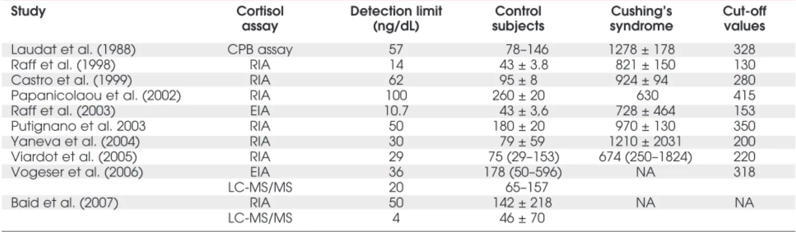
Diagnostic ranges vary between reports because of the different assays and the comparison groups used to set cut-off points. Table 2 shows an overview of late-night salivary cortisol studies conducted in adult controls and CS patients. In the majority of previous papers, salivary cortisol was measured by radioim-munoassay (RIA). Recently the U.S. Food and Drug Administration approved a new enzyme-linked immunosorbent assay (EIA) developed by Diagnostic Systems Laboratories (DSL) for salivary cortisol (46). Using this assay, Raff et al. (47) compared in the same sample the measurement of salivary cortisol obtained by this method and also by RIA. Saliva samples, col-lected at 2300 h, were sent to their laboratory to screen patients for Cushing’s syndrome. Other set of samples was obtained at 2300 h from healthy elderly and from apparently healthy individuals. The impor-tant finding was that EIA and RIA gave different results on the same sample. The EIA showed consis-tently higher salivary cortisol values than the RIA. The RIA results were much closer to the expected value of an independently created cortisol stock solution dilut-ed in saliva. These data suggest that salivary cortisol measured by RIA reflects more accurately the free level of circulating cortisol.
Another immunoassay has been commercialized from ROCHE Elecsys immunoassay system (Salimet-rics EIA) (48). The salivary cortisol measurement using this assay was also compared with the
measure-ment by the DPC RIA (46). The authors observed that Salimetrics EIA yielded results very close to those obtained with the DPC RIA, particularly in the critical diagnostic range of 0.3–10.0 nmol/L (11–357 ng/dL). These results are in contrast to those previ-ously obtained by EIA from DSL method (47). The only relevant disadvantage of the Salimetrics EIA was that the highest calibrator concentration (49.7 nmol/L) is considerably lower and frequent dilution of high-concentration samples from patients with Cushing’s syndrome are necessary. The advantages of the Salimetrics EIA compared with the DSL EIA are: it produces results not different from those obtained with the DPC RIA in the clinically important range, and it is FDA-cleared for in vitro diagnostic use.
More recently, liquid chromatography-tandem mass spectrometry (LC-MS/MS) measurement of sali-vary cortisol has been also available commercially (49,50). RIA and LC-MS/MS measurement of sali-vary cortisol and their accuracy of reference ranges were compared, in a cross-sectional prospective study of outpatients, in healthy volunteers and obese sub-jects with cushingoid features (49). This study demon-strated an important rate of abnormal bedtime salivary cortisol, measured in two different commercial assays and evaluated with laboratory provided normative ranges. The authors suggested that although a normal bedtime salivary cortisol result is useful to exclude Cushing’s syndrome, an abnormal salivary cortisol value should not be used alone to establish the diag-nosis of Cushing’s syndrome. In a recent paper, Liu et al. (45) evaluated salivary cortisol in 206 male veterans with an enzyme immunoassay whose normal range was based on 73 healthy lean subjects. In that report, 20% of all participants and 40% of diabetic, hypertensive
Table 2. Overview of studies conducted in control subjects and Cushing’s syndrome patients using late-night (2300 h) salivary cortisol.
Study Cortisol Detection limit Control Cushing’s Cut-off
assay (ng/dL) subjects syndrome values
Laudat et al. (1988) CPB assay 57 78–146 1278 ± 178 328
Raff et al. (1998) RIA 14 43 ± 3.8 821 ± 150 130
Castro et al. (1999) RIA 62 95 ± 8 924 ± 94 280
Papanicolaou et al. (2002) RIA 100 260 ± 20 630 415
Raff et al. (2003) EIA 10.7 43 ± 3,6 728 ± 464 153
Putignano et al. 2003 RIA 50 180 ± 20 970 ± 130 350
Yaneva et al. (2004) RIA 30 79 ± 59 1210 ± 2031 200
Viardot et al. (2005) RIA 29 75 (29–153) 674 (250–1824) 220
Vogeser et al. (2006) EIA 36 178 (50–596) NA 318
LC-MS/MS 20 65–157
Baid et al. (2007) RIA 50 142 ± 218 NA NA
LC-MS/MS 4 46 ± 70
CBP: competitive binding assay, RIA: radioimmunoassay, EIA: enzyme immunoassay, LC-MS/MS: liquid chromatography-tandem mass spectrometry; NA: not available.
subjects at least 60 yr of age had an abnormal 2300-h salivary cortisol level. At the time of publication, no participant had been diagnosed with Cushing’s syn-drome. Substantially lower concentrations were found with isotope dilution LC-MS/MS compared to RIA. Similar results were also observed comparing LC-MS/MS and the Roche Cobas cortisol immunoassay results (49,50).
Because salivary cortisol measurement is increa-sing in popularity, it is important to be aware of the different results generated by the commercially avail-able methods and to interpret the published reference intervals appropriately. Therefore, the normal refer-ence ranges are assay-dependent and should be vali-dated for each laboratory. Nevertheless, late-night sali-vary cortisol measurement can be recommended as a first-line diagnostic test for CS in both low-risk and high-risk (pseudo-Cushing states) patients. Given its convenience and diagnostic accuracy (table 1), with sensitivity and specificity of between 95% and 98%, this test may profitably be added to traditional screening procedures, such as UFC and 1 mg DST.
So far, the majority of bedtime salivary cortisol measurement data were obtained by RIA. A critical analysis of these RIA data (table 2) indicates that among several available studies the cut-off value of late-night salivary cortisol levels, reliable to segregate pseudo Cushing from CS, was 250 ± 104 ng/dL (mean ± SD, ranging from 130 to 415, 95%CI 153–346 ng/dL). Therefore, in the presence of bed-time salivary cortisol levels higher than 350 ng/dL, the diagnosis of CS is quite confident. On the other hand, in the presence of values lower than 150 ng/dL, the CS diagnosis is unlikely. However, if the bedtime salivary cortisol levels are in the gray area (> 150 and < 350 ng/dL), it is suggested that salivary cortisol sampling and other screening tests should be repeated. Until widely acceptable thresholds are generated, we suggest that each center should establish its own refer-ence range and cut-off points (12,23,31).
Other second-line diagnostic tests to screen for CS
The dexamethasone/corticotropin-releasing-hormone (DST-CRH) test was designed to take advantage of the physiologic impact of glucocorticoid-negative feedback on the hypothalamic–pituitary– adrenal axis, as well as the sensitivity of the pituitary adrenal system to hypothalamic stimulation with CRH. There are occasional patients in whom the screening tests described above are equivocal (figure 1). The com-bined DST-CRH test has been used in these
condi-tions. The DST-CRH test was first shown by the NIH center to be highly accurate in distinguishing CS from pseudo-CS (51), mainly mild Cushing’s disease from normal physiology (52).
The test is performed by giving dexamethasone orally 0.5 mg every 6 h for 48 h, starting at 1200 h, and then administering ovine-sequence CRH (1 µg/kg) iv at 0800 h (2 h after the last dose of dexamethasone). The plasma cortisol value 15 min after CRH is greater than 1.4 µg/dL (38 nmol/liter) in patients with CS, but remains suppressed in normal individuals and in patients with pseudo-CS (51). Criteria for a normal ACTH response have not been firmly established. The DST-CRH test is expensive and time-consuming. Cau-tion is needed in interpreting this test since it requires highly sensitive cortisol assays and the precision of the majority of cortisol assays in routine use at the this cut-off level is poor. Furthermore, abnormal DST-CRH testing has been found in patients with anorexia nervosa (53) suggesting that non-Cushing’s related increases in hypothalamic-pituitary adrenal axis activity will lead to false positive results.
Because desmopressin (1-deamino-8D-arginine vasopressin, DDAVP), a vasopressin analogue, stimu-lates ACTH release in patients with CS but not in the majority of normal, obese, and depressed subjects, it has been used to investigated its ability to discriminate CS from pseudo-Cushing states (54,55). In these studies, a plasma peak ACTH increase equal to or greater than 6 pmol/L (27 pg/mL), indicated by ROC analysis, allowed the best discrimination between patients with CS and the other groups, with a sensitivity of 86.8%, a specificity of 90.7%, and a diagnostic accuracy of 89%. Therefore, when doubt remains about diagnosis, the DST-CRH test and the
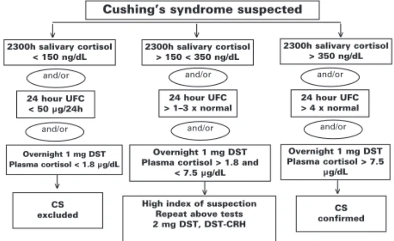
Figure 1.Algorithm for establishing the diagnosis of Cush-ing’s syndrome. DST: dexamethasone suppression test, UFC: urinary free cortisol, DST-CRH: dexamethasone-corti-cotropin-releasing hormone test.
Cushing’s syndrome suspected
2300h salivary cortisol < 150 ng/dL
2300h salivary cortisol > 150 < 350 ng/dL
2300h salivary cortisol > 350 ng/dL
24 hour UFC < 50 µg/24h
24 hour UFC > 1–3 x normal
24 hour UFC > 4 x normal
Overnight 1 mg DST Plasma cortisol < 1.8 µg/dL
Overnight 1 mg DST Plasma cortisol > 7.5
µg/dL
CS
excluded confirmedCS
High index of suspection Repeat above tests 2 mg DST, DST-CRH Overnight 1 mg DST Plasma cortisol > 1.8 and
< 7.5 µg/dL
and/or and/or and/or
and/or and/or and/or
↓ ↓ ↓
desmopressin test can be used. However, their diag-nostic accuracy needs further validation.
In conclusion, different approaches have been developed over the years, but all of them have demon-strated some limitations, and none of the proposed screening tests can detect all cases of CS. The three commonly performed diagnostic tests for CS — UFC, low doses DST, and late-night plasma or salivary corti-sol — are complementary. Figure 1 summarizes a sug-gested approach to screening for Cushing’s syndrome. In patients with mild or intermittent hypercortisolism, any of these tests may yield “normal” results and be mis-leading. Because of the high sensitivity and ease with which repeated measurements can be performed, late-night salivary cortisol appears to be the most useful screening test. UFC and DST should be performed to provide further confirmation of the diagnosis.
REFERENCES
1. Orth DN. Cushing’s syndrome. N Engl J Med 1995 ;332:791-803.
2. Newell-Price J, Trainer P, Besser GM, Grossman AB. The diagnosis and differential diagnosis of Cushing’s syndrome and pseudo-Cushing’s states. Endocr Rev 1998;19:647-72. 3. Atkinson AB, McCance DR, Kennedy L, Sheridan B. Cyclical
Cushing’s syndrome first diagnosed after pituitary sur-gery: a trap for the unwary. Clin Endocrinol 1992; 36:297-379.
4. Arnaldi G, Mancini T, Kola B, Appolloni G, Freddi S, Concet-toni C, et al. Cyclical Cushing’s syndrome in a patient with a bronchial neuroendocrine tumor (typical carcinoid) express-ing ghrelin and growth hormone secretagogue receptors. J Clin Endocrinol Metab 2003;88:5834-40.
5. Gunther DF, Bourdeau I, Matyakhina L, Cassarino D, Kleiner DE, Griffin K, et al. Cyclical Cushing syndrome presenting in infancy: an early form of primary pigmented nodular adreno-cortical disease, or a new entity? J Clin Endocrinol Metab 2004;89:3173-82.
6. Arnaldi G, Angeli A, Atkinson AB, Bertagna X, Cavagnini F, Chrousos GP, et al. Diagnosis and complications of Cushing’s syndrome: a consensus statement. J Clin Endocrinol Metab 2003;88:5593-602.
7. Leibowitz G, Tsur A, Chayen SD, Salameh M, Raz I, Cerasi E. Pre-clinical Cushing’s syndrome: an unexpected frequent cause of poor glycaemic control in obese diabetic patients.
Clin Endocrinol 1996;44:717-22.
8. Contreras LN, Cardoso E, Lozano MP, Pozzo J, Pagano P, Claus-Hermbeg H. Deteccion de sindrome de Cushing preclınico en pacientes con sobrepeso y diabetes mellitas tipo 2. Medicina 2000;60:326-30.
9. Reincke M, Nieke J, Krestin GP, Saeger W, Allolio B, Winkel-mann W. Preclinical Cushing’s syndrome in adrenal “inci-dentalomas”: comparison with adrenal Cushing’s syndrome.
J Clin Endocrinol Metab 1992;75:826-32.
10. Terzolo M, Pia A, Ali A, Osella G, Reimondo G, Bovio S. Adrenal incidentaloma: a new cause of the metabolic syn-drome? J Clin Endocrinol Metab 2002;87:998-1003. 11. Catargi B, Rigalleau V, Poussin A, Ronci-Chaix N, Bex V,
Vergnot V, et al. Occult Cushing’s syndrome in type-2 dia-betes. J Clin Endocrinol Metab 2003;88:5808-13. 12. Newell-Price J, Bertagna X, Grossman AB, Nieman LK.
Cush-ing’s syndrome. Lancet 2006;367(9522):1605-17.
13. Kaye TB, Crapo L. The Cushing syndrome: an update on diag-nostic tests. Ann Intern Med 1990;112:434-44.
14. Boscaro M, Barzon L, Sonino N. The diagnosis of Cushing’s syndrome: atypical presentations and laboratory shortcom-ings.Arch Intern Med 2000;160:3045-53.
15. Murphy BE. Urinary free cortisol determinations: what they measure. Endocrinologist 2002;12:143-50.
16. Turpeinen U, Markkanen H, Valimaki M, Stenman UH. Deter-mination of urinary free cortisol by HPLC. Clin Chem 1997;43:1386-91.
17. Lin CL, Wu TJ, Machacek DA, Jiang NS, Kao PC. Urinary free cortisol and cortisone determined by high performance liquid chromatography in the diagnosis of Cushing’s syndrome. J Clin Endocrinol Metab 1997;82:151-5.
18. Findling JW, Raff H. Newer diagnostic techniques and prob-lems in Cushing’s disease.Endocrinol Metab Clin North Am 1999;28:191-210.
19. Gorges R, Knappe G, Gerl H, Ventz M, Stahl F. Diagnosis of Cushing’s syndrome: re-evaluation of midnight plasma corti-sol vs. urinary free corticorti-sol and low-dose dexamethasone suppression test in a large patient group. J Endocrinol Invest 1999;22:241-9.
20. Viardot A, Huber P, Puder JJ, Zulewski H, Keller U, Muller B. Reproducibility of nighttime salivary cortisol and its use in the diagnosis of hypercortisolism compared with urinary free cortisol and overnight dexamethasone suppression test. J Clin Endocrinol Metab 2005;90:5730-6.
21. Papanicolaou DA, Yanovski JA, Cutler GB, Chrousos GP, Nie-man LK. A single midnight cortisol measurement distin-guishes Cushing’s syndrome from pseudo-Cushing’s states.
J Clin Endocrinol Metab 1998;83:1163-7.
22. Papanicolaou DA, Mullen N, Kyrou I, Nieman LK. Nighttime salivary cortisol: a useful test for the diagnosis of Cushing’s syndrome.J Clin Endocrinol Metab 2002;87:455-521. 23. Findling JW, Raff H. Cushing’s Syndrome: important issues in
diagnosis and management. J Clin Endocrinol Metab 2006;91:3746-53.
24. Liddle GW. Tests of pituitary-adrenal suppressibility in the diagnosis of Cushing’s syndrome. J Clin Endocrinol Metab 1960;20:1539-60.
25. Isidori AM, Kaltsas GA, Mohammed S, Morris DG, Jenkins P, Chew SL. Discriminatory value of the low-dose dexametha-sone suppression test in establishing the diagnosis and dif-ferential diagnosis of Cushing’s syndrome. J Clinical Endocrinol Metab 2003;88:5299-306.
26. Odagiri E, Naruse M, Terasaki K, Yamaguchi N, Jibiki K, Takagi S, et al. The diagnostic standard of preclinical Cushing’s syn-drome: evaluation of the dexamethasone suppression test using various cortisol kits. Endocrine J 2004;51:295-302. 27. Chriguer RS, Elias LL, da Silva IM Jr, Vieira JG, Moreira AC,
de Castro M. Glucocorticoid sensitivity in young healthy indi-viduals: in vitro and in vivo studies. J Clin Endocrinol Metab 2005;90:5978-84.
28. Crapo L. Cushing’s syndrome: a review of diagnostic tests.
Metabolism 1979;28:955-77.
29. Findling JW, Raff H, Aron DC. The low-dose dexamethasone suppression test: a reevaluation in patients with Cushing’s syndrome.J Clin Endocrinol Metab 2004;89:1222-6. 30. Newell-Price J, Trainer P, Perra L, Wass J, Grossman A,
Bess-er M. A single sleeping midnight cortisol has 100% sensitivi-ty for the diagnosis of Cushing’s syndrome. Clin Endocrinol 1995;43:545-50.
31. Putignano P, Toja P, Dubini A, Pecori Giraldi F, Corsello SM, Cavagnini F. Midnight salivary cortisol versus urinary free and midnight serum cortisol as screening tests for Cushing’s syndrome. J Clin Endocrinol Metab 2003;88:4153-7. 32. Reimondo G, Allasino B, Bovio S, Paccotti P, Angeli A,
Terzo-lo M. Evaluation of the effectiveness of midnight serum corti-sol in the diagnostic procedures for Cushing’s syndrome. Eur J Endocrinol 2005;153:803-9.
33. Giraldi FP, Ambrogio AG, De Martin M, Fatti LM, Scacchi M, Cavagnini F. Specificity of first-line tests for the diagnosis of Cushing’s syndrome. Assessment in a large series. J Clin Endocrinol Metab 2007;[Epub ahead of print]
35. Umeda T, Hiramatsu R, Iwaoka T, Shimada T, Miura F, Sato T. Use of saliva for monitoring unbound free cortisol levels in serum. Clin Chim Acta 1981;110:245-53.
36. Santiago LB, Jorge SM, Moreira AC. Longitudinal evaluation of the development of salivary cortisol circadian rhythm in infancy.Clin Endocrinol 1996;44:157-61.
37. Luthold WW, Marcondes JAM, Wajchenberg BL. Salivary cor-tisol for the evaluation of Cushing’s syndrome. Clin Chim Acta 1985;151:33-9.
38. Laudat MH, Cerdas S, Fournier C, Guiban D, Guilhaume B, Luton JP. Salivary cortisol measurement: a practical approach to assess pituitary-adrenal function. J Clin Endocrinol Metab 1988;66:343-8.
39. Raff H, Raff JL, Findling JW. Late-night salivary cortisol as a screening test for Cushing’s syndrome. J Clin Endocrinol Metab 1998;83:2681-6.
40. Castro M, Elias PCL, Quidute ARP, Halah FP, Moreira AC. Out-patient screening for Cushing’s syndrome: the sensitivity of the combination of circadian rhythm and overnight dexam-ethasone suppression salivary cortisol tests. J Clin Endocrinol Metab 1999;84:878-82.
41. Trilck M, Flitsch J, Ludecke DK, Jung R, Petersenn S. Salivary cortisol measurement — a reliable method for the diagnosis of Cushing’s syndrome. Exp Clin Endocrinol Diabetes 2005;113:225-30.
42. Yaneva M, Mosnier-Pudar H, Dugue M-A, Grabar S, Fulla Y, Bertagna X. Midnight salivary cortisol for the initial diagnosis of Cushing’s syndrome of various causes. J Clin Endocrinol Metab 2004;89:3345-51.
43. Martinelli CE, Sader SL, Oliveira EB, Daneluzzi JC, Moreira AC. Salivary cortisol for screening of Cushing’s syndrome in children. Clin Endocrinol 1999;51:67-71.
44. Gafni RI, Papanicolaou DA, Nieman LK. Nighttime salivary cortisol measurement as a simple, noninvasive, outpatient screening test for Cushing’s syndrome in children and ado-lescents.J Pediatr 2000;137:30-5.
45. Liu H, Bravata DM, Cabaccan J, Raff H, Ryzen E. Elevated late-night salivary cortisol levels in elderly male type 2 diabetic veterans. Clin Endocrinol 2005;63:642-9.
46. Raff H, Homar PJ, Skoner DP. New enzyme immunoassay for salivary cortisol [Letter].Clin Chem 2003;49:203-4. 47. Raff H, Homar PJ, Burns EA. Comparison of two methods for
measuring salivary cortisol [Letter]. Clin Chem 2002;48:207-8.
48. Chiu SK, Collier CP, Clark AF, Wynn-Edwards KE. Salivary cortisol on ROCHE Elecsys immunoassay system: pilot bio-logical variation studies. Clin Biochem 2003;36:211-4. 49. Baid SK, Sinaii N, Wade M, Rubino D, Nieman LK.
Radioim-munoassay and tandem mass spectrometry measurement of bedtime salivary cortisol levels: a comparison of assays to establish hypercortisolism. J Clin Endocrinol Metab 2007;92:3102-7.
50. Vogeser M, Durner J, Seliger E, Auernhammer C. Measure-ment of late-night salivary cortisol with an automated immunoassay system. Clin Chem Lab Med 2006;44:1441-5. 51. Yanovski JA, Cutler GB Jr., Chrousos GP, Nieman LK. Corti-cotropin-releasing hormone stimulation following low-dose dexamethasone administration. JAMA 1993;269:2232-8. 52. Yanovski JA, Cutler GB Jr., Chrousos GP, Nieman LK. The
dex-amethasone suppressed corticotropin-releasing hormone stimulation test differentiates mild Cushing’s disease from nor-mal physiology. J Clin Endocrinol Metab 1998;83:348-52. 53. Duclos M, Corcuff J-B, Rober P, Tabarin A. The
dexametha-sone-suppressed corticotrophin-releasing hormone stimulation test in anorexia nervosa. Clin Endocrinol 1999;51:725-31. 54. Coiro V, Volpi R, Capretti L, Caffarri G, Chiodera P.
Desmo-pressin and hexarelin tests in alcohol-induced pseudo-Cush-ing’s syndrome. J Intern Med 2000;247:667-73.
55. Moro M, Putignano P, Losa M, Invitti C, Maraschini C, Cav-agnini F. The desmopressin test in the differential diagnosis between Cushing’s disease and pseudo-Cushing states. J Clin Endocrinol Metab 2000;85:3569-74.
Address for correspondence:
Margaret de Castro
Departamento de Medicina Interna
Escola de Medicina de Ribeirão Preto — USP Av. dos Bandeirantes 3900
14049-900 Ribeirão Preto, SP Fax: (16) 3633-6695