Expression of P16 and PDGFR-Beta in gastric adenocarcinoma
Expression of P16 and PDGFR-Beta in gastric adenocarcinoma
Expression of P16 and PDGFR-Beta in gastric adenocarcinoma
Expression of P16 and PDGFR-Beta in gastric adenocarcinoma
Expression of P16 and PDGFR-Beta in gastric adenocarcinoma
Expressão do P16 e do PDGFR-Beta no adenocarcinoma gástrico
Expressão do P16 e do PDGFR-Beta no adenocarcinoma gástrico
Expressão do P16 e do PDGFR-Beta no adenocarcinoma gástrico
Expressão do P16 e do PDGFR-Beta no adenocarcinoma gástrico
Expressão do P16 e do PDGFR-Beta no adenocarcinoma gástrico
RODRIGO POZZA PINTO1; FERNANDO KREBS CIRNE LIMA2; JANE M U KULKZYNSKI3; LUIS FERNANDO MOREIRA4
A B S T R A C T A B S T R A C T A B S T R A C T A B S T R A C T A B S T R A C T
Objective Objective Objective Objective
Objective: To detect immunohistochemistry expression of p16 and PDGFR-beta on gastric adenocarcinoma. Methods:Methods:Methods:Methods:Methods: Thirty six patients submitted to surgery for gastric adenocarcinoma between 1998 and 2002 at Santa Casa de Porto Alegre Hospital have been studied. Variables investigated were: age, gender, tumour size and localization, number of dissected and metastatic nodes, histological type, surgical resection extension and pathological staging. Results:Results:Results:Results:Results: No expression of PDGFR-beta has been detected on surgical specimens. Concerning to p16, loss of expression lower than 10% and 1% has been detected respectively on 89% and 79% of the specimens studied. ConclusionConclusionConclusionConclusionConclusion: There has been no correlation among p16 loss and variables studied.
Key words Key words Key words Key words
Key words: Stomach Neoplasms . Adenocarcinoma. Genes p16. Immunohistochemistry.
INTRODUCTION
INTRODUCTION
INTRODUCTION
INTRODUCTION
INTRODUCTION
G
astric adenocarcinoma has been the main cause of cancer death during most of the 20th century, now overcame by lung cancer. Annually 750,000 new cases are diagnosed. Great geographic variations are seen and highest incidences can be found in Japan, South America, Eastern Europe and Middle East1. It is twice as frequent inmen as in women1,2, has a low incidence before the 4th
decade with a peak incidence in the 7th.1 In Brazil 23,000
new cases and 11,000 deaths are estimated to occur in 20053.
The prognosis of gastric adenocarcinoma is poor, mainly because lack of symptomatology and late diagnosis, with an overall survival of 5-15% in five years1,4. In Japan,
where this disease is endemic but diagnosis is usually done at an early stage due to wide endoscopic availability, survival rate is 50% in five years1. Complete resection of all gross
and microscopic disease is the only potentially curative treatment. However, disease recurs in 80% of patients even after curative resection1.
Oncogene p16 is implicated in pathogenesis of many human tumours and even in regulation of normal cellular growth, together with cycling, tyrosine kinases and tumour transforming and growth factors, like TGF-alpha and -beta and platelet derived growth factors (PDGF) ligands and receptors (alpha and beta). Inherited mutations of p16 are associated with hereditary melanomas. Deletions and acquired inactivation of p16 are found in 75% of pancreatic carcinomas, 40-70% of glioblastomas, 50% of oesophageal carcinomas and 20% of non-small
cell lung cancer5. Recent papers have demonstrated
relation between p16 inactivation and development of stomach cancer. Forty to ninety per cent of gastric adenocarcinomas show inactivation of p16,6-11 appearing
to have relation with cellular differentiation7,10,12. Expression
of p16 is decreased in node metastasis6. Apparently there
is no difference in expression between intestinal and diffuse-type gastric adenocarcinoma12.
Platelet derived growth factor receptor-beta (PDGFR-beta), a tyrosine kinase surface receptor, is important in growth, differentiation and cell death controls13,14.PDGFR has been found activated and mutated
in gastric stromal tumour where c-KIT, the most commonly marker found, is in wild type15,16. PDGF receptors
act over stromal origin cells and are not expressed in epithelial cells under normal physiologic conditions17.
Expression of PDGFR has been described in dermatofibromiosarcoma, chronic myelocytic leukemia and in gastrointestinal stromal tumours (GISTs),18,19 besides
other solid tumors like glioblastomas and prostate cancer13,18. PDGF-beta and its receptor have not been
studied concerning expression and response to cellular growth inhibitors on non-stromal gastric tumours.
METHODS
METHODS
METHODS
METHODS
METHODS
Thirty six patients submitted to surgery for gastric adenocarcinoma between 1998 and 2002 at Santa Casa de Porto Alegre Hospital have been studied with the aim to determine prevalence of p16 and PDGFR-beta.
From the Post-Graduation Program of Surgery, Faculty of Medicine, Federal University of Rio Grande do Sul and Hospital de Clínicas de Porto Alegre University Hospital, Porto Alegre, RS, BR.
None of the 36 patients had past history of any other malignant tumours (except to skin squamous and basal cell tumours), of pre-operative chemo or radiation therapy, which would exclude them from the study.
Variables investigated were: age, gender, tumour size and localization, number of dissected and metastatic nodes, histological type, extent of surgical resection and pathological staging. Clinical and pathological data were collected from patient charts as well as from surgical reports.
Surgical specimens analysis (haematoxilin-eosin, HE) included assessment of depth of tumour invasion on the gastric wall, nodal metastasis, histological grade and histological type (intestinal or diffuse, according to Lauren’s classification).
Immunohistochemistry Immunohistochemistry Immunohistochemistry Immunohistochemistry Immunohistochemistry
Specimens were processed according to routine of Pathology Department of Clinicas Hospital of Porto Ale-gre. Mouse monoclonal antibody anti-p16ink4a
(DakoCytomation, Carpinteria, California, USA) and rabbit polyclonal antibody anti-PDGFR beta Ab-1 (DakoCytomation, Carpinteria, California) were used to IH valuation, diluted on PBS saline solution at 1:100 and 1:75, respectively. Positivity was determined with ABS method (streptavidin-biotin-peroxidase complex; LABS+System HRP, DakoCytomation, Carpinteria, California, USA).
Slide sample analysis Slide sample analysis Slide sample analysis Slide sample analysis Slide sample analysis
Up to 20 high-power fields (400X) of each sample were captured to computer (Image Pro-Plus, v 4.5.1.2.2, Media Cybernetics). On computer screen, after insertion of a grid over pictures, total number of cancer cells and total number of immunoreactive cancer cells were counted. Nuclear brown staining over cytoplasmic staining was considered to be positive for p16. Lung cancer samples were used as outer positive controls. Percentage of p16 positivity was calculated dividing the number of stained cancer cells by the total number of cancer cells, and then, multiplied by 100. Two thresholds were regarded as loss of expression: <10% and <1%. A qualitative scale was determined to PDGFR-beta, according to staining intensity: (1) as no staining, (2) weak, (3) moderately and (4) strong staining. Breast cancer samples were used as outer positive controls. To be considered positive the sample should have at least 10% of moderately or strong stained cancer cells.
Statistical analysis Statistical analysis Statistical analysis Statistical analysis Statistical analysis
Qualitative data was described as media and standard deviation. Percentage and frequency were used as categorical variables. Fischer’s exact test was used comparing p16 expression to gender and histological type. Student’s t test was used comparing p16 to size, age and number of metastatic nodes. Pathological stage was compared to p16 using Chi-Square Test (P value <0.05 to all tests). Statistical
analysis was performed with Excel Microsoft Office 2003.
RESULTS
RESULTS
RESULTS
RESULTS
RESULTS
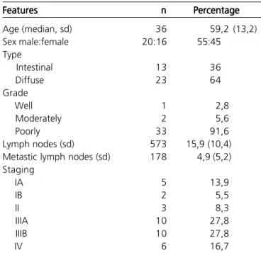
Thirty six patients with gastric adenocarcinoma were studied; 20 (55.5%) males and 16 (44.5%) females (Table 1). Overall median age was 59.2 (13.2); men median age was 59.2 (10.7) and women 59.3 (16.6). The difference was not statistically significant.
Tumour location was as follows: 2 (5.5%) cardia, 4 (11.1%) cardia and fundus, 5 (13.9%) body and fundus, 1 (2.8%) body, 2 (5.5%) body and antrum, 19 (52.8%) antrum and 3 (8.3%) pylorus. Twenty three (63.9%) patients were submitted to partial gastrectomy, 11 (33.3%) to total gastrectomy and 2 (5.5%) to oesophago-gastrectomy. The median (sd) number of lymph nodes dissected was 15.9 (10.4), among this, 5 (5.2) showed metastatic disease (30.8%). Analysis of extent of lymph node dissection data was not possible due to lack of this information in most of surgical records (Table 2).
Concerning to Lauren classification 64% was diffuse type and 36% was intestinal type. Histological grade included 91.6% of poorly differentiated, 5.6% moderately and 2.8% well-differentiated tumours. Pathological staging, according to TNM 6th edition
demonstrated 13.9%, 5.5%, 8.3%, 27.8%, 27.8%, and 16.7% for IA, IB, II, IIIA, IIIB and stage IV, respectively.
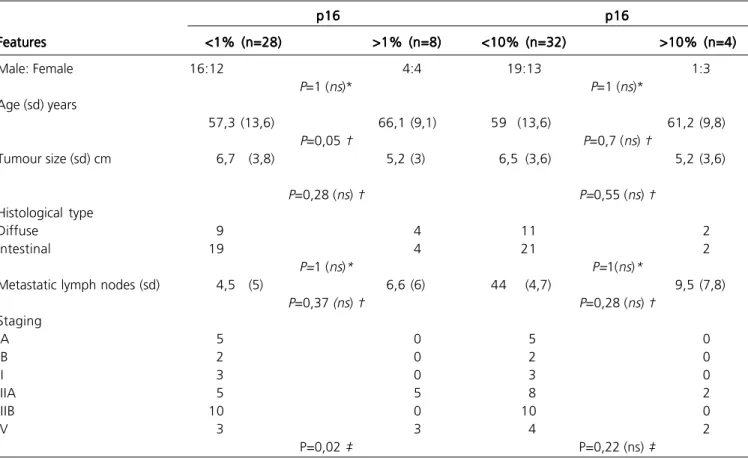
P16 analysis showed positivity lower than 10% in 32 (89%) patients and lower than 1% in 28 (78%). Positivity to p16 was compared (10% and 1%
Table 1 -Table 1 -Table 1 Table 1
-Table 1 - Patient clinical and pathological characteristics.
Features FeaturesFeatures Features
Features nnnnn PercentagePercentagePercentagePercentagePercentage
Age (median, sd) 36 59,2 (13,2)
Sex male:female 20:16 55:45
Type
Intestinal 13 36
Diffuse 23 64
Grade
Well 1 2,8
Moderately 2 5,6
Poorly 33 91,6
Lymph nodes (sd) 573 15,9 (10,4)
Metastic lymph nodes (sd) 178 4,9 (5,2) Staging
IA 5 13,9
IB 2 5,5
II 3 8,3
IIIA 10 27,8
IIIB 10 27,8
IV 6 16,7
respectively) to age, gender, tumour size, metastatic nodes, pathological staging and Lauren histological type and these results are presented on table 2. There was no statistical difference in all but age and tumour stage for a p16 loss when comparing >1% to <1% expressions (Table 2).
DISCUSSION
DISCUSSION
DISCUSSION
DISCUSSION
DISCUSSION
It was a random decision to study simultaneously p16, which loss of expression has been exhaustively studied in gastric tumours and PDGFR-beta, which association to gastric adenocarcinoma has not yet been found in medical literature. Although there is not a close relation between both markers, it could be speculated that overexpression of PDGFR-beta in cancer cells, increasing mitogenic stimuli to cell could be associated to loss of activity of p16, which is one of the important cell cycle inhibitors. Besides, promising results in treatment of these patients with tyrosine kinase inhibitors, such as imatinib (former known as STI571, Gleevec, Novartis Pharmaceutical Corp, East Hanover, NJ, USA) have been reported for gastrointestinal stromal tumours (GISTs) in which PDGFR is overexpressed13,15,20,21.
In a gastric carcinoma animal model, it was demonstrated increase in antitumour and cytotoxic effects of 5-FU and paclitaxel when combined with imatinib22.
In this study, 89% of cases had loss of expression of p16 (positivity lower than 10%) which is similar to literature data of 40%-90% loss3,6,7. Seventy eight percent of the cases
showed positivity lower than 1%. This lower threshold was chosen to determine the power of p16 loss expression and the studied variables.
Expression of PDGFR-beta was undetected in all of the 36 cases studied. The association between this marker and gastric adenocarcinoma have not been found in medical literature, although its expression (PDGFR alpha and beta) has been found in other epithelial tumours such as cholangiocarcinoma23, ovary24 and breast cancer25.
There was no statistical difference between median ages for both expressions, in contrast to american reports, where women tend to be older.1 Like other reports,6
this paper did not find relation of p16 expression with either gender or age.
Concerning to Lauren histological type, against expectations the diffuse type was predominant,1,26-28 no
statistical difference between Lauren histological type and p16 expression in the two thresholds studied was found as previously reported,12 although no agreement on this issue
have been reached7,27.
Although it is known that tumours with p16 loss tend to have a higher metastatic potential immunohistochemistry to p16 in lymph nodes was not carried out in this study, since overall p16 expression is much lower in metastatic lymph nodes than in primary lesion6.
Table 2 -Table 2 -Table 2
-Table 2 -Table 2 - P16 loss according to clinical and pathological features.
p16 p16p16 p16
p16 p16p16p16p16p16
Features FeaturesFeatures
FeaturesFeatures <1% (n=28)<1% (n=28)<1% (n=28)<1% (n=28)<1% (n=28) >1% (n=8)>1% (n=8)>1% (n=8)>1% (n=8)>1% (n=8) <10% (n=32)<10% (n=32)<10% (n=32)<10% (n=32)<10% (n=32) >10% (n=4)>10% (n=4)>10% (n=4)>10% (n=4)>10% (n=4)
Male: Female 16:12 4:4 19:13 1:3
P=1 (ns)* P=1 (ns)*
Age (sd) years
57,3 (13,6) 66,1 (9,1) 59 (13,6) 61,2 (9,8)
P=0,05 † P=0,7 (ns) †
Tumour size (sd) cm 6,7 (3,8) 5,2 (3) 6,5 (3,6) 5,2 (3,6)
P=0,28 (ns) † P=0,55 (ns) †
Histological type
Diffuse 9 4 11 2
Intestinal 19 4 21 2
P=1 (ns)* P=1(ns)*
Metastatic lymph nodes (sd) 4,5 (5) 6,6 (6) 44 (4,7) 9,5 (7,8)
P=0,37 (ns) † P=0,28 (ns) †
Staging
IA 5 0 5 0
IB 2 0 2 0
II 3 0 3 0
IIIA 5 5 8 2
IIIB 10 0 10 0
IV 3 3 4 2
P=0,02 ‡ P=0,22 (ns) ‡
R E S U M O R E S U M O R E S U M O R E S U M O R E S U M O
Objetivo: Objetivo: Objetivo: Objetivo:
Objetivo: Detectar a expressão imunoistoquímica do p16 e do PDGFR-beta no adenocarcinoma gástrico. Método: Método: Método: Método: Método: Foram estudados 36 pacientes submetidos a cirurgia para adenocarcinoma gástrico entre 1998 e 2002 no Hospital da Santa Casa de Porto Alegre. As variáveis investigadas foram: idade, sexo, tamanho e localização do tumor, número de linfonodos dissecados, número de linfonodos metastáticos, tipo histológico, extensão da ressecção cirúrgica e estadiamento patológico. Resultados: Resultados: Resultados: Resultados: Resultados: Não foi detectada expressão do PDGFR-beta nas peças cirúrgicas. Em relação ao p16, detectou-se perda de expressão menor que 10% e menor que 1% respectivamente em 89% e 79% das peças estudadas. Conclusão: . Conclusão: . Conclusão: . Conclusão: . Conclusão: Não houve correlação entre a perda de p16 e as variáveis estudadas.
Descritores: Descritores: Descritores: Descritores:
Descritores: Neoplasias gástricas. Adenocarcinoma. Genes, p16. Imunoistoquímica.
REFERENCES
REFERENCES
REFERENCES
REFERENCES
REFERENCES
1. De Vita VT Jr, Hellman S, Rosenberg AS. Cancer – Principles and Practice of Oncology. 6th Edition. Philadelphia: Lippincott Willians
and Wilkins; 2001.
2. AICR – American Institute for Cancer Research. World Cancer Research Fund. Food, nutrition and the prevention of cancer: a global perspective. Washington, DC: Banta Book Group; 1997. 3. INCA – Instituto Nacional do Câncer. Estimativa 2005 – Incidência
de Câncer no Brasil. Rio de Janeiro, 2005.
4. Brien TP, Depowski MD, Sheehan CE, Ross JS, McKeenna BJ. Prognostic factors in gastric cancer. Mod Pathol 1998; 11:870-7. 5. Wistuba II, Gazdar AF, Minna JD. Molecular genetics of small cell lung carcinoma. Semin Oncol. 2001 Apr;28(2 Suppl 4):3-13
6. He XS, Su Q, Chen ZC, et al. Expression, deletion and mutation of p16 gene in human gastric cancer. World J Gastroenterol 2001 Aug; 7(4): 515-21.
7. Zhao GH, Li TC, Shi LH, et al. Relationship between inactivation of p16 gene and gastric carcinoma. World J Gastroenterol 2003 May; 9(5): 905-9.
8. Kanyama Y, Hibi K, Nakayama H, et al. Detection of p16 promoter hypermethylation in serum of gastric cancer patients. Cancer Sci 2003 May; 94(5): 418-20.
9. Ficorella C, Cannita K, Ricevuto E, et al. P16 hypermethylation contributes to the characterization of gene inactivation profiles in primary gastric cancer. Oncol Rep 2003 Jan-Feb; 10(1): 169-73. 10. Tang S, Luo H, Yu J, et al. Relationship between alterations of
p16(INK4a) and p14(ARF) genes of CDKN2A locus and gastric cancer. Chin Med J 2003 Jul; 116(7): 1083-7.
11. Lee HS, Lee HK, Kim HS, et al. Tumor suppressor gene expression correlates with gastric cancer prognosis. J Pathol 2003 May; 200(1): 39-46.
12. Rocco A, Schandl L, Nardone G, et al. Loss of expression of tumor suppressor p16(INK4) protein in human primary gastric cancer is related to the grade of differentiation. Dig Dis 2002; 20(1): 102-5. 13. George D. Platelet-derived growth factor receptors: a therapeutic target in solid tumors. Semin Oncol 2001 Oct; 28(5 Suppl 17): 27-33.
14. Heldin CH, Westermark B. Mechanism of action and in vivo role of platelet-derived growth factor. Physiological Reviews 1999 Oct; 79(4): 1283- 316.
15. Koh JS, Trent J, Chen L, et al. Gastrointestinal stromal tumors: overview of pathologic features, molecular biology, and therapy with imatinib mesylate. Histol Histopathol 2004 Apr; 19(2): 565-74.
16. Hirota S, Ohashi A, Nishida T, et al. Gain-of-function mutations of platelet-derived growth factor receptor alpha gene in gastrointestinal stromal tumors. Gastroenterology 2003 Sep; 125(3): 660-7.
17. Liu YC, Chen SC, Chang C, et al. Platelet-derived growth factor is an autocrine stimulator for the growth and survival of human esophageal carcinoma cell lines. Exp Cell Res 1996 Nov 1; 228(2): 206-11.
18. Ashman LK. The biology of stem cell factor and its receptor c-kit. Int J Biochem Cell Biol 1999; 31: 1037-51.
19. Esposito I, KleeffJ, Bischoff SC, et al. The stem cell factor c-kit system and mast cells in human pancreatic cancer. Lab Invest 2002; 82: 1481-92.
20. Shinomura Y, Kinoshita K, Tsutsui S, Hirota S. Pathophysiology, diagnosis and treatment of gastrointestinal stromal tumors. J Gastroenterol 2005; 40: 775-80.
21. von Mehren M. Recent advances in the management of gastrointestinal stromal tumors. Current Oncol Rep 2003; 5(4): 288-94.
22. Kim R, Emi M, Arihiro K, Tanabe K, Uchida Y, Toge T. Chemosensitization by STI571 targeting the platelet derived growth factor/platelet derived growth factor receptor- signaling pathway in the tumor progression and angiogenesis of gastric carcinoma. Cancer 2005; 103(9): 1800-9.
23. Holcombe RF, Gu M, Imagawa D, Milovanovic T. Expression of Kit and platelet derived growth factors alpha and beta in cholangiocarcinoma, and case report of therapy with imatinib mesylate (STI571). Anticancer Drugs 2003; 14(8): 651-7.
24. Wilczynski SP, Chen YY, Chen W, Howell SB, Shively JE, Alberts DS. Expression and mutational analysis of tyrosine kinase receptors c-kit, PDGFR-alpha and PDGFR-beta in ovarian cancers. Human Pathol 2005; 36(3): 242-9.
25. Carvalho I, Milanezi F, Martins A, Reis RM, Schmidt F. Overexpression of platelet-derived growth factor receptor alpha in breast cancer is associated with tumor progression. Brest Cancer Res 2005; 7(5)R788-95.
26. Chow WH, Blot WJ, Vaughan TL, et al. Body mass index and the risk of adenocarcinomas of the esophagus and gastric cardia. J Natl Cancer Inst 1998; 90:150.
27. César AC, Silva AE, Tajara EH. Fatores genéticos e ambientais envolvidos na carcinogênese gástrica. Arq Gastroenterol 2002 Out-Dez; 39(4): 253-59.
28. Marigo C, Okuyama MH, Santos GS. Tipos histológicos e mortali-dade por câncer gástrico em São Paulo. 1997 Cad Saúde Pub 13 supl 1.
In pathological staging 72% of patients was at advanced stage III or IV as a result of late diagnosis, though this was not significant (p16 loss and staging) in this study. A great loss of p16 expression on both studied thresholds was demonstrated, although there were no
Received in 08/10/2008
Accepted for publication in 15/12/2008 Conflict of interest: None
Financial source: None
How to cite: How to cite:How to cite: How to cite:How to cite:
Pinto RP, Lima FKC, Kulkzynski JM, Moreira LF. Expression of p16 and PDGFR-beta in gastric adenocarcinoma. Rev Col Bras Cir.
[peri-ódico na Internet] 2009; 36(3). Disponível em URL: http:// www.scielo.br/rcbc