Solid pseudopapillary tumor of the pancreatic: evaluation of
Solid pseudopapillary tumor of the pancreatic: evaluation of
Solid pseudopapillary tumor of the pancreatic: evaluation of
Solid pseudopapillary tumor of the pancreatic: evaluation of
Solid pseudopapillary tumor of the pancreatic: evaluation of
clinical, radiological and surgical profiles
clinical, radiological and surgical profiles
clinical, radiological and surgical profiles
clinical, radiological and surgical profiles
clinical, radiological and surgical profiles
Tumor sólido pseudopapilar do pâncreas: avaliação do perfil clínico, radiológico
Tumor sólido pseudopapilar do pâncreas: avaliação do perfil clínico, radiológico
Tumor sólido pseudopapilar do pâncreas: avaliação do perfil clínico, radiológico
Tumor sólido pseudopapilar do pâncreas: avaliação do perfil clínico, radiológico
Tumor sólido pseudopapilar do pâncreas: avaliação do perfil clínico, radiológico
e cirúrgico
e cirúrgico
e cirúrgico
e cirúrgico
e cirúrgico
LEONARDO SIMÃO COELHO GUIMARÃES – ASCBC-AM1; ANA MARIA SAMPAIODE MELO2; MANOEL RIOS RUIZ2; JUCILANADOS SANTOS VIANA3;
RUBEM ALVESDA SILVA JUNIOR – TCBC-AM4
A B S T R A C T A B S T R A C T A B S T R A C T A B S T R A C T A B S T R A C T
Objective Objective Objective Objective
Objective: To describe the clinical, radiological and surgical features of solid pseudopapillary tumor of the pancreas. MethodsMethodsMethodsMethodsMethods: We conducted a retrospective, observational study evaluating the multi-institutional clinical, radiological and surgical patients with a diagnosis of solid pseudopapillary tumor of the pancreas undergoing surgical treatment. ResultsResultsResultsResultsResults: We identified eight patients in three hospitals in the state of Amazonas, seven females, six under the age of 30. The neoplasia predominated in the head of the pancreas. Five patients underwent pancreaticoduodenectomy, one enucleation, one distal pancreatectomy, and one was considered unresectable. ConclusionConclusionConclusionConclusion: Solid pseudopapillary tumor of the pancreas predominated in young female patients, predominantlyConclusion located in the pancreatic head.
Key words: Key words: Key words: Key words:
Key words: Evaluation. Pancreas. Neoplasms. Pancreatic neoplasms. Pancreatectomy.
Work performed at the University Hospital Getúlio Vargas of the Federal University of Amazonas – HUGV-UFAM, Manaus, Amazonas State – AM, Brazil.
1. Supervisor, General Surgery Residency, HUGV-UFAM; 2. Surgeon, Service of Digestive Surgery HUGV-UFAM; 3. Medical School Graduate, Amazonas State University – UEA; 4. Head, Surgery Department, HUGV-UFAM.
INTRODUCTION
INTRODUCTION
INTRODUCTION
INTRODUCTION
INTRODUCTION
T
he solid pseudopapillary tumor of the pancreas (SPTP) is a rare neoplasm, with increasingly frequent reports since its first description as a papillary cystic tumor in 1959 by Frantz1. It is also known as Hamoudi tumor, Frantz tu-mor, solid and cystic tutu-mor, papillary cystic neoplasm and papillary epithelial neoplasm2. In 1996, the World Health Organization (WHO) classified these tumors as solid pseudopapillary tumor of the pancreas. Due to its rarity, there are no large published series in a single institution, multicenter studies being necessary for its deeper understanding.The SPTP comprises 0.3% to 2.7% of all pancreatic tumors3. It predominantly affects young women, with peak incidence between 20 and 30 years of age2,4, with slow growth, being rarely metastatic and displaying high curability with complete resection of the lesion5.
Our goal is to report a series of eight cases of solid pseudopapillary tumor of the pancreas, operated in three hospitals which are reference for hepatobiliary and pancreatic surgery in the State of Amazonas, emphasizing clinical, radiological, pathological and surgical features.
METHODS
METHODS
METHODS
METHODS
METHODS
We conducted a retrospective observational study in three tertiary hospitals in the State of Amazonas (Getu-lio Vargas University Hospital of the Federal University of Amazonas – HUGV-UFAM, Foundation Oncology Control Center of the State of Amazonas (FCECON) and Santa Julia Hospital - HSJ).
The study included patients who underwent pancreatic resection with histopathology and immunohistochemical diagnosis of SPTP, from January 2000 to May 2011. We reviewed data from medical records and evaluated clinical presentation and radiological, surgical, histopathological and immunohistochemical findings.
Postoperative complications were evaluated according to the classification of Dindo et al.6.
RESULTS
RESULTS
RESULTS
RESULTS
RESULTS
We identified eight patients with SPTP, three patients operated on HUGV-UFAM, four patients on FCECON and one operated in HSJ.
Only one patient was male, six patients were younger than 30 years and the most common tumor location was the head/uncinate process of the pancreas, which occurred in six individuals. Abdominal pain was the most common symptom found.
Three patients were diagnosed after complications directly related to the pathology, which were mild acute pancreatitis, upper gastrointestinal bleeding and acute cholangitis.
All patients had normal tumor markers (CEA and CA 19-9) with preoperative diagnosis of SPTP in the seven female patients due to clinical and radiological features. In the male patient, the preoperative hypothesis was adenocarcinoma.
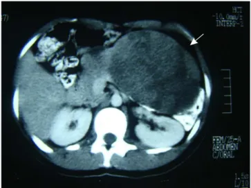
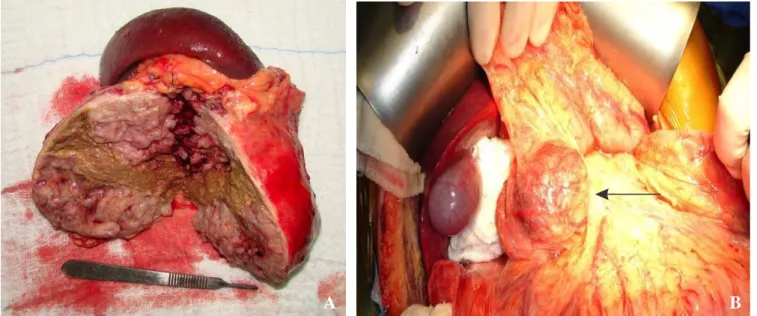
During clinical investigation, four patients underwent CT, one MRI and three underwent both. The main radiological features were: complex, encapsulated, heterogeneous mass, with regular borders, solid and cystic component, and hemorrhagic or necrotic foci. Lesion size ranged from 1.8 to 11.7cm, and in five patients the diameter of the lesion was greater than 5cm. Figures 1, 2 and 3 show the main radiological and surgical aspects of this neoplasm.
No patient had distant metastasis. Seven patients had the tumor in the head and uncinate process, five patients underwent pancreaticoduodenectomy (PD), in one enucleation was performed and in one the tumor was considered unresectable due to invasion of mesenteric vessels, being treated with biliodigestive anastomosis and tumor biopsy.
Of the five patients who underwent pancreaticoduodenectomy (PD), four were with preservation of the pylorus, while one underwent gastroduodenopancreatectomy due to small surgical margin of the pylorus. A single patient with extensive tumor of body and tail of the pancreas was submitted
Table 1 Table 1 Table 1 Table 1
-Table 1 - Characteristics of solid pseudopapillary tumors of the pancreas.
C a s e C a s e C a s e C a s e
C a s e A g eA g eA g eA g eA g e G e n d e rG e n d e rG e n d e rG e n d e rG e n d e r ClinicClinicClinicClinicClinic L o c a t i o nL o c a t i o nL o c a t i o nL o c a t i o nL o c a t i o n S i z eS i z eS i z eS i z eS i z e Local Invasion /Local Invasion /Local Invasion /Local Invasion /Local Invasion / O p e r a t i o nO p e r a t i o nO p e r a t i o nO p e r a t i o nO p e r a t i o n ( y e a r s )
( y e a r s ) ( y e a r s ) ( y e a r s )
( y e a r s ) P r e s e n t a t i o nP r e s e n t a t i o nP r e s e n t a t i o nP r e s e n t a t i o nP r e s e n t a t i o n ( c m )( c m )( c m )( c m )( c m ) M e t a s t a s i sM e t a s t a s i sM e t a s t a s i sM e t a s t a s i sM e t a s t a s i s
1 14 F Abdominal pain Head 6,0x5,0 Absent Enucleação
2 15 F Abdominal pain Uncinate process 4,0x3,0 Absent PPPD
3 19 F Palpable mass Head 8,7x9,7 Absent Whipple
4 23 F Abdominal pain, UGB Neck 1,8x2,0 Absent PPPD
5 25 F Abdominal pain Tail 11,7x8,3 Absent BTP
6 29 F Abdominal pain, Head 8,0x7,0 Absent PPPD
palpable mass
7 36 F Abdominal pain Head 6,8x6,5 Absent PPPD
8 54 M Abdominal pain, Head 10,2x9,0 Vascular invasion unresectable
palpable mass, jaundice
Source: Medical records of patients from the Getúlio Vargas University Hospital of the Federal University of Amazonas, Foundation Oncology Control Center of the State of Amazonas and Santa Julia Hospital.
DP: Pancreaticoduodenectomy; BTP: Body-tail pancratectomy; PPPD: Pylorus preserving pancreaticoduodenectomy; UGB: upper gastrointestinal bleeding.
Figure 2 Figure 2 Figure 2 Figure 2
-Figure 2 - Solid pseudopapillary tumor in head/uncinate process of the pancreas.
Figure 1 Figure 1 Figure 1 Figure 1
body-caudal pancreatectomy with splenectomy. Reconstructions after pancreaticoduodenectomy with preservation of the pylorus were with single bowel loop anastomosed to the pancreas, common hepatic duct and pylorus. The technique used for reconstruction after gastroduodenopancreatectomy was Roux-en-Y with the excluded bowel loop anastomosed to the stomach. The technique pancreaticojejunal anastomosis was duct-to-mucosa and the gastric and biliary anastomoses to the jejunum were end-to-side, single layer, extramucosal, with 3.0 prolene sutures.
Surgical complications occurred in five patients, four classified as grade I and II according to Dindo et al.6, and one grade V, who died.
The length of stay ranged from 5 to 21 days. Two PD patients with preservation of the pylorus evolved with delayed gastric emptying, requiring nasogastric tube decompression for ten days. Low output pancreatic fistula
occurred in two patients, with good response to conservative treatment. One of the patients undergoing PD, in whom a silastic catheter was inserted intraluminally in the hepaticojejunal anastomosis, evolved with later choledocholithiasis and recurrent acute cholangitis, being subjected to new biliodigestive anastomosis after extraction of the calculi and the catheter.
The histopathological analysis of each specimen was held at the Pathology Department of each institution. Macroscopically, lesions were characterized as well-circumscribed and presenting a fibrotic pseudocapsule. The neoplasis had cystic and solid areas in variable proportions. Microscopically, lesions showed epithelial neoplasm with solid areas, cells with granular and eosinophilic cytoplasm, with round or ovoid nuclei, occasional mitotic figures and cellular structure in papillary arrangements, suggestive of SPTP. All parts were sent for immunohistochemistry study. The results are shown in table 2.
Figure 3 -Figure 3 -Figure 3 Figure 3
-Figure 3 - SPTP located in the tail (left) and head (right) of pancreas.
Table 2 -Table 2 -Table 2 Table 2
-Table 2 - Immunohistochemistry.
M a r k e r M a r k e rM a r k e r M a r k e r
M a r k e r Case 1Case 1Case 1Case 1Case 1 Case 2Case 2Case 2Case 2Case 2 Case 3Case 3Case 3Case 3Case 3 Case 4Case 4Case 4Case 4Case 4 Case 5Case 5Case 5Case 5Case 5 Case 6Case 6Case 6Case 6Case 6 Case 7Case 7Case 7Case 7Case 7 Case 8Case 8Case 8Case 8Case 8
Alpha 1-antitrypsin + +
Neuron Specific Enolase + +
Vimentin + + + +
synaptophysin +
-chromogranin A
-progesterone + + + +
CD 10 + + NC
Beta-catenin + + + +
CD 56 +
Source: Medical records of patients from the Getúlio Vargas University Hospital of the Federal University of Amazonas, Foundation Oncology Control Center of the State of Amazonas and Santa Julia Hospital.
Non-conclusivo (NC); negative (-); positive (+)
The follow-up of patients in the late postoperative period ranged from three months to eleven years. None of the six living patients undergoing resection showed any recurrence. One patient with unresectable tumor underwent chemotherapy (5-Fluorouracil and Leucovorin) and radiotherapy (4.500Gy), with reduction of tumor volume from 10.2x9.0cm to 3.0x3.0cm, which made the lesion resectable according to an angiotomographic study. The patient, however, refused the surgical procedure and was kept in outpatient treatment; he remains asymptomatic and without signs of metastatic disease for 11 years. Among the remaining patients, one had exocrine pancreatic insufficiency, being under regular replacement of pancreatic enzymes. The other patients remain asymptomatic.
DISCUSSION
DISCUSSION
DISCUSSION
DISCUSSION
DISCUSSION
The SPTP is a rare malignant tumor of the pancreas of unknown origin that affects young women aged from 20 to 30 years4,7. Santini et al.8 indicate that it probably arises from pluripotent pancreatic cells, as there is no definitive evidence of its pancreatic origin, whether endocrine or exocrine. Kosmahl et al.9 suggest that the tumor originates from the incorporation of primitive ovarian cells within the pancreatic parenchyma during the seventh week of embryogenesis, though this does not justify the presence of malignancy in males.
This neoplasm occurs almost exclusively in young women, and several hypotheses have been proposed to explain this distribution. The hormonal factor has been mostly implicated, due to the presence of progesterone receptors in the tumor and lesion progression during pregnancy10. Kosmahl et al.9 identified the presence of progesterone receptors in more than 90% of 59 SPTPs through immunohistochemistry. Our study also showed a predominance of females. Seven patients were women and the research of progesterone receptor in the tumor, carried out in four patients, was positive in all of them.
Regarding age, six patients were less than 30 years, three of them under 20 years, similar to what is described in the literature.
In young, female patients with a solid, cystic, or solid-cystic pancreatic mass, the possibility of this tumor should always be raised. Whenever possible, there should be complete resection of the primary tumor and metastases, if present.
Many patients present with irrelevant signs and symptoms. When symptomatic, they may have recurrent episodes of pancreatitis, chronic abdominal pain and a palpable mass. Large tumors can compress the stomach, duodenum and bile ducts, resulting in early satiety, vomiting or jaundice2,11. In a study that evaluated 37 patients, it was found that abdominal pain was the most common symptom, affecting 84% of subjects7. A similar result was found in our study, where from the eight patients, seven had
abdo-minal pain and five palpable mass. Four had abdoabdo-minal pain as an isolated symptom, and one had a palpable mass alone. Other symptoms found were hematemesis, vomiting and melena. Only one of our patients had a history of jaundice.
The most common location of SPTP in the literature is the tail, and the second most common site is the head3. In our series, only one patient had a lesion in the tail of the pancreas, five had it in the pancreatic head, one in the uncinate process and one in the neck of the pancreas, differing from the literature and rendering surgical treatment more aggressive, pancreaticoduodenectomy having been performed in five of our patients.
The SPTP is often described as an indolent and not aggressive tumor. However, local invasion and hematogenous metastasis may occur2,12. The most common sites of distant metastases are the liver, lymph nodes and peritoneum3,5. Reddy et al.7 studied 37 patients and detected that 11% developed metastases. Of these, two had lymph node metastasis, one had liver metastases, and one patient developed systemic disease about eight years after a potentially curative resection. In our study, only the male patient presented invasion of mesenteric vessels and unresectability.
The SPTP is usually a solitary, well-defined, heterogeneous, hypovascular lesion, with peripheral enhancement with intravenous contrast and with clear separation from the adjacent pancreatic parenchyma by a fibrous capsule, with solid and cystic components. Irregular calcification is present in 30% of patients8,13.
Sometimes it invades extrapancreatic organs, as well as displaying liver and peritoneal metastases8,13. The primary tumor is almost invariably ovoid and with large dimensions at diagnosis (usually greater than 6cm, reaching up to 30cm), probably due to its indolent growth. The magnetic resonance imaging provides better morphologic evaluation of bleeding and can help characterize the lesion14.
Macroscopically, the mass appears with complex texture, with solid and cystic components and hemorrhagic and/or necrotic content, with internal debris8,9,13.
Microscopically, Frantz tumor presents as an encapsulated lesion with cystic and solid areas. The solid component shows a pseudopapillary structure. Eosinophilic cytoplasm is often observed in the tumor’s cells and cytoplasmic vacuoles may also be evident. The nucleus is round to oval, with a uniform appearance. The stroma can be hyaline or myxoid8,9.
CD10 positivity is characteristic of SPTP sand is less useful in the differential diagnosis, as it is positive in about 10% of invasive ductal adenocarcinomas and neuroendocrine tumors15.
Alpha-1-antitrypsin and alpha-1-antichymotrypsin indicate exocrine pancreatic origin, while neuron-specific enolase and synaptophysin are characteristic of neuroendocrine tumors16.
The presence of progesterone receptors is another feature of Frantz tumor16. In addition to negative chromogranin, synaptophysin and enolase, which virtually rule out pancreatic islets malignancy, the absence of cytokeratin excludes acinar carcinoma16,17.
Our results concerning immunohistochemistry are shown in table 2. Due to realization in various services outside Amazonas State, different immunohistochemistry profiles were used, demonstrating positivity of 100% when performed for vimentin (4 patients), progesterone (4 patients), beta-catenin (4 patients), consistent with the literature.
Surgical excision of the SPTP is the treatment of choice5,12. Since it is usually a benign tumor of low malignant potential, the prognosis after surgical resection is excellent3. Tipton et al.2 mention that resection, even of distant metastases, may be included in the treatment when the patient has favorable clinical status. In contrast, lymph node metastasis is rare, and lymphadenectomy, unnecessary18.
Radiation therapy is a viable alternative for the treatment of unresectable tumors and can offer improved quality of life, especially regarding pain relief4.
In our series, seven patients underwent resection procedures and one patient underwent chemotherapy with 5-fluorouracil and radiotherapy at a total dose of 4.500Gy due to having been considered unresectable. This patient has been followed for 13 years and is asymptomatic without metastatic disease, demonstrating reduction of the lesion size from 10.2 x 9.0 cm to 3.0 x 3.5 cm. This made the lesion resectable, though the patient refused surgery.
With an average of 85 months follow-up, Kang et al.3 detected a median survival of 175 months, with only one death. Aside from this, no cases of tumor-related death or recurrences were observed during follow-up. Our results were similar, with 100% of patients with resectable disease and alive, without signs of recurrence. Only one patient had postoperative complications, dying on the 21st day after surgery. One patient had exocrine pancreatic insufficiency, presenting with steatorrhea, making regular use of pancreatic enzymes. The patient with unresectable tumor is asymptomatic and without signs of distant metastasis at 13 years.
In conclusion, the solid pseudopapillary tumor of the pancreas preponderated in young female patients, predominantly located in the pancreatic head.
R E S U M O R E S U M O R E S U M O R E S U M O R E S U M O
Objetivo Objetivo Objetivo Objetivo
Objetivo: descrever perfil clínico-cirúrgico do tumor sólido pseudopapilar do pâncreas. MétodosMétodosMétodosMétodos: estudo observacional retrospec-Métodos tivo multi-institucional avaliando as características clínicas, radiológicas e cirúrgicas dos pacientes com diagnóstico de tumor sólido pseudopapilar do pâncreas submetidos a tratamento cirúrgico. ResultadosResultadosResultadosResultadosResultados: foram identificados oito pacientes em três hospitais no estado do Amazonas, sendo sete do sexo feminino, seis com menos de 30 anos. A neoplasia predominou na cabeça do pâncreas. Cinco pacientes foram submetidos à duodenopancreatectomia, um à enucleação, um à pancreatectomia corpocaudal e o último foi considerado irressecável. ConclusãoConclusãoConclusãoConclusãoConclusão: o tumor sólido pseudopapilar do pâncreas predominou em pacientes jovens do sexo feminino, com localização predominante na cabeça do pâncreas.
Descritores: Descritores: Descritores: Descritores:
Descritores: Avaliação. Pâncreas. Neoplasias. Neoplasias pancreáticas. Pancreatectomia.
REFERENCES
REFERENCES
REFERENCES
REFERENCES
REFERENCES
1. Frantz VK. Papillary tumors of the pancreas: benign or malignant tumors of the pancreas. In: Frantz UK, editor. Atlas of tumor pathology, 1st series, Fascicles 27 and 28. Washington, DC: Armed Forces Institute of Pathology; 1959. p.32-3.
2. Tipton SG, Smyrk TC, Sarr MG, Thompson GB. Malignant potential of solid pseudopapillary neoplasm of the pancreas. Br J Surg. 2006;93(6):733-7.
3. Kang CM, Kim KS, Choi JS, Kim H, Lee WJ, Kim BR. Solid pseudopapillary tumor of the pancreas suggesting malignant potential. Pancreas. 2006;32(3):276-80.
4. Zauls JA, Dragun AE, Sharma AK. Intensity-modulated radiation therapy for unresectable solid pseudopapillary tumor of the pancreas. Am J Clin Oncol. 2006;29(6):639-40.
5. Seo HE, Lee MK, Lee YD, Jeon SW, Cho CM, Tak WY, et al. Solid-pseudopapillary tumor of the pancreas. J Clin Gastroenterol. 2006;40(10):919-22.
6. Dindo D, Demartines N, Clavien PA. Classification of surgical complications: a new proposal with evaluation in a cohort of 6336 patients and results of a survey. Ann Surg. 2004;240(2):205-13. 7. Reddy S, Cameron JL, Scudiere J, Hruban RH, Fishman EK, Ahuja
N, et al. Surgical management of solid-pseudopapillary neoplasms of the pancreas (Franz or Hamoudi tumors): a large single-institutional series. J Am Coll Surg. 2009;208(5):950-7; discussion 957-9.
8. Santini D, Poli F, Lega S. Solid-papillary tumors of the pancreas: histopatology. JOP. 2006;7(1):131-6.
10. Morales A, Ruiz-Molina JM, Estéves HO, Robles-Díaz G, Díaz-Sánchez V. Papillary-cystic neoplasm of the pancreas. A sex-steroid dependent tumor. Int J Pancreatol. 1998;24(3):219-25. 11. Brugge WR, Lauwers GY, Sahani D, Fernandez-del Castillo C,
Warshaw AL. Cystic neoplasm of the pancreas. N Eng J Med. 2004;351(12):1218-26.
12. Vollmer CM Jr, Dixon E, Grant DR. Management of a solid pseudopapillary tumor of the pancreas with liver metastases. HPB. 2003;5(4):264-7.
13. Roggin KK, Chennat J, Oto A, Noffsinger A, Briggs A, Matthews JB. Pancreatic cystic neoplasm. Curr Probl Surg. 2010;47(6):459-510.
14. Coleman KM, Doherty MC, Bigler SA. Solid-pseudopapillary tumor of the pancreas. Radiographics. 2003;23(6):1644-8.
15. Nguyen NQ, Johns AL, Gill AJ, Ring N, Chang DK, Clarkson A, et al. Clinical and immunohistochemical features of 34 solid pseudopapillary tumors of the pancreas. J Gastroenterol Hepatol. 2011;26(2):267-74.
16. Serra S, Chetty R. Revision 2: an immunohistochemical approach and evaluation of solid pseudopapillary tumour of the pancreas. J Clin Pathol. 2008;61(11):1153-9.
17. Romics L Jr, Oláh A, Belágyi T, Hajdú N, Gyurus P, Ruszinkó V. Solid pseudopapillary neoplasm of the pancreas--proposed algorithms for diagnosis and surgical treatment. Langenbecks Arch Surg. 2010;395(6):747-55.
18. Klimstra DS, Wenig BM, Heffess CS. Solid-pseudopapillary tumor of the pancreas: a typically cystic carcinoma of low malignant potential. Semin Diagn Pathol. 2000;17(1):66-80.
Received on 15/07/2012
Accepted for publication 22/10/2012 Conflict of interest: none
Source of funding: none
How to cite this article: How to cite this article: How to cite this article: How to cite this article: How to cite this article:
Guimarães LSC, Melo AMS, Ruiz MR, Viana JS, Silva Júnior RA. Tumor sólido pseudopapilar do pâncreas: avaliação do perfil radiológico e cirúrgico. Rev Col Bras Cir. [periódico na Internet] 2013;40(5). Disponí-vel em URL: http://www.scielo.br/rcbc