ABSTRACT
Sao Paulo Med J. 2007;125(6):329-32.
ORIGINAL AR
TICLE
Márcia Cristina Bastos Boëchat
Kátia Silveira da Silva
Juan Clinton Llerena Jr
Paulo Roberto Mafra Boëchat
Cholelithiasis and biliary sludge
in Down’s syndrome patients
Instituto Fernandes Figueira, Fundação Oswaldo Cruz, Rio de Janeiro,
Rio de Janeiro, Brazil.
CONTEXT AND OBJECTIVE: Although studies have demonstrated increased frequency of gall-bladder abnormalities among Down’s syndrome (DS) patients in some countries, there is only one paper on this subject in the Brazilian literature. The aim of this study was to demonstrate the prevalence, clinical characteristics and evolu-tion of lithiasis and biliary sludge among DS patients in a maternity and children’s hospital in Rio de Janeiro.
DESIGN AND SETTING:This was a cross-sectional study followed by a retrospective cohort study on all individuals with an ultrasound diagnosis of gallbladder abnormalities.
METHODS: 547 DS patients (53.2% male, 46.8% female) attending the Instituto Fernandes Figueira in 2001 underwent abdominal ultrasound ex-amination at ages of between one day and three years (mean: fi ve months). Clinical and ultrasound data were analyzed.
RESULTS: In 50 patients (9.1%), the ultrasound demonstrated gallbladder abnormalities (6.9% lithiasis and 2.1% biliary sludge). Spontaneous resolution was observed in 66.7% of the patients with biliary sludge and 28.9% with lithiasis. Cholecystectomy was carried out on 26.3% of the patients with gallstones.
CONCLUSION: The results from this study and comparison with the literature suggest that DS pa-tients are at risk of developing lithiasis and biliary sludge and should be monitored throughout the neonatal period, even if there are no known risk factors for gallstone formation. Most frequently, these gallbladder abnormalities occur without symptoms and spontaneously resolve in most non-symptomatic patients. DS patients should be monitored with serial abdominal ultrasound, and cholecystectomy is indicated for symptomatic cases or when cholecystitis is present.
KEY WORDS: Down syndrome. Gallbladder. Lithiasis. Cholecystitis. Cholecystectomy. INTRODUCTION
Before the 1980s, few cases of chole-lithiasis in children, relating to hemolytic anemia, had been described in the medical literature.1-3 With the increased use of
ab-dominal ultrasound, cholelithiasis has been diagnosed more frequently in children, and the reported prevalence is between 0.13% and 0.5% in different series.4-7 Several
con-ditions are considered to be risk factors for biliary lithiasis among neonates, infants and children: hemolytic disease, cystic fi brosis, ileal resection, hypercholesterolemia, con-genital hepatobiliary anomalies, concon-genital heart disease, prematurity, phototherapy, sepsis, parenteral nutrition, diuretics and antibiotics, particularly ceftriaxone.8-11
Some reports12-16 have showed that
biliary abnormalities like lithiasis and bil-iary sludge occur more frequently among Down’s syndrome (DS)* patients. However, only one of these reports15 relates to
Brazil-ian patients.
OBJECTIVE
The objective of this study was to deter-mine the prevalence, clinical characteristics and evolution of biliary abnormalities in a group of 547 DS patients who were followed up at a maternity and children’s hospital in the City of Rio de Janeiro, Brazil.
MATERIAL AND METHODS
The patients in this study came from the register of 547 DS children who attended the Department of Medical Genetics of Instituto Fernandes Figueira, Fundação Oswaldo Cruz (IFF-Fiocruz) in the year 2001. All of these children underwent routine abdominal ultra-sound when they began their treatment at the institute. Their ages ranged from one day to three years (mean: fi ve months). At the time
of the fi rst ultrasound examination, all the patients were non-symptomatic and did not present any known risk factors for gallstones or biliary sludge.
For each patient, the following items were reviewed: sex, presence or absence of biliary abnormalities, age at the time of diag-nosing the biliary abnormality on ultrasound, biliary abnormality type, clinical symptoms, other associated congenital anomalies, associated risk factors, type of treatment and clinical follow-up. All the individuals with an ultrasound diagnosis of gallbladder abnormality were monitored by abdominal ultrasound every three to six months during their fi rst year and every year thereafter. All the patients were seen and followed up by the Departments of Medical Genetics and Pediatric Surgery.
Ultrasound examinations were carried by means of a convex transducer (5 or 7 MHz) after the patients had been fasting for three to six hours. If after this period of time the gallbladder was not seen, the child remained without food for a further few hours until the examination could be adequately performed. The ultrasound diagnosis of biliary sludge and lithiasis was confi rmed by parameters that are well-established in the literature.17-18 Echogenic
material without an acoustic shadow in the gallbladder was interpreted as biliary sludge (Figure 1), while echogenic material with an acoustic shadow was considered to be a gallstone (Figure 2).
This project received approval from the institution’s Ethical Review Board, under the number CAEE 0026.0.008.000-03 (Brazil). The EpiInfo 3.2 software (February 2004; Centers for Disease Control and Prevention, CDC, United States) was used to create a database and for all statistical analyses.
330
Sao Paulo Med J. 2007;125(6):329-32.
RESULTS
Abdominal ultrasound examinations were performed before the age of two years in 94% of the patients. Ultrasound biliary abnormalities were identifi ed in 50 individuals (9.1%): 38 patients had biliary lithiasis and 12 presented biliary sludge, with prevalences of 6.9% (95% confi dence interval, CI: 5.1 - 9.6) and 1.7% (95% CI: 1.2 – 3.9), respectively. Table 1 describes the prevalence of these biliary abnormalities according to sex and age at the time of the ultrasound diagnosis.
The ages of the DS patients with biliary sludge ranged from one month to two years (mean: 5.7 months), with slight predominance in females (58.3%). Spontaneous resolution of the sludge was observed in 66.7% of these patients and the others continued to present non-symptomatic biliary sludge for a period of three years, which was monitored by serial abdominal ultrasound examinations.
The ages of the DS patients with gall-stones ranged from one day to three years (mean: nine months) at the time of the ultra-sound diagnosis, with slight predominance in males (57.9%). Spontaneous resolution of the calculi occurred in 11 children (28.9%), over a period of three months to three years after the diagnosis (mean: nine months). Chole-cystectomy was carried out on 26.3% of the patients with a mean age of 45 months; four of these patients were non-symptomatic and Table 1. Prevalence of lithiasis and biliary sludge according to sex and the age at the
time of ultrasound diagnosis
Prevalence
Age range in months Normal (n = 497) Lithiasis (n = 38) Biliary sludge (n = 12)
% (n) % (n) % (n)
0-2 (n = 135) 88.9% (120) 9.6% (13) 1.5% (2)
3-6 (n = 181) 91.1% (165) 5.0% (9) 3.9% (7)
7-12 (n = 113) 95.6% (108) 3.5% (4) 0.9% (1)
13-24 (n = 79) 91.1% (72) 6.3% (5) 2.5% (2)
25-36 (n = 39) 82.0% (32) 17.9% (7)
Sex
Female (n = 256) 46.9% (233) 42.1% (16) 58.3% (7)
Male (n = 291) 53.1% (264) 57.9% (22) 41.7% (5)
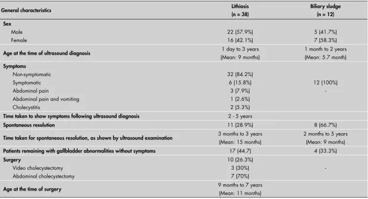
Table 2. General characteristics of Down’s syndrome patients with ultrasound diagnosis of lithiasis or biliary sludge
General characteristics Lithiasis
(n = 38)
Biliary sludge (n = 12) Sex
Male Female
22 (57.9%) 16 (42.1%)
5 (41.7%) 7 (58.3%)
Age at the time of ultrasound diagnosis 1 day to 3 years
(Mean: 9 months)
1 month to 2 years (Mean: 5.7 month)
Symptoms
Non-symptomatic Symptomatic Abdominal pain
Abdominal pain and vomiting Cholecystitis
32 (84.2%) 6 (15.8%) 3 (7.9%) 1 (2.6%) 2 (5.3%)
12 (100%)
-Time taken to show symptoms following ultrasound diagnosis 2 - 5 years
Spontaneous resolution 11 (28.9%) 8 (66.7%)
Time taken for spontaneous resolution, as shown by ultrasound examination 3 months to 3 years
(Mean: 15 months)
2 months to 5 years (Mean: 9 months)
Patients remaining with gallbladder abnormalities without symptoms 17 (44.7) 4 (33.3%)
Surgery
Video cholecystectomy Abdominal cholecystectomy
10 (26.3%) 3 (30%) 7 (70%)
-Age at the time of surgery 9 months to 7 years
(Mean: 11 months) Figure 2. Ultrasound appearance of biliary lithiasis.
G = Gallbladder, GS = Gallstones, AS = Acoustic shadow.
Figure 1. Ultrasound appearance of biliary sludge.
331
Sao Paulo Med J. 2007;125(6):329-32.
the other six were symptomatic. This surgery was indicated after a period of clinical and ultrasound follow-up (mean: 28 months). Eleven patients presented spontaneous resolution of their calculi and did not need to undergo operations. Among the remaining 27 patients, ten underwent operations (four without symptoms and six with symptoms), while 17 continued to present calculi. These 17 patients with lithiasis (44.7%) remained non-symptomatic for a longer period (mean: 45 months). Table 2 summarizes the main characteristics of the patients with biliary sludge and lithiasis.
DISCUSSION
Ultrasound investigation is considered to be the initial imaging method for diagnostic investigations of the gallbladder and bile duct and presents great sensitivity and specifi city for detecting such abnormalities,16-19 with an
accuracy of up to 96%.16
In our study, the prevalence of biliary abnormalities was 9.1% and most patients were non-symptomatic and did not present any known risk factors for gallbladder lithiasis or biliary sludge at the time of the ultrasound diagnosis. 66% of the patients were diagnosed at an age of less than 12 months. Clinical fol-low-up and serial ultrasound examinations demonstrated that most patients remained without symptoms or had spontaneous resolu-tion of the biliary abnormalities. Considering the absence of risk factors and symptoms, the high frequency in the fi rst year of life and the fact that some cases had spontaneous reso-lution of the biliary sludge or gallstones, these results are in agreement with the medical lit-erature.5,7,20,21 It has been reported9 that biliary
sludge can be a precursor for biliary lithiasis, but this was not observed in our study.
In recent years, abdominal ultrasound has been widely used in clinics for neonates and children, particularly for detecting cholelithi-asis.4-7 Clinical conditions such as biliary stasis,
parenteral nutrition and prolonged fasting are considered to be high risk factors for development of biliary sludge and calculi, in comparison with hemolytic disease. However, the majority of cases of biliary abnormalities
in children, particularly during the neonatal period, are non-symptomatic and idiopathic, with predominance among males and spon-taneous resolution within six months after the ultrasound diagnosis in most cases.9,11,21
Despite the existence of studies demon-strating increased frequency of cholelithiasis among neonates and children who present associations with particular risk factors,4-6 few
of them describe associations with DS.7,12,13,15
In the general pediatric population, cholelithi-asis prevalence has been found to be 0.13% to 0.22%.1,11 Case series relating to children with
DS have demonstrated high prevalence.15,22 In
1986, among 187 DS patients, Buchin et al.22
identifi ed only one individual (0.5%) with cholelithiasis, who was 18 years old. Subse-quently, in 1993, Llerena et al.15 described a
group of 145 patients with a 9% prevalence of biliary abnormalities: three children with biliary sludge and ten with gallstones. Nei-ther Buchin et al.22 nor Llerena et al.15 made
any reference to the patients’ ages. In 2001, Toscano et al.7 presented six cases (4.7%)
cholelithiasis among 126 DS children aged between six months and six years.
The present study confi rmed our previ-ous fi ndings15 regarding the high prevalence
of biliary abnormalities in DS patients. The pathological mechanism for this higher prevalence in DS patients than in the general pediatric population remains unknown, but it could be related to hypercholesterolemia dur-ing intrauterine life, as suggested by Bocconi et al. in 1997.23
In our series, the great age range seen at the time of the ultrasound investigation was due to two main reasons. Firstly, our hospital is a reference center for the State of Rio de Janeiro24 and therefore receives DS patients
from different regions of the state at different ages. Secondly, few DS patients were born at our hospital. In such cases, the referral for ultrasound investigation was done while the child was still at the neonatal stage.
Surgical treatment was performed on ten patients (26.3%): six with and four without symptoms. At the beginning of the 1990s, although medical reports demon-strated the possibility of spontaneous
reso-lution of cholelithiasis during the neonatal period,4 little was known about the natural
history of cholelithiasis in DS patients. At that time, patients with biliary lithiasis that did not spontaneously resolve over the next six months received indication for surgical treatment, even if they did not present any symptoms.4,25 In our hospital, ultrasound
and clinical follow-up demonstrated that most patients remained free of symptoms and, in many cases, spontaneous resolution of the gallstone or biliary sludge occurred. In the light of this experience, the manage-ment of cases of gallstones or biliary sludge, especially regarding surgical procedures, was reviewed. Following this review, the man-agement method changed such that only symptomatic children with abdominal pain, vomiting and/or cholecystitis symptoms were indicated for surgical procedures. The non-symptomatic cases were followed by clinical means and serial abdominal ultra-sound examinations. This clinical approach and ultrasound follow-up for DS patients with gallstones or biliary sludge has also been reviewed by other authors.6,7,9
CONCLUSION
Analysis and comparison of our ultra-sound data on 547 DS patients in relation to the literature showed that DS must be considered to be a risk factor for the devel-opment of biliary lithiasis and biliary sludge in children, especially in the neonatal period without any other associated risk factor. In most DS cases, biliary lithiasis and biliary sludge were non-symptomatic.
It could be concluded that gallstones and biliary sludge in DS patients mostly had fa-vorable evolution and a good prognosis, with spontaneous resolution and non-symptomatic presentation in most patients. These DS patients need to be followed up with serial abdominal ultrasound examinations, and surgical treatment should only be indicated in symptomatic cases or in the presence of cholecystitis.
332
Sao Paulo Med J. 2007;125(6):329-32.
AUTHOR INFORMATION Márcia Cristina Bastos Boëchat, MD.Pediatric radiologist, Radiology Service, Instituto Fernandes Figueira, Fundação Oswaldo Cruz, Rio de Janeiro, Rio de Janeiro, Brazil.
Kátia Silveira da Silva, PhD. Epidemiologist, Epidemiology Group, Instituto Fernandes Figueira, Fundação Oswaldo Cruz, Rio de Janeiro, Rio de Janeiro, Brazil.
Juan Clinton Llerena Júnior, PhD. Geneticist, Department of Medical Genetics, Instituto Fernandes Figueira, Fundação Oswaldo Cruz, Rio de Janeiro, Rio de Janeiro, Brazil.
Paulo Roberto Mafra Boëchat, MD. Pediatric surgeon, Department of Pediatric Surgery, Instituto Fernandes Figueira, Fundação Oswaldo Cruz, Rio de Janeiro, Rio de Janeiro, Brazil.
Address for correspondence: Márcia Cristina Bastos Boëchat
Rua Assis Brasil, 120 Conj. 402 — Copacabana Rio de Janeiro (RJ) — Brasil — CEP 22030-010 Tel (55 21) 2542-8883 — (55 21) 2554-1786 — Fax (55 21) 2542-3164
E-mail: [email protected] ocruz.br
Copyright © 2007, Associação Paulista de Medicina
RESUMO
Colelitíase e lama biliar em pacientes com síndrome de Down
OBJETIVO:Demonstrar prevalência, características clínicas e evolução de litíase e lama biliar em pacientes com síndrome de Down (SD) num hospital materno-infantil no Rio de Janeiro. Apesar de estudos revelarem aumento das anormalidades biliares em pacientes com SD em alguns países, no Brasil existe apenas um trabalho abordando o tema.
TIPO DE ESTUDO E LOCAL:Estudo transversal seguido por um estudo de coorte retrospectivo de todos os indivíduos com diagnóstico ultra-sonográfi co de anormalidades da vesícula biliar.
MÉTODOS: Foram selecionados 547 pacientes com SD (53,2% sexo masculino, 46,8% feminino) atendidos no Instituto Fernandes Figueira, Fundação Instituto Oswaldo Cruz (IFF-Fiocruz) em 2001. Todos os pacientes incluídos neste estudo foram submetidos a ultra-sonografi a abdominal quando tinham idades variando entre um dia e três anos (mediana cinco meses). Dados clínicos e ultra-sonográfi cos foram avaliados.
RESULTADOS: Em 50 (9,1%) crianças, a ultra-sonografi a demonstrou alteração da vesícula biliar (6,9% litíase e 2,2 % lama biliar). Houve resolução espontânea em 66,7% dos pacientes com lama biliar e em 28,9% dos pacientes com litíase. A colecistectomia foi realizada em 26,3% dos pacientes com cálculo biliar.
CONCLUSÃO:Resultados deste estudo e a comparação com a literatura sugerem que a SD deve ser consi-derada como fator de risco para desenvolvimento de litíase e lama biliar em crianças, sobretudo no período neonatal, sem que existam outros fatores predisponentes para formação de cálculo biliar. Na maioria das vezes, estas alterações são assintomáticas e, freqüentemente, têm evolução favorável, permanecendo desta forma ou tendo resolução espontânea. Pacientes devem ser acompanhados com ultra-sonografi as seriadas. Tratamento cirúrgico está indicado para casos sintomáticos ou na presença de colecistite.
PALAVRAS-CHAVE: Síndrome de Down. Vesícula biliar. Litíase. Colecistite. Colecistectomia. 1. Palasciano G, Portincasa P, Vinciguerra V, et al. Gallstone
prevalence and gallbladder volume in children and adolescents: an epidemiological ultrasonographic survey and relationship to body mass index. Am J Gastroenterol. 1989;84(11):1378-82. 2. Reif S, Sloven DG, Lebenthal E. Gallstones in children. Char-acterization by age, etiology, and outcome. Am J Dis Child. 1991;145(1):105-8.
3. Ruibal Francisco J, Aleo Luján E, Alvarez Mingote A, Piñero Martínez E, Gómez Casares R. Colelitiasis en la infancia. Análisis de 24 pacientes y revisión de 123 casos publicados en España. [Childhood cholelithiasis. Analysis of 24 patients diagnosed in our department and review of 123 cases published in Spain]. An Esp Pediatr. 2001;54(2):120-5.
4. Holcomb GW 3rd. Laparoscopic cholecystectomy. Semin Pediatr Surg. 1993;2(3):159-67.
5. Kumar R, Nguyen K, Shun A. Gallstones and common bile duct calculi in infancy and childhood. Aust N Z J Surg. 2000;70(3):188-91.
6. Wesdorp I, Bosman D, de Graaff A, Aronson D, van der Blij F, Taminiau J. Clinical presentations and predisposing factors of cholelithiasis and sludge in children. J Pediatr Gastroenterol Nutr. 2000;31(4):411-17.
7. Toscano E, Trivellini V, Andria G. Cholelithiasis in Down’s syndrome. Arch Dis Child. 2001;85(3):242-3.
8. Bor O, Dinleyici EC, Kebapci M, Aydogdu SD. Ceftriaxone-associated biliary sludge and pseudocholelithiasis during child-hood: a prospective study. Pediatr Int. 2004;46(3):322-4.
9. Lobe TE. Cholelithiaisis and cholecystitis in children. Semin Pediatr Surg. 2000;9(4):170-6.
10. Palanduz A, Yalçin I, Tonguç E, et al. Sonographic assessment of ceftriaxone-associated biliary pseudolithiasis in children. J Clin Ultrasound. 2000;28(4):166-8.
11. Rescorla FJ. Cholelithiasis, cholecystitis, and common bile duct stones. Curr Opin Pediatr. 1997;9(3):276-82.
12. Aughton DJ, Gibson P, Cacciarelli A. Cholelithiasis in infants with Down syndrome. Three cases and literature review. Clin Pediatr (Phila). 1992;31(11):650-2.
13. Aynaci FM, Erduran E, Mocan H, Okten A, Sarpkaya AO. Cholelithiasis in infants with Down syndrome: report of two cases. Acta Paediatr. 1995;84(6):711-2.
14. Elías Pollina J, Garate J, Martin Bejarano E, et al. Colelitiasis en la infancia. Propuestas de un estudio multicéntrico. [Cholelithi-asis in childhood. Proposals based on a multicentric study]. Cir Pediatr. 1992;5(2):96-100.
15. Llerena JC Jr, Boy R, Barbosa Neto J, et al. Abdominal ultrasound scan in Down syndrome patients: high frequency of nonsymptomatic biliary tract disease. Am J Med Genet. 1993;46(5):612.
16. Hanbidge AE, Buckler PM, O’Malley ME, Wilson SR. From the RSNA refresher courses: imaging evaluation for acute pain in the right upper quadrant. Radiographics. 2004;24(4):1117-35. 17. Hayden KC Jr, Swischuk LE. Liver and biliary tract. In: Hayden
KC Jr, Swischuk LE, editors. Pediatric ultrasonography. Balti-more: Williams & Wilkins; 1987. p. 186-93.
18. Siegel MJ. Liver and biliary tract. In: Siegel MJ, editor. Pediatric sonography. 1st edition. New York: Raven Press Ltd.; 1991. p. 154-7.
19. Yang HL, Li ZZ, Sun YG. Reliability of ultrasonography in diagno-sis of biliary lithiadiagno-sis. Chin Med J (Engl). 1990;103(8):638-41. 20. Debray D, Pariente D, Gauthier F, Myara A, Bernard O.
Cholelithiasis in infancy: a study of 40 cases. J Pediatr. 1993;122(3):385-91.
21. St-Vil D, Yazbeck S, Luks FI, Hancock BJ, Filiatrault D, Youssef S. Cholelithiasis in newborns and infants. J Pediatr Surg. 1992;27(10):1305-7.
22. Buchin PJ, Levy JS, Schullinger JN. Down’s syndrome and gastrointestinal tract. J Clin Gastroenterol. 1986;8(2):111-4. 23. Bocconi L, Nava S, Fogliani R, Nicolini U. Trisomy 21 is
as-sociated with hypercholesterolemia during intrauterine life. Am J Obstet Gynecol. 1997;176(3):540-3.
24. Horovitz DD, de Mattos RA, Llerena JC Jr. Medical genetic services in the state of Rio de Janeiro, Brazil. Community Genet. 2004;7(2-3):111-6.
25. Bailey PV, Connors RH, Tracy TF Jr, Sotelo-Avila C, Lewis JE, Weber TR. Changing spectrum of cholelithiasis and cholecystitis in infants and children. Am J Surg. 1989;158(6):585-8.
Sources of funding: None
Confl ict of interest: None
Date of fi rst submission: October 10, 2006
Last received: November 7, 2007
Accepted: November 14, 2007