ABSTRACT
ORIGINAL AR
Omero Benedicto Poli Neto
Francisco José Candido dos Reis
Antonio Alberto Nogueira
among postmenopausal women
Department of Gynecology and Obstetrics, Faculdade de Medicina de Ribeirão
Preto (FMRP), Universidade de São Paulo (USP), São Paulo, Brazil
CONTEXT AND OBJECTIVES:Endometrial cancer is the most prevalent type of malignant neoplasia of the genital tract. The objective of this study was to calculate the sensitivity, specifi city, accuracy and positive and negative predictive values for diagnostic hysteroscopy, in comparison with histopathological tests, for all lesions of the endometrial cavity.
DESIGN AND SETTING:Retrospective descriptive study at the public tertiary-level university hospital of Faculdade de Medicina de Ribeirão Preto, Universidade de São Paulo.
METHODS: Diagnostic hysteroscopy was indi-cated in the following instances: endometrial thickness > 4 mm in asymptomatic patients; postmenopausal bleeding; and irregular endometrium or endometrium difficult to as-sess from ultrasound, with or without vaginal bleeding. Ultrasound evaluations were car-ried out no more than three months prior to hysteroscopy.
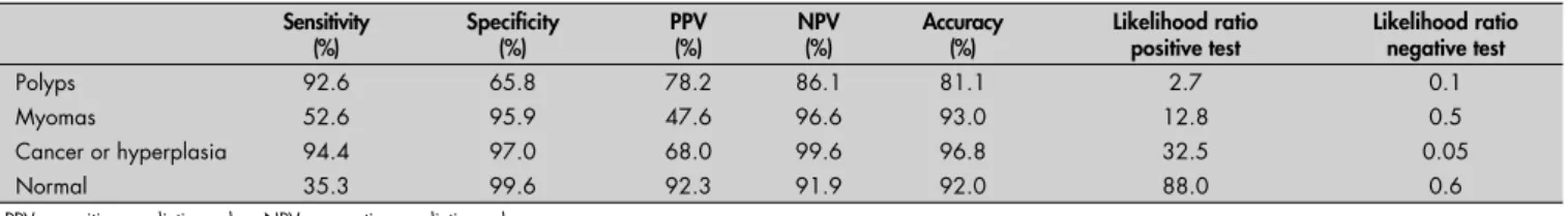
RESULTS:There were 510 patients, with a mean age of 61.1 ± 2.0 years and mean time elapsed since the menopause of 12.7 ± 2.5 years. Endometrial biopsies were performed on 293 patients (57.5%). Histopathological analysis showed that 18 patients presented endometrial carcinoma or typical or atypical hyperplasia, and none of them presented endometrial thickness of less than 8 mm. No signifi cant differences were found between the median thicknesses of the various benign lesions (p > 0.05). In our data, the sensitivity, specifi city, accuracy and positive and negative predictive values for cancer or hyperplasia were 94.4%, 97.0%, 96.8%, 68% and 99.6%, respectively.
CONCLUSIONS:Our results suggest that hys-teroscopy is valuable as a diagnostic tool for malignant/hyperplastic and benign lesions, except for submucous myomas, for which the sensitivity was only 52.6%.
KEY WORDS: Hysteroscopy. Ultrasonics. Post-menopause. Biopsy. Endometrial carcinoma.
INTRODUCTION
Endometrial cancer, the most prevalent type of malignant neoplasia of the genital tract in Western countries,1 also presents
the best prognosis, since approximately 70% of the cases are detected in their early stages.2 Between 80 and 95% of such
pa-tients present vaginal bleeding as the fi rst symptom,3 thus enabling prompt diagnosis
and therapeutic intervention.
Because of the early manifestation of symptoms of this neoplasia, the need for a screening program is debatable. One of the most widely investigated tests used for this purpose is transvaginal ultrasound. Despite the fact that some authors have defended its application to asymptomatic patients,4,5
oth-ers have failed to show any benefi ts from this procedure, since it appears to present a positive predictive value of only 2%.6-8
Nevertheless, since vaginal bleeding is a common symptom among postmenopausal women, ultrasound has been a useful screening method for determining which symptomatic patients should undergo invasive collection of endometrial samples. In this situation, endometrial thickening, as measured by ultrasonography, correlates better with the presence of endometrial lesions9-11 than among
asymptomatic women.
Depending on the availability of each type of procedure, biopsies can be performed using either image guidance techniques, such as hysteroscopy with direct viewing of the uterine cavity, or various random sample col-lection methods, such as Pipelle endometrial sampling, Vabra catheter aspiration, Novak curettage or traditional uterine curettage.
Diagnostic hysteroscopy plays a major role in assessing bleeding among postmenopausal women due to its high sensitivity and spe-cifi city for diagnosing endometrial lesions.12-14
This method has increasingly been used as
an alternative to uterine curettage,15,16 with
the advantage of enabling directed or guided biopsies of small lesions.
OBJECTIVES
The primary objective of this study was to calculate the sensitivity, specifi city, accuracy, positive and negative predictive values and likelihood ratios for diagnostic hysteroscopy, in comparison with histopathological tests, for all lesions of the endometrial cavity. The secondary objective was to determine the mean thicknesses of benign lesions of the endometrial cavity, comparing them to those found in cases of endometrial carcinoma and endometrial hyperplasia.
MATERIAL AND METHODS
Type of study
Type of study
This was a retrospective descriptive study.
Setting
Setting
It was carried out at the public terti-ary-level university hospital of Faculdade de Medicina de Ribeirão Preto of Universidade de São Paulo.
Subjects
Subjects
The study reviewed the results from 510 diagnostic hysteroscopy procedures that were performed in the gynecological endos-copy clinic of the university hospital between January 2002 and March 2004. The inclusion criteria were that the patients had to have pre-sented amenorrhea for at least one year, and not have taken hormone replacement therapy within the preceding six months.
Procedures
Procedures
asymp-tomatic patients; postmenopausal bleeding (the duration of bleeding was defi ned as the length of time, in months, from the detection of symptoms until the surgery for staging); and irregular endometrium or endometrium that was diffi cult to assess on ultrasound, with or without vaginal bleeding.
Ultrasound evaluations were carried out no more than three months prior to hysteroscopy, using a 5 to 7.5 MHz transducer and the HDI®
3000 imaging apparatus (high defi nition imag-ing; Advanced Technology Laboratories [ATL] Ultrasound, Bothell, Washington, United States). On these ultrasound images, which encompassed a longitudinal section of the uterus, including both endometrial layers and excluding the surrounding hypoechoic halo, the greatest endometrial thickness obtained was recorded. When there was a liquid fi lm between the two endometrial layers, they were measured separately and the values were summed.
All the hysteroscopy procedures were performed in outpatient settings and most were performed without anesthetic, which was only administered if the patient requested it because of pain during the examination. A
Storz® optical hysteroscope with a 5 mm
diam-eter (Karl Storz Endoscopy America, Culver City, California, United States) was used. To distend the uterine cavity, carbon dioxide was introduced at a fl ow rate of 50 ml/min and a maximum pressure of 200 mmHg. When indicated, biopsies were carried out under hysteroscopic viewing. For focal lesions cover-ing less than 25% of the endometrial cavity, Storz® 2 mm biopsy forceps were used, whereas
Novak curettes were used for diffuse lesions. The material was fi xed in 10% formalin, pre-pared for paraffi n embedding and stained with hematoxylin and eosin (H&E), after which it was sent for histological analysis.
Even when presenting normal hys-teroscopic results, patients with endometrial thickening and bleeding underwent Novak curette biopsies.
Statistical methods
Statistical methods
The statistical analysis was conducted using the GraphPad Prism 4.0® 32-bit
ex-ecutable software (GraphPad Software Inc., San Diego, CA, USA). The Mann-Whitney or Kruskal-Wallis test, together with Dunn’s
post-test, were used for variables that did not follow normal distribution or presented an F test with p < 0.05. The unpaired Student’s t test was applied for variables with normal distribution. The signifi cance level was set at p < 0.05.
This study was approved by the Research Ethics Committee of our institution.
RESULTS
The mean age of the patients was 61.1 ± 2.0 years and the mean time elapsed since the menopause was 12.7 ± 2.5 years. Among the 510 patients studied, 12 were undergoing treatment with tamoxifen. The indications for hysteroscopy are presented in Table 1.
Of these 510 patients, 293 (57.5%) underwent endometrial biopsies, of which seven (2.4%) produced a quantity of mate-rial that was insuffi cient for histopathological analysis. None of the remaining 217 patients (42.5%) underwent biopsies, since their uter-ine cavities were normal and did not present any suspicious areas.
The histopathological analysis showed that 18 patients presented endometrial car-cinoma or typical or atypical hyperplasia, or both. None of them presented endometrial thickness of less than 8 mm.
No signifi cant differences were found between the median thicknesses of the vari-ous benign lesions (p> 0.05). However, the median thicknesses of endometria and benign lesions differed signifi cantly from the median thicknesses of carcinomas and hyperplasias (p < 0.05 for myomas and functional en-dometria; p< 0.01 for polyps and endome-trial atrophy).
Table 2 correlates the diagnostic hyster-oscopy fi ndings with the histopathological fi ndings. The diagnosis based on hysteroscopy results was erroneous in only one case of atypical hyperplasia, which was seen as being consistent with an endometrial polyp (present-ing typical vessels).
The sensitivity, specifi city, accuracy, positive and negative predictive values and likelihood ra-tios for various endometrial cavity abnormalities are listed in Table 3. Cases with biopsy samples that were insuffi cient or unrepresentative were excluded from the calculations.
Table 1. Indications for diagnostic hysteroscopy among 510 procedures in an university hospital
Indication n %
Asymptomatic endometrial thickening 399 78.2
Bleeding with endometrial thickening 58 11.4
Endocervical polyps 17 3.3
Bleeding without thickening 11 2.2
Other 22 4.3
Heterogeneous/poorly-defi ned endometrium 03 0.6
Total 510 100
Table 2. Results from diagnostic hysteroscopy compared with the histopathological examination among 510 procedures performed in an university hospital
Hysteroscopy Anatomopathology
n (%) Active or normal
endometrium Polyp Atrophy
Hormonal imbalance
Cancer/
hyperplasia Myoma Other
Polyps (n = 193) 13 151 10 2 1 8 8
Myomas (n = 21) 4 4 2 - - 10 1
Cancer/hyperplasia
(n = 25) 1 4 1 2 17 -
-Normal (n = 13)* 8 - 4 - - 1
-Other (n = 34) 4 4 5 2 - - 19
*Nine cases of hysteroscopy-suggested atrophy and four cases of active endometrium.
Table 3. Sensitivity, specifi city, accuracy, positive and negative predictive values and likelihood ratios for hysteroscopy in comparison with the histopathological diagnosis
Sensitivity (%)
Specifi city (%)
PPV (%)
NPV (%)
Accuracy (%)
Likelihood ratio positive test
Likelihood ratio negative test
Polyps 92.6 65.8 78.2 86.1 81.1 2.7 0.1
Myomas 52.6 95.9 47.6 96.6 93.0 12.8 0.5
Cancer or hyperplasia 94.4 97.0 68.0 99.6 96.8 32.5 0.05
Normal 35.3 99.6 92.3 91.9 92.0 88.0 0.6
Among the symptomatic patients for whom hysteroscopy did not detect any lesions and who then underwent Novak curettage bi-opsies, there were no cases of biopsy-diagnosed carcinoma or hyperplasia.
Only two asymptomatic patients were diagnosed with carcinoma or endometrial hy-perplasia. In the fi rst of these cases, the patient presented complex hyperplasia with atypia, was treated clinically and remained asymptomatic. After two months, hysteroscopy was repeated and no suspicious areas that would warrant biopsy were found. An endometrial sample was collected using Novak curettage and was subjected to histopathological analysis, from which the result was a diagnosis of hypotrophic endometrium with metaplastic areas. This patient remains under clinical follow-up evalu-ations and has annual ultrasound examinevalu-ations. The second of these two cases consisted of endometrial adenocarcinoma in stage IaG1. This patient underwent hysterectomy, bilateral salpingoophorectomy and pelvic lymphadenec-tomy. No radiotherapy was administered.
DISCUSSION
Endometrial evaluation among postmeno-pausal women is a topic of ongoing debate in the literature. There is a trend towards inves-tigating intracavitary uterine lesions only in patients with postmenopausal bleeding when the endometrial thickness, as measured by ultrasound, is > 4 mm.14-19 Other authors have
recommended systematic collection of biopsies from symptomatic patients,20 regardless of
en-dometrial thickness, because of reports of can-cer in patients presenting ultrasound-measured endometrial thickness ≤ 5 mm.5,12,21
The question is at what time, based on ultrasound measurements of endometrial thick-ness and the patient’s history of vaginal bleeding, endometrial sample collection is indicated. In the present study, no cancer or hyperplasia was found in patients presenting endometrial thicknesses < 8 mm on ultrasound, with or without bleeding. We found that the median endometrial thickness of carcinomas and hyperplasias was considerable (17 mm) and signifi cantly greater than the thick-nesses of all other intracavitary lesions. Other authors have demonstrated similar fi ndings, re-porting mean thicknesses of carcinomas ranging from 18.2 mm to 23.05 mm.10,17,18,22
It is worth noting that all the benign lesions or functional endometria in our patients, including submucous myomas (which, although usually referred to as such in ultrasound reports, do not constitute real endometrial lesions), presented the same me-dian endometrial thickness (10 mm).
With regard to which method is best for performing endometrial biopsies, Ben-Yehuda et al.23 defended the use of curettage as a
standard diagnostic procedure. Nevertheless, based on the classic study conducted by Word et al.,24 it is known that this procedure fails
to diagnose one in every ten lesions of the endometrial cavity. This is ascribed to the fact that, in more than half of all such cases, less than 50% of the uterine cavity is curetted, even by experienced surgeons,25 which can
make the diagnosis diffi cult, especially in cases of focal uterine lesions. In addition, curettage presents higher rates of morbidity and mor-tality than do other methods of endometrial sampling, as well as resulting in higher hospital costs due to the need for anesthesia.23,25
In an attempt to improve diagnostic qual-ity and to lower the morbidqual-ity, biopsies can be performed using less invasive procedures, such as the Vabra aspirating catheter, Pipelle or Novak curette. In comparison studies, the ac-curacy of traditional curettage has proven to be less than or equal to the accuracy of these other procedures,26,27 although these other methods
produce insuffi cient samples in up to 28% of the cases,28 thus making it indispensable to also
use auxiliary assessment methods.
Within this context, diagnostic hysteros-copy has been increasingly used, thereby pro-viding evidence that allows it to be concluded that histological sample collection is not es-sential when the hysteroscopic appearance is normal.15,16 Considering the high specifi city
found for diagnostic hysteroscopy, our results corroborate this point.
In the present study, none of the sympto-matic patients presenting normal hysteroscopy results who subsequently underwent Novak curettage biopsies were diagnosed with car-cinomas or hyperplasias. This reinforces the advantage of hysteroscopy in detecting lesions in the endometrial cavity with high sensitiv-ity (94.4%), specifi csensitiv-ity (97%) and accuracy (96.8%). As shown by Garuti et al., blind endometrial sampling is safe in excluding hy-perplasias and carcinomas, thus allowing it to be assumed that no cases were actually missed.29
Although endometrial cancer or hy-perplasia did not remain undetected in our study, this phenomenon has been described by others. Deckardt et al. found 10 cases of undetected endometrial carcinoma among 1,286 patients who underwent hysteroscopy without biopsy, and in these cases the diag-nosis was made by subsequent dilatation and curettage.21 Perhaps if we had studied a larger
sample our results would have been different. However, another possible explanation for the
fi ndings of Deckardt et al. is that they analyzed perimenopausal and postmenopausal patients together.21 Since functional endometrium
is the pattern found around the time of the menopause, endometrial lesions possibly more often remain unrecognized than in the atrophic postmenopausal endometrium.
Another advantage found was that repre-sentative/suffi cient material for analysis was acquired in 97.6% of the cases. This indicated the superiority of both hysteroscopy alone and hysteroscopy accompanied by Novak curettage biopsy over other random sampling methods used in isolation.27,28
The hysteroscopic impression was errone-ous in one case of hyperplasia, which was taken to be a benign polyp. Nevertheless, the correct diagnosis was made when the hysteroscopy-directed biopsy was performed. This case illus-trates the debate found in a recently published study of 323 hyperplasias in which the criteria for hysteroscopic diagnosis were found to be inaccurate, thereby providing justifi cation for histopathologically assessing every irregularly lined or thickened endometrium.30
Among the 399 asymptomatic patients who underwent hysteroscopy, one had com-plex hyperplasia with atypia and another had endometrial adenocarcinoma, together ac-counting for only 0.5% of the examinations performed. This is consistent with the fi ndings of Gambacciani et al.,31 who conducted 148
hysteroscopy procedures on asymptomatic patients with endometrial thickening and also found only one case of hyperplasia and one of adenocarcinoma. Therefore, a high number of invasive examinations were performed unnec-essarily. In view of the existing evidence in the literature showing that the use of transvaginal ultrasound does not improve the diagnosis among asymptomatic patients,8,32 our study
reinforces the opinion that performing this procedure is of questionable utility as a screen-ing method among asymptomatic women.
CONCLUSION
Our results suggest that hysteroscopy is valuable as a diagnostic tool for malignant/hy-perplasia lesions, as well as for benign lesions, with the exception of submucous myomas, for which the sensitivity was only 52.6%.
1. Münstedt K, Grant P, Woenckhaus J, Roth G, Tinneberg HR. Cancer of the endometrium: current aspects of diagnostics and treatment. World J Surg Oncol. 2004;2:24.
2. Gatta G, Lasota MB, Verdecchia A. Survival of European women with gynaecological tumours, during the period 1978-1989. EUROCARE Working Group. Eur J Cancer. 1998;34(14 Spec No):2218-25.
3. Morrow CP, Di Saia PJ, Townsend DE. Current management of endometrial carcinoma. Obstet Gynecol. 1973;42(3):399-406. 4. Osmers R, Völksen M, Schauer A. Vaginosonography
for early detection of endometrial carcinoma? Lancet. 1990;335(8705):1569-71.
5. Varner RE, Sparks JM, Cameron CD, Roberts LL, Soong SJ. Transvaginal sonography of the endometrium in postmenopau-sal women. Obstet Gynecol. 1991;78(2):195-9. 6. Langer RD, Pierce JJ, O’Hanlan KA, et al. Transvaginal
ultra-sonography compared with endometrial biopsy for the detection of endometrial disease. Postmenopausal Estrogen/Progestin Interventions Trial. N Engl J Med. 1997;337(25):1792-8. 7. Fleischer AC, Wheeler JE, Lindsay I, et al. An assessment of the
value of ultrasonographic screening for endometrial disease in postmenopausal women without symptoms. Am J Obstet Gynecol. 2001;184(2):70-5.
8. Gerber B, Krause A, Müller H, et al. Ultrasonographic detection of asymptomatic endometrial cancer in postmenopausal patients offers no prognostic advantage over symptomatic disease dis-covered by uterine bleeding. Eur J Cancer. 2001;37(1):64-71. 9. Goldstein SR, Nachtigall M, Snyder JR, Nachtigall L. Endome-trial assessment by vaginal ultrasonography before endomeEndome-trial sampling in patients with postmenopausal bleeding. Am J Obstet Gynecol. 1990;163(1 Pt 1):119-23.
10. Granberg S, Wikland M, Karlsson B, Norström A, Friberg LG. Endometrial thickness as measured by endovaginal ultrasonog-raphy for identifying endometrial abnormality. Am J Obstet Gynecol. 1991;164(1 Pt 1):47-52.
11. Karlsson B, Granberg S, Wikland M, Ryd W, Norström A. Endovaginal scanning of the endometrium compared to cytology and histology in women with postmenopausal bleeding. Gynecol Oncol. 1993;50(2):173-8.
12. Litta P, Merlin F, Saccardi C, et al. Role of hysteroscopy with endome-trial biopsy to rule out endomeendome-trial cancer in postmenopausal women with abnormal uterine bleeding. Maturitas. 2005;50(2):117-23. 13. de Wit AC, Vleugels MP, de Kruif JH. Diagnostic hysteroscopy:
a valuable diagnostic tool in the diagnosis of structural intra-cavital pathology and endometrial hyperplasia or carcinoma? Six years of experience with non-clinical diagnostic hysteroscopy. Eur J Obstet Gynecol Reprod Biol. 2003;110(1):79-82. 14. Birinyi L, Daragó P, Török P, et al. Predictive value of hysteroscopic
examination in intrauterine abnormalities. Eur J Obstet Gynecol Reprod Biol. 2004;115(1):75-9.
15. Loffer FD. Hysteroscopy with selective endometrial sampling compared with D&C for abnormal uterine bleeding: the value of a negative hysteroscopic view. Obstet Gynecol. 1989;73(1):16-20. 16. Gimpelson RJ, Rappold HO. A comparative study between panoramic
hysteroscopy with directed biopsies and dilatation and curettage. A re-view of 276 cases. Am J Obstet Gynecol. 1988;158(3 Pt 1):489-92. 17. Karlsson B, Granberg S, Wikland M, et al. Transvaginal ultrasonography
of the endometrium in women with postmenopausal bleeding--a Nor-dic multicenter study. Am J Obstet Gynecol. 1995;172(5):1488-94. 18. Smith-Bindman R, Kerlikowske K, Feldstein VA, et al.
En-dovaginal ultrasound to exclude endometrial cancer and other endometrial abnormalities. JAMA. 1998;280(17):1510-7. 19. Gupta JK, Chien PF, Voit D, Clark TJ, Khan KS.
Ultrasono-graphic endometrial thickness for diagnosing endometrial pathology in women with postmenopausal bleeding: a meta-analysis. Acta Obstet Gynecol Scand. 2002;81(9):799-816. 20. Tabor A, Watt HC, Wald NJ. Endometrial thickness as a test
for endometrial cancer in women with postmenopausal vaginal bleeding. Obstet Gynecol. 2002;99(4):663-70.
21. Deckardt R, Lueken RP, Gallinat A, et al. Comparison of trans-vaginal ultrasound, hysteroscopy, and dilatation and curettage in the diagnosis of abnormal vaginal bleeding and intrauterine pathology in perimenopausal and postmenopausal women. J Am Assoc Gynecol Laparosc. 2002;9(3):277-82.
22. Giusa-Chiferi MG, Gonçalves WJ, Baracat EC, de Albuquerque Neto LC, Bortoletto CC, de Lima GR. Transvaginal ultrasound, uterine biopsy and hysteroscopy for postmenopausal bleeding. Int J Gynaecol Obstet. 1996;55(1):39-44.
23. Ben-Yehuda OM, Kim YB, Leuchter RS. Does hysteroscopy improve upon the sensitivity of dilatation and curettage in the diagnosis of endometrial hyperplasia or carcinoma? Gynecol Oncol. 1998;68(1):4-7.
24. Word B, Gravlee LC, Wideman GL. The fallacy of simple uterine curettage. Obstet Gynecol. 1958;12(6):642-8.
25. Stock RJ, Kanbour A. Prehysterectomy curettage. Obstet Gynecol. 1975;45(5):537-41.
26. Grimes DA. Diagnostic dilation and curettage: a reappraisal. Am J Obstet Gynecol. 1982;142(1):1-6.
27. Stovall TG, Solomon SK, Ling FW. Endometrial sampling prior to hysterectomy. Obstet Gynecol. 1989;73(3 Pt 1):405-9. 28. Stovall TG, Ling FW, Morgan PL. A prospective, randomized
com-parison of the Pipelle endometrial sampling device with the Novak curette. Am J Obstet Gynecol. 1991;165(5 Pt 1):1287-90. 29. Garuti G, Cellani F, Colonnelli M, Garzia D, Gonfi antini C,
Luerti M. Hysteroscopically targeted biopsies compared with blind samplings in endometrial assessment of menopausal women taking tamoxifen for breast cancer. J Am Assoc Gynecol Laparosc. 2004;11(1):62-7.
30. Garuti G, Cellani F, Garzia D, Colonnelli M, Luerti M. Accuracy of hysteroscopic diagnosis of endometrial hyperplasia: a retrospective study of 323 patients. J Minim Invasive Gynecol. 2005;12(3):247-53. 31. Gambacciani M, Monteleone P, Ciaponi M, Sacco A, Genazzani AR.
Clinical usefulness of endometrial screening by ultrasound in asymp-tomatic postmenopausal women. Maturitas. 2004;48(4):421-4. 32. Obermair A, Hanzal E, Schreiner-Frech I, et al. Infl uence of
de-layed diagnosis on established prognostic factors in endometrial cancer. Anticancer Res. 1996;16(2):947-9.
Sources of funding: Not declared Confl icts of interest: None
Date of fi rst submission: December 20, 2006 Last received: November 26, 2007 Accepted: November 27, 2007
AUTHOR INFORMATION
Camila Toffoli Ribeiro, MD. Postgraduate student, Department of Gynecology and Obstetrics, Faculdade de Medicina de Ribeirão Preto (FMRP), Universidade de São Paulo (USP), Ribeirão Preto, São Paulo, Brazil
Júlio César Rosa-e-Silva, MD. Attending physician, De-partment of Gynecology and Obstetrics, Faculdade de Medicina de Ribeirão Preto (FMRP), Universidade de São Paulo (USP), Ribeirão Preto, São Paulo, Brazil Marcos Felipe Silva-de-Sá, MD. Professor, Department of
Gynecology and Obstetrics, Faculdade de Medicina de Ribeirão Preto (FMRP), Universidade de São Paulo (USP), Ribeirão Preto, São Paulo, Brazil
Ana Carolina Japur de Sá Rosa-e-Silva, MD. Attending physi-cian, Department of Gynecology and Obstetrics, Faculdade de Medicina de Ribeirão Preto (FMRP), Universidade de São Paulo (USP), Ribeirão Preto, São Paulo, Brazil Omero Benedicto Poli Neto, MD. Professor, Department of
Gynecology and Obstetrics, Faculdade de Medicina de Ribeirão Preto (FMRP), Universidade de São Paulo (USP), Ribeirão Preto, São Paulo, Brazil
Francisco José Candido dos Reis, MD. Professor, Department of Gynecology and Obstetrics, Faculdade de Medicina de Ribeirão Preto (FMRP), Universidade de São Paulo (USP), Ribeirão Preto, São Paulo, Brazil
Antonio Alberto Nogueira, MD. Professor, Department of Gynecology and Obstetrics, Faculdade de Medicina de Ribeirão Preto (FMRP), Universidade de São Paulo (USP), Ribeirão Preto, São Paulo, Brazil
Address for correspondence: Julio César Rosa-e-Silva
Faculdade de Medicina de Ribeirão Preto da Univer-sidade de São Paulo
Av. Bandeirantes, 3.900
Ribeirão Preto (SP) — Brasil — CEP 14049-900 Tel. (+ 55 16) 3602-2231 — Fax. (+ 55 16) 3633-0946 E-mail: [email protected]
Copyright © 2007, Associação Paulista de Medicina
RESUMO
Histeroscopia como procedimento padrão para avaliação de lesões endometriais em mulheres na pós-menopausa
CONTEXTO E OBJETIVO:O câncer endometrial é o tipo mais prevalente de neoplasia maligna do trato genital. Os objetivos deste estudo foram: calcular a sensibilidade, especifi cidade e acurácia, bem como valor preditivo positivo e negativo das histeroscopias diagnósticas em comparação com a análise histopa-tológica de todas as lesões da cavidade endometrial.
DESENHO E LOCAL:Estudo retrospectivo e descritivo no setor de endoscopia ginecológica do hospital uni-versitário, terciário e público da Faculdade de Medicina de Ribeirão Preto, Universidade de São Paulo. MÉTODOS: A histeroscopia diagnóstica foi indicada nas seguintes situações: espessura endometrial > 4 mm em pacientes assintomáticas, sangramento na pós-menopausa, endométrio irregular ou endométrio de difícil avaliação pelo ultra-som, com ou sem sangramento vaginal. A avaliação ultra-sonográfi ca foi realizada não mais que três meses antes da histeroscopia.
RESULTADOS:A idade média das pacientes foi 61,1 ± 2,0 anos, e a duração média do período pós-meno-pausa foi de 12,7 ± 2,5 anos. Das 510 pacientes, 293 (57,5%) foram submetidas à biópsia endometrial, no estudo histopatológico, 18 pacientes apresentavam carcinoma endometrial, hiperplasia típica ou atípica e nenhuma delas apresentava espessura endometrial maior que 8 mm. Nenhuma diferença signifi cativa foi encontrada entre as espessuras medianas das várias lesões benignas (p > 0,05). A sensibilidade (94,4%), especifi cidade (97%), acurácia (96,8%) e valores preditivos positivo e negativo (68% e 99,6% respectivamente) foram altos em nosso estudo.
CONCLUSÃO:Nossos resultados sugerem que a histeroscopia diagnóstica apresenta boa validade como ferramenta diagnóstica para lesões malignas e hiperplasias, bem como para lesões benignas, com exceção dos leiomiomas submucosos, para o qual a sensibilidade foi de somente 52,6%.