Iná Silva SantosI Raúl A Mendoza SassiII Gicele Costa MintenI Giovana Costa TuerlinckxI Neiva C J ValleI
Sandro S de OliveiraIII Jose BoccioIV
Domingo Andrés BarradoIV Samanta Gaertner MarianiI Joaquim Freitas CarricondeI
I Universidade Federal de Pelotas. Pelotas,
RS, Brasil
II Fundação Universidade Federal do Rio
Grande. Rio Grande, RS, Brasil
III Universidade Católica de Pelotas. Pelotas,
RS, Brasil
IV Universidad de Buenos Aires. Buenos Aires,
Argentina Correspondence: Gicele Costa Minten
R. Marechal Deodoro 1160, 3º piso, Centro 96020-220 Pelotas, RS, Brasil
E-mail: [email protected] Recebido: 25/07/2008
Revisado: 31/10/2008 Aprovado: 03/11/2008
Validity of an epidemiologic
instrument for
H. pylori
screening among dyspeptic
patients
ABSTRACT
OBJECTIVE: To validate an epidemiological score for identifying dyspeptic patients at high risk of being H. pylori positive.
METHODS: Cross-sectional study including 434 users of primary health care units in the city of Pelotas, Southern Brazil, aged 18-45 years, and with symptoms of non-investigated dyspepsia, between 2006 and 2007. Dyspepsia was diagnosed according to Roma-II. The gold standard for H. pylori infection was the 13C-urea-breath-test. The association between presence of H. pylori and
independent variables was assessed through Logistic Regression. The score was built based on adjusted odds ratios. Sensitivity, specifi city, and predictive values of different cutoffs were calculated.
RESULTS: Prevalence of H. pylori dyspeptic subjects was 73.5% (95% CI: 69.3; 77.7). Prevalence was directly associated with age and number of siblings during childhood, and inversely associated with schooling; these variables were used in the construction of the score. The score ranged from 3 to 9 points. Scores 7, 8, and 9 had sensitivity of 36.6%, 22.3%, and 11.1%, and positive predictive values of 87.8%, 90.9%, and 92.1%, respectively. Without the score, 3 in every 4 dyspeptic patients would have received H. pylori erradication therapy. This proportion would have been lower with the score (one in three, six, and 11, for the cutoff points betwewen 7 and 9, respectively), albeit at the expense of a high rate of false-negatives.
CONCLUSIONS: The score was not valid for selectively identifying dyspeptic individuals candidate to eradication therapy for H. pylori. Contrary to the recommendation in developed countries, the test-and-treat strategy seems inappropriate for use in developing settings due to the high prevalence of H. pylori infection.
DESCRIPTORS: Helicobacter pylori. Helicobacter Infections, diagnosis. Dyspepsia. Validity of Tests. Sensitivity and Specifi city. Cross-Sectional Studies.
INTRODUCTION
to overlap with symptoms of gastroesophageal refl ux, the clinical relevance of such classifi cation is still a matter of debate.1 Identifi cation of patients requiring
further investigation to discard the possibility of severe structural diseases such as peptic ulcer or cancer is a key issue in dyspepsia management, especially given the low reliability of purely clinical diagnosis.1
International recommendations propose two strate-gies for managing patients with dyspepsia: “test-and-endoscopy” and “test-and-treat”.9 The latter, unlike
the former, does not require endoscopy in order to provide eradication therapy to dyspeptic patients with positive tests for Helicobacter pylori. In primary care settings in developed countries, the “test-and-treat” strategy has been recommended as a substitute for endoscopy for identifying dyspeptic patients that are positive for H. pylori.9
Both strategies recommend that patients with symptoms of persistent dyspepsia be tested for H. pylori using a non-invasive method. Included in these strategies are dyspeptic patients under 45 years of age, except for those whose symptoms are predominantly of gastroesophageal refl ux disease, under treatment with non-steroidal antiinfl ammatory drugs, or with signs or symptoms indicative of malignancy or peptic disease complications (e.g., progressive dysphagia, early satiety, involuntary loss of over 10% of body weight, persistent vomiting, anorexia, melena, enterorrhagia, odinophagia, hematemesis, palpable abdominal mass, prior peptic disease, family history of cancer, prior stomach surgery, and jaundice).
However, regardless of the means used to identify pres-ence of H, pylori (serological test, labeled urea breath test, or endoscopic biopsy), the cost of investigation and eradication therapy in developing countries are high, especially given the high prevalence of H. pylori among the general population.13 Therefore, most of the patients
in primary healthcare units (PHUs) of the Brazilian Unifi ed Healthcare System (SUS) would either remain undiagnosed or untreated. Thus, identifying those dyspeptic patients that are most likely to be infected with
H. pylori may prove extremely useful for patients and for SUS alike. In order to implement a strategy of this sort, it will be necessary, as a fi rst step, to design and validate an instrument that allows for identifi cation of dyspeptic patients likely to be infected by H. pylori.
The aim of the present study was to validate a screening instrument designed to identify dyspeptic subjects at higher risk of being H. pylori-positive.
METHODS
We carried out a cross-sectional study of all 31 PHUs of the urban area of the municipality of Pelotas, Southern
Brazil, between August 2006 and September 2007. The study’s target population comprised adult PHU users suffering from dyspeptic symptoms. We recruited subjects aged 18 to 45 years (to avoid subjects at higher risk of gastric cancer) from the facility’s waiting room, in addition to their companions (provided that they were also self-reported users of the Pelotas public health network). Patients were recruited regardless of the reason for their appointment. Dyspepsia was defi ned using the Rome II criteria2 – based on the “presence of
persistent or recurrent pain or discomfort centered in the upper abdomen, not exclusively relieved by defeca-tion, and not associated with the onset of a change in stool frequency or stool form, i.e., not associated with irritable bowel.” The Rome II criteria were also used to defi ne “uninvestigated dyspepsia,” “investigated dyspepsia,” and “functional dyspepsia.” “Uninvesti-gated dyspepsia” refers to patients with symptoms of dyspepsia who were not examined to exclude ulcerous peptic disease or upper digestive tract malignancies. “Investigated dyspepsia” refers to patients who under-went evaluation of upper digestive tract structure. “Functional dyspepsia” is a clinical syndrome in which patients were investigated, but presence of peptic ulcer, upper digestive tract malignancies, and gastroesopha-geal refl ux was not detected.
Sample size was calculated assuming 90% sensitivity and specifi city, prevalence of positivity for H. pylori of 50%, 95% confi dence intervals and 80% power, using independent groups. This would require a sample size of 125 patients in each group (H. pylori-positive and H. pylori-negative according to the gold standard); with an additional 10% for losses and refusals, total sample size would have to be of 140 patients in each group. Given that prevalence of H. pylori among dyspeptic individuals in Pelotas is 75%,13 recruiting 140 H.
pylori-negative dyspeptic patients would require a total of approximately 560 patients.
The study was carried out in four consecutive medical appointment sessions within one week for each PHU. Patients were approached while in the waiting room, and those aged 18-45 years were invited to participate in the study. Of the 6,910 adults who visited the PHUs, 2,161 were outside the age range, 11 had some form of physical or mental disability, and one had undergone prior digestive tract surgery. Among the remaining 4,749 patients, there were 101 losses and refusals to participate prior to administration of the screening test (2.1%).
those who gave positive answers (often, very often, almost always, or always), we showed a picture of an abdomen, divided into quadrants by one vertical and one horizontal line crossing at the navel. We
classi-fi ed as dyspeptic those patients who reported pain or discomfort in either of the two upper quadrants. When the subject indicated more than one quadrant, we asked which of the indicated quadrants was most bothersome, and patients who indicated one of the upper quadrants were considered as positive. We excluded individuals suffering from either heartburn, regurgitation, or both, without the remaining symptoms of dyspepsia (these were considered as suffering from gastroesophageal refl ux disease); patients with history of gastric tract surgery; and patients who reported concomitant intes-tinal symptoms (diarrhea or constipation).
To all dyspeptic subjects, we administered a structured questionnaire, which included questions on demo-graphic, socioeconomic, behavioral, biological, and family-related variables. Age was collected in full years on the day of the interview. Color was classifi ed by the interviewer as white, black, or mixed.
Regarding socioeconomic characteristics, schooling was obtained in full years of study with passing. We investigated household status throughout life (rural vs. urban) and number of siblings during childhood. Subjects were classifi ed according to smoking into smokers, former smokers, and never smokers. We obtained information on daily frequency of coffee intake and weekly frequency of intake of alcoholic beverages, fried foods, and raw vegetables. Participants were weighed using portable scales (Seca– UNICEF) with 100 g precision and measured using an aluminum anthropometer with 0.1 cm precision. Body mass index (BMI) was calculated by dividing weight (kg) by height (m) squared. Regarding family history of disease, we investigated history of peptic ulcer and stomach cancer among parents and their siblings.
The gold-standard for diagnosing H. pylori infection was the breath test with 13C-labeled urea (13C-UBT).
Two teams of two interviewers each were trained to administer questionnaires and to carry out breath tests. The interview and all other procedures were carried out within the PHU itself. Since breath tests required at least six hours of fasting, tests were scheduled for the morning after the day of the interview. A change of > 3.5‰ in the delta over baseline values was considered positive.14 Analysis of expired air was carried out at
the Isótopos Estáveis Aplicados à Biologia e Medicina Laboratory of Universidad de Buenos Aires, Argentina, using a Finnigan BreathMAT GC-mass spectrometer. For quality control purposes, a short version of the questionnaire was administered by a research assistant, by telephone, to approximately 10% of subjects, chosen by systematic random selection.
To construct our score, we initially carried out a logistic regression analysis to identify variables inde-pendently associated with H. pylori infection. Since we selected several participants from each PHU, we assumed a strong intra-PHU correlation, that is, users of a same PHU would tend to be more similar to each other than to patients of another PHU. Due to these characteristics, crude and adjusted odds ratios (OR) were calculated taking clustering into account. All variables were considered in multiple regression analysis, and those associated with H. pylori infec-tion with p≤0.20 were kept in the model as potential confounders. Risk scores for H. pylori infection were constructed based on adjusted OR. The weights of each item in the score were defi ned by rounding the adjusted OR to the closest integer. We then calculated the sensitivity and specifi city of each point in the score and constructed a ROC curve. All analyses were carried out using Stata software v. 9.0.
The present study was approved by the Research Ethics Committee of the Federal University of Pelotas and by the Municipal Secretariat of Health and Social Well-Being. Prior to responding to the questionnaire, all participants signed a term of informed consent. Test results were forwarded to the PHUs.
RESULTS
Of the 4,648 surveyed patients, 434 were dyspeptic, corresponding to a prevalence of 9.3% (95%CI: 7.8;11.4). Of these, only 11.1% were at the PHU because of dyspepsia. Seven dyspeptic patients did not undergo 13C-UBT testing (1.6% losses/refusals);
among the remaining patients, prevalence of H. pylori
infection was 73,5% (95%CI: 69,3;77.7).
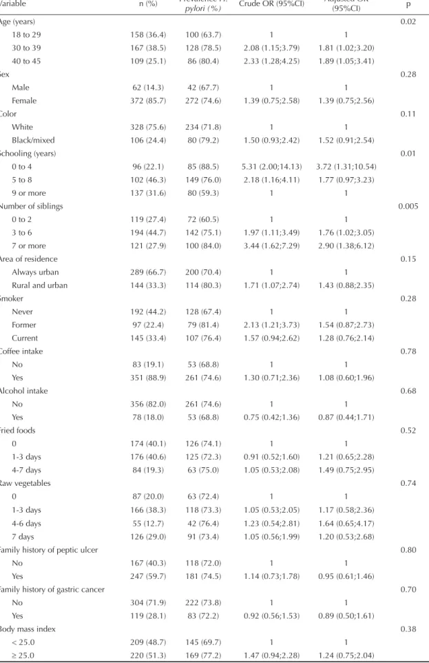
Table 1. Prevalence of Helicobacter pylori among users of primary healthcare units aged 18-45 years with uninvestigated dyspepsia. Municipality of Pelotas, Southern Brazil, 2006-2007 (n= 434).
Variable n (%) Prevalence H.
pylori (%) Crude OR (95%CI)
Adjusted OR
(95%CI) p
Age (years) 0.02
18 to 29 158 (36.4) 100 (63.7) 1 1
30 to 39 167 (38.5) 128 (78.5) 2.08 (1.15;3.79) 1.81 (1.02;3.20)
40 to 45 109 (25.1) 86 (80.4) 2.33 (1.28;4.25) 1.89 (1.05;3.41)
Sex 0.28
Male 62 (14.3) 42 (67.7) 1 1
Female 372 (85.7) 272 (74.6) 1.39 (0.75;2.58) 1.39 (0.75;2.56)
Color 0.11
White 328 (75.6) 234 (71.8) 1 1
Black/mixed 106 (24.4) 80 (79.2) 1.50 (0.93;2.42) 1.52 (0.91;2.54)
Schooling (years) 0.01
0 to 4 96 (22.1) 85 (88.5) 5.31 (2.00;14.13) 3.72 (1.31;10.54)
5 to 8 102 (46.3) 149 (76.0) 2.18 (1.16;4.11) 1.77 (0.97;3.23)
9 or more 137 (31.6) 80 (59.3) 1 1
Number of siblings 0.005
0 to 2 119 (27.4) 72 (60.5) 1 1
3 to 6 194 (44.7) 142 (75.1) 1.97 (1.11;3.49) 1.76 (1.02;3.05)
7 or more 121 (27.9) 100 (84.0) 3.44 (1.62;7.29) 2.90 (1.38;6.12)
Area of residence 0.15
Always urban 289 (66.7) 200 (70.4) 1 1
Rural and urban 144 (33.3) 114 (80.3) 1.71 (1.07;2.74) 1.43 (0.88;2.35)
Smoker 0.28
Never 192 (44.2) 128 (67.4) 1 1
Former 97 (22.4) 79 (81.4) 2.13 (1.21;3.73) 1.54 (0.87;2.73)
Current 145 (33.4) 107 (76.4) 1.57 (0.94;2.62) 1.28 (0.76;2.14)
Coffee intake 0.78
No 83 (19.1) 53 (68.8) 1 1
Yes 351 (88.9) 261 (74.6) 1.30 (0.71;2.36) 1.08 (0.60;1.96)
Alcohol intake 0.68
No 356 (82.0) 261 (74.6) 1 1
Yes 78 (18.0) 53 (68.8) 0.75 (0.42;1.36) 0.87 (0.44;1.71)
Fried foods 0.52
0 174 (40.1) 126 (74.1) 1 1
1-3 days 176 (40.6) 125 (72.3) 0.91 (0.52;1.60) 1.21 (0.65;2.28)
4-7 days 84 (19.3) 63 (75.0) 1.05 (0.53;2.08) 1.49 (0.75;2.95)
Raw vegetables 0.74
0 87 (20.0) 63 (72.4) 1 1
1-3 days 166 (38.3) 118 (73.3) 1.05 (0.53;2.05) 1.17 (0.58;2.36)
4-6 days 55 (12.7) 42 (76.4) 1.23 (0.54;2.81) 1.64 (0.65;4.17)
7 days 126 (29.0) 91 (73.4) 1.05 (0.56;1.99) 1.20 (0.53;2.68)
Family history of peptic ulcer 0.80
No 167 (40.3) 118 (72.0) 1 1
Yes 247 (59.7) 181 (74.5) 1.14 (0.73;1.78) 0.95 (0.61;1.46)
Family history of gastric cancer 0.70
No 304 (71.9) 222 (73.8) 1 1
Yes 119 (28.1) 83 (72.2) 0.92 (0.56;1.53) 0.89 (0.50;1.61)
Body mass index 0.38
< 25.0 209 (48.7) 145 (69.7) 1 1
Table 1 also shows that prevalence of H. pylori infec-tion increased with age, from 63.7% among subjects under 30 and 80.4% among those aged 40-45 years. Prevalence of infection was inversely associated with schooling. Prevalence was 88.5% among subjects with up to four years of schooling, 76% among those with 5-8 years, and 59.3% among those with ≥9 years. There was a direct association with number of siblings during childhood, from 60.5% among subjects with up to two siblings, to 75.1% among those with 3-6 siblings, and 84% among those with ≥ 7 siblings. For the remaining variables, associations with presence of
H. pylori were not signifi cant.
In multiple regression analysis (Table 1), age, schooling and number of siblings during childhood remained asso-ciated with prevalence of H. pylori infection, and were thus included in the score. ORs for infection with H. pylori among dyspeptic subjects aged 40-45 and 30-39 years were, respectively, 89% and 81% higher than those of dyspeptic subjects aged 18-29 years (reference group). Subjects with up to four years of schooling showed an OR of 3.7 compared to those with nine or more years of schooling. Among those with seven or more siblings during childhood, odds of infection were 190% higher than among those with up to two siblings.
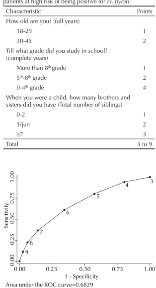
Table 2 lists the variables included in the score, along with their respective weights. The remaining variables are listed in the order in which questions were asked in the interview. OR for all subjects older than 29 years were similar in multiple regression analysis; therefore, for reasons of simplicity, age was dichotomized into 18-29 and 30-45 years. Variables with the greatest weight in the score were low schooling (0-4th grade,
with 4 points) and large family during childhood (3 points for those reporting seven or more siblings). In total, the score can range from 3 to 9 points.
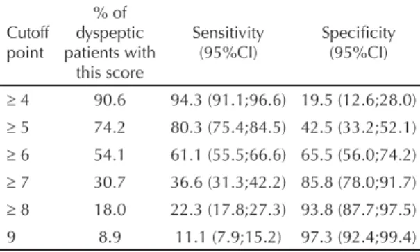
Table 3 presents sensitivity and specifi city values for different cutoff points. Sensitivity decreased from 94.3% at the ≥4 cutoff to 11.1% at the =9 cutoff. As a consequence, specifi city increased from 19.5% at ≥4 to 97.3% at =9. The ROC curve (Figure) shows that the cutoff point ≥6 yielded the best combination of sensi-tivity and specifi city (61.1% and 65.5%, respectively). Approximately 54% of dyspeptic subjects scored 6 points or more, indicating that, upon administration of the score, over half of the patients would be treated for H.pylori (Table 3). However, the positive predic-tive value of the ≥6 cutoff point (83.1%; 77.7 to 87.7) (Table 4), did not exceed by much the pre-test prob-ability of infection (74%).
Using as a cutoff scores ≥7, ≥8, and 9, 87.8%, 90.9%, and 92.1% of dyspeptic patients, respectively, were true
H. pylori positive (Table 4). With these cutoff points, treatment would be indicated for 30.7%, 18%, and 8.9% of patients. However, the sensitivity associated with these cutoff points was low for a screening test (36.6%, 22.3%, and 11.1%), and would therefore lead to a high rate of false-negative results (H. pylori-positive patients erroneously identifi ed as negatives).
DISCUSSION
The prevalence of uninvestigated dyspepsia detected in the present study (9.3%; 95%CI: 7.8;11.4) was similar to that observed in a population-based cross-sectional study carried out in Pelotas in 2005,a which detected a
Figure. ROC curve for the performance of different cutoff points of the epidemiological score for identifying dyspeptic patients. Municipality of Pelotas, Southern Brazil, 2006-2007.
0.00
0.25
0.50
0.75
1.00
Sensitivity
0.00 0.25 0.50 0.75 1.00 1 - Specificity
Area under the ROC curve=0.6829 9
7
8
6
5
4 3
Table 2. Epidemiological score for identifying dyspeptic patients at high risk of being positive for H. pylori.
Characteristic Points How old are you? (full years)
18-29 1
30-45 2
Till what grade did you study in school? (complete years)
More than 8th grade 1
5th-8th grade 2
0-4th grade 4
When you were a child, how many brothers and sisters did you have (Total number of siblings)
0-2 1
3/jun 2
≥7 3
Total 3 to 9
a Oliveira SS. Prevalência de dispepsia segundo Roma II e fatores associados: um estudo de base populacional [doctorate thesis]. Pelotas:
prevalence of 11.5% (95%CI: 10.6;12.4). Even though the latter study included subjects of a wider age range (20 years or older), prevalence of dyspepsia has been reported to be lower in the extremes of adult age.2
Therefore, it is possible that the present study included subjects from higher-prevalence age groups. Further-more, it is less likely that symptomatic individuals aged over 45 years remain uninvestigated, given the greater likelihood of malignancies among this population. Popu-lation-based studies carried out in England,4 Taiwan,6
and Denmark5 have reported decreasing prevalence of
uninvestigated dyspepsia with increasing age.
In addition to the self-limited character of symptomatic episodes of dyspepsia, the decision to consult a physi-cian depends on a range of factors, from the intensity of symptoms to the anxiety generated by fear of severe disease.3,7 In the present study, 11.1% of dyspeptic
individuals were visiting the health facility for that very reason, corresponding to a fraction of approximately 1% (95%CI: 0.7;1.3) of the total demand among 18 to 45-year-olds attending PHUs in the urban area of Pelotas. This rate is lower than the 4% described in a one-year period by McCormick et al,11 in 1995 in
England, and closer to that reported by Majundar et al,8
in a cohort of 5,064 adults over 18 years old followed for 18 months in the United States (2%-4%).
Though there is no indication of association between
H. pylori infection and dyspepsia, current directives recommend the identifi cation and eradication of H.
pylori among patients under 45 years and with persis-tent dyspepsia.9 The major reason for investigating
young dyspeptic patients is to exclude the presence of peptic ulcer,12 since upper digestive-tract cancer is
exceedingly rare among dyspeptic patients under 45 years. The strategy of screening for H. pylori among dyspeptic patients under 45 years and treating positive cases is therefore based on two assumptions: low risk of malignancy in this age group and the probability of treating virtually all patients with peptic ulcer. The disadvantage of such a strategy is related to the fact that certain H. pylori-positive patients that do not have peptic ulcer will be treated without necessity. However, there is evidence that presence of the bacteria is an important predictor of ulcerous disease, especially among smokers. McColl et al10 investigated 327
consec-utive dyspeptic patients referred to further investiga-tion, carrying out urea breath tests prior to endoscopy. Among H. pylori-negative patients, 2% had duodenal ulcer, 3% had gastric ulcer, and 17% had esophagitis. Corresponding prevalences among H. pylori-positives were 40%, 13%, and 12%, respectively.
In areas such as Pelotas, the probability of a dyspeptic patient being H. pylori-positive prior to any tests is high (prevalence of infection = 74%). Non-selective admin-istration of 13C-UBT would therefore lead to indication
of eradicating treatment for three out of four dyspeptic patients seen in the context of primary care. Clinical identifi cation of patients at highest risk of being H. pylori-positive, and the selective referral of these patients for investigation, could result in a reduction in costs with unnecessary tests and treatments. However, the use of three patient variables (age, schooling, and number of childhood siblings), in the form of a simple score that could be administered during a medical appointment, was not valid for identifying dyspeptic patients at higher risk of H. pylori infection in this sample of young dyspeptic patients recruited from PHUs in Pelotas. Cutoff points that would allow for accurate identifi cation of presence of the bacteria with a probability (positive predictive value) higher than the pre-test probability would be of great use for indicating which patients should be preferentially investigated. Scores 7 through 9 fulfi ll this condition, but gains are very modest. For
Table 3. Sensitivity and specifi city of different cutoff points of the score among dyspeptic patients. Municipality of Pelotas, Southern Brazil, 2006-2007. (n= 427)
Cutoff point
% of dyspeptic patients with
this score
Sensitivity (95%CI)
Specifi city (95%CI)
≥ 4 90.6 94.3 (91.1;96.6) 19.5 (12.6;28.0) ≥ 5 74.2 80.3 (75.4;84.5) 42.5 (33.2;52.1) ≥ 6 54.1 61.1 (55.5;66.6) 65.5 (56.0;74.2) ≥ 7 30.7 36.6 (31.3;42.2) 85.8 (78.0;91.7) ≥ 8 18.0 22.3 (17.8;27.3) 93.8 (87.7;97.5) 9 8.9 11.1 (7.9;15.2) 97.3 (92.4;99.4)
Table 4. Positive and negative predictive value (PV) and accuracy of different cutoff points for the score among dyspeptic patients. Municipality of Pelotas, Southern Brazil, 2006-2007. (n= 427)
Thus, management of uninvestigated dyspepsia among young patients seen in PHUs should consist of symp-tomatic treatment of the syndrome, with upper digestive endoscopy being reserved for specifi c cases, such as patients with certain alarm symptoms.
Finally, the “test-and-treat” strategy recommended in developed countries was found to be inadequate for use in populations in which prevalence of infection is very high, such as that of the present study.
ACKNOWLEDGEMENTS
We would like to thank the International Agency of Atomic Energy (IAEA) of Vienna, Austria, for providing
13C-labeled urea for breath tests (Projeto ARCAL LIV-
RLA 6/054); and the Pelotas Municipal Secretariat of Health and Social Wellbeing, for making their facilities and professionals available to the present study. example, score ≥7 increases this probability by 13.8
percentage points (or 19%), from a pre-test probability of 74% to a post-test probability of 87.8%). Applying this cutoff point to a population of dyspeptic patients with prevalence of infection similar to that of the present study would signify recommending H. pylori testing for 30.7% of patients (one out of three). With cutoff points
≥8 and ≥9, respectively, only 18% and 8.9% dyspeptic patients (one in six and one in 11) would be referred to diagnostic testing, with a probability of over 90% of accurately detecting presence of H. pylori. However, the sensitivity of these cutoff points was exceedingly low for a screening instrument, with a consequent increase in the number of false-negatives, which would be ques-tionable from the ethical standpoint. Low sensitivity would mean that, when administered to patients with characteristics similar to those of the present sample, a high proportion of carriers would fail to be identifi ed as such, and consequently remain untested and untreated.
1. Bytzer P, Talley NJ. Dyspepsia. Ann Intern Med.
2001;134(9 Pt 2):815-22.
2. Drossman DA, editor. Rome II: the functional gastrointestinal disorders – diagnosis, pathophysiology, and treatment: a multinational consensus. 2nd edition.
McLean, VA: Degnon Associates; 2000.
3. Howell S, Talley NJ. Does fear of serious disease predict consulting behaviour amongst patients with dyspepsia in general practice? Eur J Gastroenterol Hepatol. 1999;11(8):881-6. DOI: 10.1097/00042737-199908000-00012
4. Jones RH, Lydeard SF, Hobbs FD, Kenkre JE, Williams EL, Jones SJ, et al. Dyspepsia in England and Scotland.
Gut. 1990;31(4):401-5. DOI: 10.1136/gut.31.4.401
5. Kay L, Jørgensen T. Epidemiology of upper dyspepsia in a random population. Prevalence, incidence, natural history, and risk factors.
Scand J Gastroenterol.1994;29(1):2-6. DOI: 10.3109/00365529409090428
6. Lu CL, Lang HC, Chang FY, Chen CY, Luo JC, Wang SS, et al. Prevalence and health/social impacts of functional dyspepsia in Taiwan: a study based on the Rome criteria questionnaire survey assisted by endoscopic exclusion among a physical check-up population. Scand J Gastroenterol. 2005;40(4):402-411. DOI: 10.1080/00365520510012190
7. Lydeard S, Jones R. Factors affecting the decision to consult with dyspepsia: comparison of consulters and non-consulters. J R Coll Gen Pract.
1989;39(329):495-8.
8. Majumdar SR, Soumerai SB, Farraye FA, Lee M, Kemp JA, Henning JM, et al. Chronic acid-related
disorders are common and underinvestigated. Am J Gastroenterol. 2003;98(11):2409-14. DOI: 10.1111/ j.1572-0241.2003.07706.x
9. Malfertheiner P, Megraud F, O’Morain C, Bazzoli F, El-Omar E, Graham D, et al. Current concepts in the management of Helicobacter pylori infection: the Maastricht III Consensus Report. Gut. 2007;56(6):772-781. DOI: 10.1136/gut.2006.101634
10. McColl KE, el-Nujumi A, Murray L, el-Omar E, Gillen D, Dickson A, et al. The Helicobacter pylori breath test: a surrogate marker for peptic ulcer disease in dyspeptic patients. Gut. 1997;40(3):302-6.
11. McCormick A, Fleming D, Charlton J. Morbidity statistics from general practice. Fourth National Study 1991-1992. London: Offi ce of Population Censuses and Surveys; 1995.
12. Moayyed P. Helicobacter pylori test and treat strategy for young dyspeptic patients: new data. Gut.
2002;50(Suppl 4):iv47-50. DOI: 10.1136/gut.50. suppl_4.iv47
13. Santos IS, Boccio J, Santos AS, Valle NCJ, Halal CS, Bachilli MC, Lopes RD. Prevalence of Helicobacter pylori infection and associated factors among adults in Southern Brazil: a population-based cross-sectional study. BMC Public Health. 2005;5:118. DOI: 10.1186/1471-2458-5-118
14. Zubillaga M, Oliveri P, Panarello H, Buzurro M, Adami J, Goldman C, et al. Stable isotope techniques for the detection of Helicobacter pylori infection in clinical practice. 13C-urea breath test in different experimental
conditions. Acta Physiol Pharmacol Ther Latinoam.
1999;49(2):101-7. REFERENCES