Linear competitive inhibition of human
tissue kallikrein by 4-aminobenzamidine
and benzamidine and linear mixed
inhibition by 4-nitroaniline and aniline
Departamentos de 1Análises Clínicas e Toxicológicas, Faculdade de Farmácia, 2Engenharia Q uímica, Escola de Engenharia, and 3Bioquímica e Imunologia,
Instituto de Ciências Biológicas, Universidade Federal de Minas Gerais, Belo Horizonte, MG, Brasil
M.O . Sousa1,
T.L.S. Miranda2, E.B. Costa1,
E.R. Bittar3, M.M. Santoro3
and A.F.S. Figueiredo1
Abstract
Hydrolysis of D-valyl-L-leucyl-L-arginine p-nitroanilide (7.5-90.0 µM) by human tissue kallikrein (hK1) (4.58-5.27 nM) at pH 9.0 and 37oC was studied in the absence and in the presence of increasing concentrations of 4-aminobenzamidine (96-576 µM), benzamidine (1.27-7.62 mM), 4-nitroaniline (16.5-66 µM) and aniline (20-50 mM). The kinetic parameters determined in the absence of inhibitors were: Km = 12.0 ± 0.8 µM and kcat = 48.4 ± 1.0 min-1. The data indicate that the inhibition of hK1 by 4-aminobenzamidine and benzamidine is linear competitive, while the inhibition by 4-nitroaniline and aniline is linear mixed, with the inhibitor being able to bind both to the free enzyme with a dissociation constant Ki yielding an EI complex, and to the ES complex with a dissociation constant Ki, yielding an ESI complex. The calculated Ki values for 4-aminobenzamidine, benzami-dine, 4-nitroaniline and aniline were 146 ± 10, 1,098 ± 91, 38.6 ± 5.2 and 37,340 ± 5,400 µM, respectively. The calculated Ki values for 4-nitroaniline and aniline were 289.3 ± 92.8 and 310,500 ± 38,600 µM, respectively. The fact that Ki>Ki indicates that 4-nitroaniline and aniline bind to a second binding site in the enzyme with lower affinity than they bind to the active site. The data about the inhibition of hK1 by 4-aminobenzamidine and benzamidine help to explain previous observations that esters, anilides or chloromethyl ketone derivatives of Na-substituted arginine are more sensitive substrates or inhibitors of hK1 than the corresponding lysine compounds.
Co rre spo nde nce
A.F.S. Figueiredo
Departamento de Análises Clínicas e Toxicológicas
Faculdade de Farmácia, UFMG Caixa Postal 689
30123-970 Belo Horizonte, MG Brasil
Fax: + 55-31-339-7666 E-mail: afsf@ farmacia.ufmg.br
Presented at the XXVIII Annual Meeting of the Brazilian Society of Biochemistry and Molecular Biology, Caxambu, MG, Brazil, May 22-25, 1999.
This work is part of a PhD thesis to be presented by M.O . Sousa to the Graduate course in Biochemistry and Immunology, ICB, UFMG.
Research supported by FAPEMIG (No. CBS 1173/95) and CNPq (No. 521259/95-9). T.S. Miranda was the recipient of a Recent Doctor fellowship from CNPq (No. 300716/95-8). E.B. Costa was the recipient of a Scientific Initiation fellowship from CNPq (No. 521259/95-9). E.R. Bittar is the recipient of a Graduate Student fellowship from CNPq.
Received November 26, 1999 Accepted O ctober 11, 2000
Ke y wo rds
·Kinetics of human tissue kallikrein inhibition ·Tissue kallikrein ·4-Nitroaniline ·Aniline ·Benzamidine ·4-Aminobenzamidine ·Enzyme inhibition
Intro ductio n
The kallikreins (EC 3.4.21.8) are serine proteases present in glandular cells, neutro-phils and biological fluids. They are divided into two main groups, i.e., plasma (EC 3.4.21.34) and tissue (EC 3.4.21.35)
prin-cipal known biological function is the highly selective hydrolysis of plasma high and low molecular mass kininogens at two different peptide bonds (Met379-Lys380 and Arg389
-Ser390), with residues numbered on the basis
of the structure of prekallikrein (3), to stoi-chiometrically release the vasoactive and spasmogenic decapeptide kallidin (Lys-bradykinin) (2). Human tissue kallikrein (hK1) (4) also hydrolyzes various synthetic substrates such as Na-substituted arginine and lysine derivatives (amides, esters and fluorogenic peptides) (5-8), Cbz-Tyr-OpNP (9,10) and D-Pro-Phe-Phe-Nan (8). Like other serine proteinases, hK1 is inhibited by diiso-propylfluorophosphate, chloromethyl ke-tones of arginine and lysine, and basic pan-creatic trypsin inhibitor (BPTI) (also known as aprotinin, Trasylol® or Kunitz pancreatic
trypsin inhibitor) (6,11). In contrast, soy-bean trypsin inhibitor, a potent inhibitor of trypsin, plasma kallikrein and other serine proteinases, does not inhibit hK1 (6).
The inhibition of hK1 by BPTI is not a simple competitive inhibition as first reported (6,10), but is competitive inhibition of the parabolic type, with two inhibitor molecules binding to one enzyme molecule, forming a ternary enzymatic complex (12). It is note-worthy that the second BPTI molecule binds to the enzyme with higher affinity, suggest-ing that this second bindsuggest-ing site was prob-ably created or positively modulated as a consequence of the binding of the first BPTI molecule (12). However, the nature of the second binding site for this inhibitor in hK1 is still unknown (12).
There are controversial reports in the literature about the inhibition of hK1 es-terase activity by benzamidine (BzA). BzA has been reported to be a competitive inhib-itor of the hK1-catalyzed hydrolysis of Tos-Arg-OMe with a Ki value of 6.42 mM, but
does not inhibit the hK1-catalyzed hydroly-sis of Cbz-Tyr-OpNP even at a level of 2 mM (9). On the other hand, BzA has been re-ported to be a competitive inhibitor of the
hK1-catalyzed hydrolysis of Cbz-Tyr-OpNP with a Ki value of 0.2 mM (10). However,
there are no reports concerning the inhibi-tion of the amidase activity of hK1 by 4-aminobenzamidine (4-ABzA), BzA, aniline (An) and 4-nitroaniline (4-NAn).
The purpose of the present study was to examine in depth the kinetics of the inhibi-tion of hK1 amidase activity by 4-ABzA, BzA, 4-NAn and An in order to identify the precise mechanism of inhibition and to de-termine the number of binding sites and their accurate inhibition constants (Ki).
Our results clearly demonstrate that inhibition of hK1 amidase activity by 4-ABzA and BzA is a linear competitive inhi-bition with only one inhibitor molecule bind-ing to one enzyme molecule, formbind-ing a bi-nary enzymatic complex EI. On the other hand, our results also demonstrate that inhibition of hK1 amidase activity by An and 4-NAn is of the linear mixed type, with the inhibitors being able to bind both to the free enzyme to yield a binary enzymatic complex EI, and to the ES complex to yield a non-reactive ternary enzymatic complex ESI.
Mate rial and Me tho ds
Mate rial
An, 4-NAn, 4-ABzA·2HCl, BzA·HCl and BPTI were purchased from Sigma Chemical Co. (St. Louis, MO, USA). D-Valyl-L-leucyl-L-arginine 4-nitroanilide (D-Val-Leu-Arg-Nan) was obtained from Chromogenix AB, (Mölndal, Sweden).
All other reagents were of reagent grade from Sigma.
Enzym e
Enzym e assay
Kallikrein amidase activity was assayed spectrophotometrically (6) at 410 nm to moni-tor the release of 4-NAn (e410 = 8800 M-1 cm-1).
A 1-cm path-length cuvette containing 50 µl of 45.8-52.7 nM hK1 (Mr 31,000) (12) in 200
mM glycine/NaOH buffer, pH 9.0, containing 0.05% (w/v) NaN3 and 100 µl of buffer or 100
µl of an adequate dilution of a stock inhibitor solution (10 mM 4-ABzA, 100 mM BzA, 1.66 mM 4-NAn and 100-300 mM An) in the same buffer was placed in the thermostated cell compartment of a Shimadzu UV 160 A spec-trophotometer at 37oC and pre-incubated for 5
min. The concentrations of 4-ABzA (e292= 15,280 M-1 cm-1) (13), BzA (e225 = 9390 M-1
cm-1) (13) and 4-NAn solutions were
deter-mined spectrophotometrically. Recently dis-tilled aniline (d = 1.022 g/cm3) was used. The
ranges of inhibitor concentrations were se-lected in order to obtain 20-80% inhibition. Then, 350 µl (10.7-128.6 µM) of D-Val-Leu-Arg-Nan in 200 mM glycine/NaOH buffer, pH 9.0, containing 0.05% (w/v) NaN3 was
added, and the increase in absorbance at 410 nm with time was continuously recorded for 3 min. The slope of the time-dependent absorbance curve extrapolated to zero time was converted to µM of released 4-NAn per minute.
Bovine ß-trypsin (Bß-TR) (2.91 nM) was also assayed spectrophotometrically at 410 nm with the substrate D-Val-Leu-Arg-Nan (200-600 µM) in 100 mM Tris-HCl buffer, pH 8.1, containing 20 mM CaCl2 and 0.05% (w/v)
NaN3, in the absence and in the presence of An
(10-40 mM) in order to check for inhibition. Total substrate concentration was determined from the amount of 4-NAn released after com-plete hydrolysis by excess Bß-TR. In the inhi-bition assays with 4-NAn the reference cu-vette contained the same amount of 4-NAn as the sample cuvette.
Tre atm e nt o f kine tic data
The kinetic data of the inhibition of hK1
by 4-ABzA and BzA and the kinetic data of Bß-TR inhibition by An can be described, according to Plowman (14), as competitive inhibition.
On the other hand, the kinetic data of the inhibition of hK1 by 4-NAn and An can be described by the following scheme:
Ks@ Km kcat
E + S ES E + P
I Ki I Ki
EI ESI
which, according to Cornish-Bowden (15), is the simplest formal mechanism for mixed inhibition. According to the author, the in-hibitor (I) can bind both to the free enzyme (E) to give a complex EI with dissociation constant Ki, and to the ES complex to give an
unreactive ESI complex with dissociation constant Ki. As shown in this scheme, both
inhibitor-binding reactions are dead-end re-actions and are therefore equilibria. As both EI and ESI exist, however, it is difficult to see why S should not bind directly to EI to give ESI. If this reaction is included in the mechanism the rate equation becomes much more complicated, because terms in S2 and I2
appear in it. These terms cancel only if all of the binding reactions are equilibria, i.e., if the substrate- and product-release steps are all fast compared with the reaction that con-verts ES into products. In practice, however, the predicted deviations from simple kinet-ics are difficult to detect experimentally, and one cannot use adherence to simple kinetics as evidence that Km, Ki and Ki are true
dissociation constants.
The normalized initial rate (v) will be given by the following equation:
Eq. 1
vi kcat
.
[S]= v =
According to Cornish-Bowden (15), when linear mixed inhibition occurs, both kcatapp, and
kcatapp/Kmapp vary with the inhibitor
concentra-tion according to the following equaconcentra-tions:
kcat
kcatapp = Eq. 2
1 + [I]/Ki
which can be rearranged to
1/kcatapp = 1/kcat + [I]/(kcat
.
Ki) Eq. 3Additionally,
Km (1 + [I]/Ki)
Kmapp = Eq. 4
1 + [I]/Ki
and
kcat/Km
kcatapp/Kmapp = Eq. 5
1 + [I]/Ki
which can be rearranged to
Eq. 6 Kmapp/kcatapp = Km/kcat + [Km/(kcat
.
Ki)].
[I]The kinetic parameters (Km, Kmapp, kcat,
kcatapp and Ki for 4-ABzA, BzA, 4-NAn and
An) were calculated based on unweighted non-linear regression analysis of the data fit
to the appropriate Michaelis-Menten equa-tions, while the Ki values for 4-NAn and An
were determined by fitting the experimental data to Equation 1. Whenever used, the lin-ear replots (Equations 3 and 6) were shown only for diagnostic purposes; they were not used to calculate the Ki and Ki values.
Re sults
The hK1-catalyzed hydrolysis of D-Val-Leu-Arg-Nan followed Michaelis-Menten kinetics under the assayed substrate concen-tration range (7.5-90.0 µM). Figure 1 shows the 1/v vs 1/[S] plot for the hydrolysis of D-Val-Leu-Arg-Nan (7.5-90.0 µM) catalyzed by hK1 (4.58 nM) in the absence and in the presence of 4-ABzA (1.27-7.62 mM). The inset shows the replot of the slopes of the lines from Figure 1 (Kmapp/kcatapp vs 4-ABzA
concentration) according to Plowman (14). Each point is the mean of 4 determinations. Similar results were obtained with BzA.
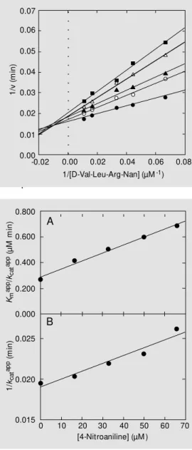
Figure 2 shows the 1/v vs 1/[S] plot for the hydrolysis of D-Val-Leu-Arg-Nan (7.5-90.0 µM) catalyzed by hK1 (5.27 nM) in the ab-sence and in the preab-sence of 4-NAn (16.5-66 µM). Each point is the mean of 4 determina-tions. Similar results were obtained with An.
Figure 3 shows the replots of Kmapp/kcatapp
(panel A) and 1/kcatapp (panel B) vs 4-NAn
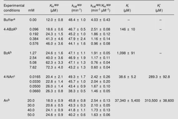
concentration according to Cornish-Bowden (15) (Equations 6 and 3, respectively). The straight lines obtained are consistent with linear mixed inhibition. Statistical analysis of these data using the GraphPad program at the 95% confidence level showed that both lines have slopes that are significantly dif-ferent from zero, and the statistical test for departure from linearity gave a negative (non-significant) result. Similar results were ob-tained with An. The kinetic parameters for hK1-catalyzed hydrolysis of D-Val-Leu-Arg-Nan in the absence and in the presence of 4-ABzA, BzA, 4-NAn and An are shown in Table 1.
The Bß-TR-catalyzed hydrolysis of D-Val-Figure 1 - Linew eaver-Burk plot
for the hydrolysis of D-Val-Leu-Arg-Nan by hK1 in the absence and in the presence of 4-ABzA. Inset, Kmapp/kcatappvs [4-ABzA]. Experimental conditions: 200 m M glycine/NaOH, pH 9.0, 37oC, 5-min incubation. hK1
con-centration: 4.58 nM . 4-ABzA concentrations: closed circles, 0 mM ; open circles, 0.096 mM ; closed triangles, 0.192 mM ; open triangles, 0.384 mM , and closed squares, 0.576 mM . Each point in the plot is the mean of quadruplicate determinations. M ore details are described in M aterial and M ethods.
1/
v
(m
in
)
0.25
0.20
0.10
0.05
0.00 0.15
Km
ap
p/k
ca
t
ap
p (µ
M
m
in
)
1.2
0.8
0.4
0.0
0.00 0.20 0.40 0.60 [4-ABzA] (mM )
-0.20 -0.10 0.00
1/[D-Val-Leu-Arg-Nan] (µM-1)
0.10 0.20
○○○○○○○
Leu-Arg-Nan followed Michaelis-Menten ki-netics in the substrate concentration range assayed (200-600 µM) (data not shown).
The Michaelis-Menten plot for the hy-drolysis of D-Val-Leu-Arg-Nan (200-600 µM) catalyzed by Bß-TR (2.91 nM) in the absence and in the presence of An (10-40 mM) showed enzyme inhibition (data not shown). The double-reciprocal plot for the An data showed convergent lines crossing at the same point on the 1/v axis, indicating competitive inhibition (data not shown). The replot of the slopes of the lines obtained from the double-reciprocal plot (Kmapp/kcatapp)
vs An concentration according to Plowman (14) showed a straight line with r2 = 0.952
(data not shown), indicating that An is a linear competitive inhibitor of Bß-TR ami-dase activity in the concentration range tested. No hint of a possible second binding site for An can be discerned in the experimental data obtained. The kinetic parameters (Km, kcat,
kcat/Km and Ki) for the Bß-TR-catalyzed
hy-drolysis of D-Val-Leu-Arg-Nan in the ab-sence and in the preab-sence of An, determined according to Plowman (14), were 191.7 ± 38.2 µM, 1604 ± 107 min-1, 8.4 ± 1.8 min-1
µM-1 and 10.8 ± 1.0 mM, respectively.
D iscussio n
During the characterization of our hK1 preparation, we decided to check the inhibi-tion of its amidase activity by 4-ABzA (pKa2
= pKa of the amidinium group = 12.39), BzA
(pKa = 11.41), 4-NAn (pKa = 1.0) (16) and
An (pKa = 4.60) (16). At pH 9.0, the
opti-mum pH for the hK1-catalyzed hydrolysis of D-Val-Leu-Arg-Nan, 4-ABzA and BzA showed a positive charge, while 4-NAn and An had no apparent electric charge, but di-pole moments (µ) of 6.10 D and 1.53 D (16), respectively. 4-ABzA and BzA were chosen because they are good models of the side chains of arginine and lysine (17) in the usual human tissue kallikrein substrates (6,7). Aniline was also chosen because of its
struc-Km
ap
p/k
ca
t
ap
p (µ
M
m
in
)
0.800
0.400
0.015 0.600
0.200
0.025 0.000
0.020
0 10 20 30 40 50 60 70
[4-Nitroaniline] (µM )
1/
kca
t
ap
p (m
in
)
A
B
Figure 3 - Replots of Kmapp/ kcatapp (panel A) and of 1/kcatapp
(panel B) against 4-nitroaniline concentration. Regression coef-ficients: for panel A - intercept: 0.29000 ± 0.01405; slope: 0.006152 ± 0.000347; r2:
0.9906; for panel B - intercept: 0.01898 ± 0.00057; slope: 0.00009593 ± 0.00001406; r2:
0.9395. M ore details are de-scribed in M aterial and M eth-ods.
1/
v
(m
in
)
0.07
0.06
0.04
0.01
0.00 0.05
-0.02 0.02 0.04
1/[D-Val-Leu-Arg-Nan] (µM-1)
0.06 0.08
0.03
0.02
0.00
○○○○○○○○○○○○○○○○
○
Figure 2 - Linew eaver-Burk plot for the hydrolysis of D-Val-Leu-Arg-Nan by hK1 in the absence and in the presence of 4-NAn. Experimental conditions: 200 m M glycine/NaOH, pH 9.0, 37oC, 3-min incubation. hK1
concentration: 4.58 nM . 4-NAn concentrations: closed circles, 0 µM ; open circles, 16.5 µM ; closed triangles, 33.0 µM ; open triangles, 50.0 µM , and closed squares, 66.0 µM . Each point in the plot is the mean of quadru-plicate determinations. M ore details are described in M aterial and M ethods.
tural relationship to the side chain of the tyrosine constituent of the synthetic sub-strate CBz-Tyr-OpNP, which was demon-strated to be the hK1 substrate (9,10). On the other hand, 4-NAn was chosen not only because of its structural relationship to An, but also because it shows a larger dipole moment than the dipole moment of An (16); it is also one of the products of the hydrolysis of the chosen substrate.
hK1 inhibitio n by 4-ABzA and BzA
The data in Table 1 are consistent with com-petitive inhibition since in the presence of 4-ABzA and BzA the Kmapp values increase
with inhibitor concentration, although kcat
values remain approximately constant (15). The present results are consistent with the model in which hK1 and 4-ABzA or BzA can form enzyme-inhibitor complexes with a stoichiometry of 1:1. No hint of a possible second binding site for these inhibitors can be discerned in the experimental data ob-tained. Similar results were reported for the 4-ABzA and BzA inhibitions of the amidase activity of trypsin, a well-known serine pro-teinase, which showed that 4-ABzA and BzA are potent competitive inhibitors with Ki
val-ues of 8.25 µM and 18.4 µM, respectively (17).
Comparison of the Ki values for 4-ABzA
(pKa2 = 12.39) (146 ± 10 µM) and BzA (pKa
= 11.41) (1,098 ± 91 µM) reveals that 4-ABzA is a 7.5-fold more potent hK1 inhibi-tor than BzA.
Table 1 - Kinetic parameters of human tissue kallikrein.
a200 mMGlycine/NaOH, pH 9.0, at 37oC; enzyme concentration: b4.58 nM , c5.27 nM .
Experimental Kmapp kcatapp kcatapp/Kmapp Ki Ki’
conditions mM (µM ) (min-1) (min-1 µM-1) (µM ) (µM )
Buffera 0.00 12.0 ± 0.8 48.4 ± 1.0 4.03 ± 0.43 -
-4-ABzAb 0.096 18.6 ± 0.6 46.7 ± 0.5 2.51 ± 0.08 146 ± 10
-0.192 24.3 ± 1.5 45.2 ± 1.0 1.86 ± 0.12
0.384 41.3 ± 4.6 47.9 ± 2.4 1.16 ± 0.14
0.576 46.0 ± 3.6 44.1 ± 1.6 0.96 ± 0.08
BzAb 1.27 24.6 ± 1.6 47.1 ± 1.1 1.91 ± 0.05 1,098 ± 91
-2.54 40.0 ± 3.6 46.9 ± 1.9 1.17 ± 0.11
5.08 62.3 ± 3.3 47.1 ± 1.3 0.76 ± 0.04
7.62 72.3 ± 4.0 43.2 ± 1.3 0.60 ± 0.04
4-NAnc 0.0165 20.4 ± 2.1 49.3 ± 1.7 2.42 ± 0.26 38.6 ± 5.2 289.3 ± 92.8
0.0330 22.8 ± 1.4 45.7 ± 1.0 2.04 ± 0.20
0.0500 26.0 ± 1.4 43.4 ± 0.9 1.67 ± 0.10
0.0660 26.3 ± 0.8 38.3 ± 0.5 1.46 ± 0.05
Anb 20.0 18.0 ± 0.9 45.8 ± 0.8 2.54 ± 0.13 37,340 ± 5,400 310,500 ± 38,600
30.0 20.6 ± 0.5 43.3 ± 0.3 2.10 ± 0.05
40.0 24.1 ± 0.9 41.8 ± 1.1 1.73 ± 0.13
50.0 24.6 ± 0.9 40.2 ± 0.6 1.63 ± 0.06
Comparison of the Ki values for hK1
inhibition by 4-ABzA (146 ± 10 µM) and BzA (1098 ± 91 µM) with the Ki for trypsin
inhibition by 4-ABzA (8.25 µM) and BzA (18.4 µM) (17), respectively, reveals that 4-ABzA and BzA bind much more weakly to hK1 than to trypsin. These results agree with a published report stating that BzA binds much more weakly to hK1 (Ki ~15 mM) and
pK1 (Ki ~1 mM) than to trypsin (Ki = 20 µM)
(18).
The present results can explain previous observations regarding the hK1 substrate specificity, which indicate that arginine es-ter or anilide derivatives are more sensitive substrates for the enzyme than the corre-sponding lysine compounds (19). Similarly, our data explain previous observations about the reactivity of arginine and lysine chloromethyl ketones in inactivating hK1 which revealed that the enzyme was 10-fold more reactive with the Arg chloromethyl ketones than with the Lys ones (11). Thus, since the pKa value of the guanidinium group
localized on the side chain of Arg is 12.5 and the pKa value of the amino group localized
on the side chain of Lys is 10.0, it is easy to explain why hK1 has a significant prefer-ence for Arg over Lys residues at the P1
position (20) of their substrates (21). As previously reported, the stability of ion pairs increases with the difference in pKa of the
groups involved (22). In this way, ion pairs formed by a given anion (for instance, car-boxylate) with Arg will be more stable than those formed by Lys (22). There is evidence that this is true in many circumstances of biological interests (23,24). The interactions between hK1 and 4-ABzA and BzA seem to follow the same rule.
Comparison of the 4-ABzA and BzA inhi-bition of the amidase activities of human tis-sue kallikrein (this work) and trypsin (17) reveals that with these small molecules the inhibition mechanism of these two serine pro-teinases is similar - both enzymes are inhibited by a linear competitive mechanism.
hK1 inhibitio n by 4-NAn and An
The Michaelis-Menten plot for the hy-drolysis of D-Val-Leu-Arg-Nan (7.5-90 µM) catalyzed by hK1 (4.58-5.27 nM) in the ab-sence and in the preab-sence of 4-NAn (pKa =
1.00) (16.5-66 µM) and An (pKa = 4.60)
(20-50 mM) showed enzyme inhibition (data not shown). The double-reciprocal plot for the 4-NAn data (Figure 2) showed convergent lines crossing approximately at the same point in the second quadrant, indicating lin-ear mixed inhibition (15). Similar results were obtained with An. The data for 4-NAn and An were also analyzed by the Dixon plot (1/v vs [I]) and by the Cornish-Bowden plot ([S]/v vs [I]) (15), respectively. The straight lines obtained from the Dixon plots inter-sected approximately at the same point in the second quadrant, while the straight lines ob-tained from the Cornish-Bowden plots inter-sected approximately at the same point in the third quadrant (data not shown). These re-sults also indicate linear mixed inhibition (15). According to Segel (25), the Dixon plots for partial and most mixed-type inhibi-tion systems are curved. However, when the ESI complex is not catalytically active, the plot is linear. The data for 4-NAn and An in Table 1 are also not consistent with competi-tive inhibition. As both Kmapp and kcatapp vary
with inhibitor concentration, linear mixed inhibition is suggested (15).
Thus, in order to further clarify the inhi-bition type, we decided to replot the values of Kmapp/kcatapp and 1/kcatapp vs [I],
respec-tively, according to Equations 6 and 3, re-spectively (Figure 3, panels A and B, shows the results obtained with 4-NAn). Similar results were obtained with An. As both Kmapp
and kcatapp vary with [I], linear mixed
inhibi-tion was indicated (15). The Ki values for
4-NAn (38.6 ± 5.2 µM) and for An (37,340 ± 5,400 µM) and the Ki values for 4-NAn
corresponding data in a Michaelis-Menten plot.
Comparison of the Ki values for hK1
inhibition by 4-NAn (pKa = 1.00) (38.6 ± 5.2
µM) and 4-ABzA (pKa2 = 12.39) (146 ± 10
µM), respectively, reveals that 4-NAn binds 3.8-fold more strongly to the active center of hK1 than 4-ABzA. Since at pH 9.0 the amidinium group of 4-ABzA is bearing a full positive charge, while the amino group of 4-NAn shows a positive charge induced by intramolecular transfer of electrons from the amino group to the nitro group by isovalent resonance (16,26), it would be reasonable to expect that 4-ABzA would interact better with the S1 subsite of the active center of
hK1 (20) than 4-NAn. However, the data obtained do not concur with this reasoning. As a speculation, we may assume that 4-NAn, after binding to the S1 subsite of the
active center of hK1 (20), possibly could participate in an additional enzyme-inhibitor interaction of the dipole-dipole type, involv-ing the negative charge of the oxygen atom of the nitro group at the C-4 position of the aromatic ring, induced by resonance (16), with some group in the neighborhood of the active center region of hK1, while 4-ABzA, which shows an uncharged-NH2 group, also
at the C-4 position of the aromatic ring, would not participate. The additional dipole-dipole interaction would reinforce the bind-ing of 4-NAn to the active center of hK1.
A similar interaction was suggested by Mares-Guia et al. (26) to explain their results about the electronic effects in the interaction of para-substituted benzamidines with tryp-sin. According to these authors, their data can be interpreted in terms of an enzyme-inhibitor interaction of the dipole-dipole type. A dipole will appear in the inhibitor as a consequence of an intramolecular charge transfer from the substituent to the ring or vice-versa. Their data concur with the model in which intramolecular charge transfer ren-ders positive the electron-donating substitu-ent, thereby giving origin to a dipole that is
able to interact with a site in the enzyme. According to the authors, the hydroxyl group of the reactive Ser183 is the most probable
candidate for the dipole of the enzyme that interacts with the dipole at the para position in substituted benzamidines. On the other hand, electron-withdrawing groups generate a dipole of opposite polarity that decreases binding by a dipole-dipole repulsion.
We do not know which group is a pos-sible candidate for the dipole of the enzyme that interacts with the dipole in 4-NAn.
Comparison of the Ki values for 4-NAn
(38.6 ± 5.2 µM) and for An (37,340 ± 5,400 µM) reveals that 4-NAn binds 967-fold more strongly to the active center of hK1 than An. This result is partially consistent with the larger dipole moment of 4-NAn (6.10 D) over An (1.53 D) (16). However, the larger dipole moment of 4-NAn over An is not sufficient to explain the data obtained. As a speculation, we may state that, after binding to the S1 subsite of the active center of hK1
(20) through its =NH2+ group, An could not
participate in an additional dipole-dipole in-teraction as 4-NAn can.
Comparison of the Ki values for 4-NAn
(289.3 ± 92.8 µM) and for An (310,500 ± 38,600 µM) reveals that 4-NAn binds 1073-fold more strongly to a second binding site on hK1 than An. The second binding site for these molecules is not known, but it is quite probable that the negative charge of the oxy-gen atom of the nitro group of the 4-NAn molecule is more available for an additional dipole-dipole interaction with it than the negative charge at the C-4 position of the An molecule.
The fact that Ki>Ki indicates that both
4-NAn and An bind to a second binding site in the hK1 molecule with lower affinity than they bind to the S1 subsite of the hK1 active
center.
and also whether it is different from the second binding site for BPTI.
4-NAn is a special case since it is present in the substrate molecule D-Val-Leu-Arg-Nan where it shows neither a positive nor a negative charge. In the ES complex, the Nan (4-NAn) group is accommodated at the S1
position (20) of the hK1 active center. After substrate hydrolysis the released 4-NAn be-comes a dipolar molecule. As a dipolar mol-ecule 4-NAn is able to bind to the anionic site of hK1 as a competitive inhibitor through the positive charge on the amino group, and is also able to bind to a second binding site in hK1, possibly through the negative charge of the oxygen atom of the nitro group. Thus, 4-NAn is a product of the reaction that is able to inhibit hK1 as a mixed inhibitor.
The presence of a second inhibitor bind-ing site in hK1 seems to be clear and may have important implications in the physi-ological activity of this enzyme.
Bß-TR inhibitio n by An
Comparison of the kcat/Km values for the
hK1- and Bß-TR-catalyzed hydrolysis of D-Val-Leu-Arg-Nan (4.03 ± 0.43 and 8.4 ± 1.8 min-1 µM-1, respectively) reveals that
D-Val-Leu-Arg-Nan is a slightly better substrate for Bß-TR than for hK1.
Additionally, comparison of the Ki
val-ues for An inhibition of hK1 (37,340 ± 5,400 µM) and of Bß-TR (10,800 ± 1,000 µM) reveals that An interacts better with the S1
subsite of the active center of Bß-TR than with the S1 subsite of the active center of
hK1.
Ackno wle dgm e nts
We thank Prof. Giovanni Gazzinelli for reading the manuscript and offering con-structive criticism.
Re fe re nce s
1. Bhoola KD, Figueroa CD & Worthy K (1992). Bioregulation of kinins: kallikreins, kininogens and kininases. Pharmacologi-cal Review s, 44: 1-80.
2. M acDonald RJ, M argolius HS & Erdös EG (1988). M olecular biology of tissue kal-likrein. Biochemical Journal, 253: 313-321.
3. Del Nery E, Chagas JR, Juliano M A, Prado ES & Juliano L (1995). Evaluation of the extent of the binding site in human tissue kallikrein by synthetic substrates w ith se-quences of human kininogen fragments. Biochemical Journal, 312: 233-238. 4. Berg T, Bradshaw RA, Carretero OA, Chao
J, Chao L, Clements JA, Fahnestock M , Fritz H, Gauthier F, M acDonald RJ, M argolius HS, M orris BJ, Richards RI & Scicli AG (1992). A common nomencla-ture for members of the tissue (glandular) kallikrein gene families. Agents and Ac-tions, 38 (Suppl): 19-25.
5. Schachter M (1980). Kallikreins (kinino-genases) - A group of serine proteases w ith bioregulatory actions. Pharmacologi-cal Review s, 31: 1-17.
6. Geiger R & Fritz H (1981). Human urinary
kallikrein. M ethods in Enzymology, 80: 466-492.
7. Antonini E, Ascenzi P, M enegatti E, Bortolotti F & Guarneri M (1982). Catalytic properties of human urinary kallikrein. Biochemistry,21: 2477-2482.
8. Chagas JR, Portaro FCV, Hirata IY, Almeida PC, Juliano M A, Juliano L & Prado ES (1995). Determinants of the un-usual cleavage specificity of lysyl-bradyki-nin-releasing kallikreins. Biochemical Jour-nal, 306: 63-69.
9. Hial V, Diniz CR & M ares-Guia M (1974). Purification and properties of a human uri-nary kallikrein (kininogenase). Biochemis-try, 13: 4311-4318.
10. Geiger R, M ann K & Bettels T (1977). Isolation of human urinary kallikrein by affinity chromatography. Journal of Clini-cal Chemistry and CliniClini-cal Biochemistry, 15: 479-483.
11. Kettner C, M irabelli C, Pierce JV & Shaw E (1980). Active site mapping of human and rat urinary kallikreins by peptidyl chloromethyl ketones. Archives of Bio-chemistry and Biophysics, 202: 420-430. 12. M iranda TLS, Ramos CHI, Freire RTS,
Souza EP, Rogana E, Santoro M M & Figueiredo AFS (1995). Kinetic mechan-ism of the inhibition of human urinary kal-likrein by basic pancreatic trypsin inhibi-tor. Brazilian Journal of M edical and Bio-logical Research, 28: 505-512.
13. Rogana E, Nelson DL & M ares-Guia M (1975). Characterization of the ultraviolet absorption spectra of para-substituted de-rivatives of benzamidine. Journal of the American Chemical Society, 97: 6844-6848.
14. Plow man KM (1972). Inhibitor studies. In: Hume DN, Stork G, King EL, Herschbach DR & People JA (Editors), Enzyme Kinet-ics. M cGraw -Hill Book Company, New York, 56-75.
15. Cornish-Bow den A (1981). Inhibitors and activators. In: Cornish-Bow den A (Editor), Fundam ent als of Enzym e Kinet ics. Butterw orth & Co., Ltd., London, 73-98. 16. Cram DJ & Hammond GS (1964). Physical
properties. In: Cram DJ & Hammond GS (Editors), Organic Chemistry. M cGraw -Hill Book Company, New York, 183-225. 17. M ares-Guia M & Shaw E (1965). Studies
Biological Chemistry, 210: 1579-1585. 18. Katz BA, Liu B, Barnes M & Springman
EB (1998). Crystal structure of recombi-nant human tissue kallikrein at 2.0 Å reso-lution. Protein Science, 7: 875-885. 19. Ascenzi P, M enegatti E, Guarneri M ,
Bortolotti F & Antonini E (1982). Catalytic properties of serine proteases. 2. Com-parison betw een human urinary kallikrein and human urokinase, bovine ß-trypsin, bovine thrombin, and bovine a -chymo-trypsin. Biochemistry, 21: 2483-2490. 20. Schechter I & Berger A (1967). On the
size of the active site in proteases. I. Pa-pain. Biochemical and Biophysical Re-search Communications, 27: 157-162.
21. Chen Z & Bode W (1983). Refined 2.5 Å X-ray crystal structure of the complex formed by porcine kallikrein A and the bovine pancreatic trypsin inhibitor. Jour-nal of M olecular Biology, 164: 283-310. 22. Weber G (1992). Transfer of proteins to
apolar media and the dynamic interactions of proteins and membranes. In: Weber G (Editor), Protein Interactions. Routledge, Chapman & Hall, Inc., New York, 161-176.
23. Riordan JF, M cElvany KD & Borders Jr CL (1976). Arginyl residues: anion recogni-tion sites in enzymes. Science, 195: 884-886.
24. Paddlan EA, Davis D, Rudikoft S & Potter
EM (1976).Structural basis for the speci-ficity of phosphorylcholine-binding immu-noglobulins. Immunochemistry, 13: 945-949.
25. Segel IH (1975). Rapid equilibrium partial and mixed-type inhibition. In: Segel IH (Editor), Enzyme Kinetics. John Wiley & Sons, New York, 161-226.
![Figure 2 shows the 1/v vs 1/[S] plot for the hydrolysis of D-Val-Leu-Arg-Nan (7.5-90.0 µM) catalyzed by hK1 (5.27 nM) in the ab-sence and in the preab-sence of 4-NAn (16.5-66 µM)](https://thumb-eu.123doks.com/thumbv2/123dok_br/15805869.649947/4.918.257.541.244.583/figure-shows-plot-hydrolysis-catalyzed-sence-preab-sence.webp)

