Rat vas deferens SERCA2 is modulated
by Ca
2
+
/calmodulin protein kinase
II-mediated phosphorylation
J.B.R. Rodriguez
1, H. Muzi-Filho
1, R.H.F. Valverde
2, L.E.M. Quintas
1, F. Noel
3,
M. Einicker-Lamas
2,4and V.M.N. Cunha
11
Programa de Farmacologia e Inflamac¸a˜o, Instituto de Cieˆncias Biome´dicas, Universidade Federal do Rio de Janeiro, Rio de Janeiro, RJ, Brasil
2
Instituto de Biofı´sica Carlos Chagas Filho, Universidade Federal do Rio de Janeiro, Rio de Janeiro, RJ, Brasil
3
Programa de Desenvolvimento de Fa´rmacos, Instituto de Cieˆncias Biome´dicas, Universidade Federal do Rio de Janeiro, Rio de Janeiro, RJ, Brasil
4Instituto Nacional de Cieˆncia e Tecnologia em Biologia Estrutural e Bioimagem, Rio de Janeiro, RJ, Brasil
Abstract
Ca2+
pumps are important players in smooth muscle contraction. Nevertheless, little information is available about these pumps in the vas deferens. We have determined which subtype of sarco(endo)plasmic reticulum Ca2+
-ATPase isoform (SERCA) is expressed in rat vas deferens (RVD) and its modulation by calmodulin (CaM)-dependent mechanisms. The thapsigargin-sensitive Ca2+
-ATPase from a membrane fraction containing the highest SERCA levels in the RVD homogenate has the same molecular mass (,115 kDa) as that of SERCA2 from the rat cerebellum. It has a very high affinity for Ca2+(Ca0.5
= 780 nM) and a low sensitivity to vanadate (IC50= 41mM). These facts indicate that SERCA2 is present in the RVD. Immunoblotting for CaM and Ca2+
/calmodulin-dependent protein kinase II (CaMKII) showed the expression of these two regulatory proteins. Ca2+
and CaM increased serine-phosphorylated residues of the 115-kDa protein, indicating the involvement of CaMKII in the regulatory phosphorylation of SERCA2. Phosphorylation is accompanied by an 8-fold increase of thapsigargin-sensitive Ca2+
accumulation in the lumen of vesicles derived from these membranes. These data establish that SERCA2 in the RVD is modulated by Ca2+
and CaM, possibly via CaMKII, in a process that results in stimulation of Ca2+
pumping activity.
Key words: Ca2+
/calmodulin-dependent kinase II; Calcium homeostasis; Calmodulin; Rat vas deferens; SERCA2; Thapsigargin
Introduction
The Ca2+ion is one of the most important intracellular
messengers in eukaryotic cells. After a triggering event, Ca2+can be quickly mobilized from either the extracellular
medium or internal stores, leading to diverse cell responses, including smooth muscle contraction (1,2). Most of the Ca2+ that enters the cytoplasm is rapidly
bound to various cytosolic buffers (1). Calmodulin (CaM) is the most relevant Ca2+ binding protein and is also a
sensor of alterations in intracellular Ca2+ concentration
(3). This protein interacts reversibly with Ca2+to form a
Ca2+/CaM complex, which can bind to different cellular
targets. Indeed, many of the effects attributed to Ca2+are
exerted through Ca2+/CaM-regulated enzymes (4). At the
end of a Ca2+-dependent cellular event, Ca2+ pumps
embedded in the plasma membrane (PMCA) and in the membrane of the sarco(endo)plasmic reticulum (SERCA) actively transport Ca2+ions from cytosol to the outside of
the cells and to the lumen of the sarco(endo)plasmic reticulum, respectively (5,6).
The vas deferens is the tubule with contractile function essential for the ejaculation of sperm and hence male fertility. Experimentally, the rat vas deferens (RVD) has been extensively used in many physiological and phar-macological studies as a typical non-vascular smooth muscle tissue (7), representing an interesting model for investigation into the mechanisms of Ca2+ homeostasis
(8). For instance, we have demonstrated that denervation of the RVD modifies the expression of several proteins
Correspondence: V.M.N. Cunha, Centro de Cieˆncias da Sau´de, UFRJ, Av. Carlos Chagas Filho, 373, Bloco J, 21941-902 Rio de Janeiro, RJ, Brasil. Fax:+55-21-2562-6659. E-mail: [email protected]
involved in intracellular Ca2+regulation, including Na+/K+
-ATPase, L-type Ca2+channels and SERCA (9,10).
The SERCA family includes 3 gene products – SERCA1, SERCA2 and SERCA3 – that are expressed in a tissue-specific manner. Alternative splicing of the SERCA2 gene results in other different protein isoforms (11). The smooth muscle SERCA2b (115 kDa) isoform is identical to SERCA2a (110 kDa) except for its carboxyl terminus, where the four terminal amino acids of SERCA2a are replaced by an extended hydrophobic sequence of 49 amino acids. This stretch of amino acids presumably constitutes an 11th transmembrane segment for SERCA2b (12), which may be responsible for small biochemical and pharmacological differences between SERCA2a and SERCA2b isoforms (13). Activation of Ca2+/CaM-dependent protein kinase II (CaMKII)
stimu-lates the activity of SERCA2a pumps by direct phosphor-ylation of the Ser38 residue in these isoforms (14,15). In addition, CaMKII can phosphorylate phospholamban (16), a well-known modulator of SERCA pumps (17), and ryanodine receptor-Ca2+release channels (18).
A 115-kDa SERCA isoform is present in the RVD (19), but its biochemical and regulatory properties have yet to be determined. We have shown (20) that, among all subcellular fractions of RVD, the nuclear fraction contains the highest content of thapsigargin-sensitive Ca2+
-ATPase activity (SERCA), with minor contamination of thapsigargin-resistant Ca2+-ATPase activity (PMCA).
Thus, this SERCA-enriched membrane fraction is more suitable for studies of sarcoplasmic reticulum components than the classical microsomal fraction. The objectives of the present study were to characterize the type of SERCA pump isoform in RVD as well as to investigate the ability of CaMKII to modulate SERCA in smooth muscle.
Material and Methods
Ethical considerations
All experimental procedures involving the animals were approved by the Committee for Ethics in Animal Experimentation of Universidade Federal do Rio de Janeiro, and were carried out in accordance with the Committee’s guidelines.
Reagents and antibodies
The primary antibodies, anti-CaM and anti-CaMKII, were purchased from Sigma Chemical Company (USA) and anti-SERCA2 and anti-SERCA1 were purchased from Calbiochem-Novabiochem Co. (USA). Rainbow molecular weight markers were provided by GE Healthcare (UK); anti-P-Ser antibody was purchased from Biomol International (USA), and peroxidase-conjugated secondary antibodies were purchased from Promega Corporation (USA).
Calmodulin was purchased from Sigma Chemical Company. A23187 and thapsigargin were purchased
from Calbiochem-Novabiochem Co. Stock solutions of 5 mM thapsigargin were prepared in 100% dimethyl sulfoxide (DMSO). At saturating concentrations of thapsi-gargin (3mM) in buffers, the final concentration of DMSO was 0.07% (v/v), a concentration that had no effect on Ca2+-ATPase activity or active Ca2+uptake.
Preparation of a SERCA-enriched membrane fraction of RVD
Preparation of membranes was carried out as previously described (20). Briefly, RVDs were removed and immersed in cold Tyrode’s solution containing 137 mM NaCl, 2.7 mM KCl, 11.9 mM NaHCO3 plus
0.36 mM NaH2PO4, pH 7.4, 5.55 mM glucose, 1.77 mM
CaCl2, and 0.40 mM MgCl2. The tissue was dissected,
homogenized and the crude homogenate was either centrifuged at 1000g for 10 min to obtain the SERCA-enriched membrane fraction, or spun at 105,000g for 60 min to obtain the whole homogenate fraction. The pellets were resuspended in Tris-HCl-buffered 0.25 M sucrose solution, pH 7.4, and stored in liquid N2until use
(20). The protein content was determined by the method of Lowry et al. (21).
Measurements of Ca2+-ATPase activity
SERCA-enriched subcellular samples of RVD (20mg protein) were incubated for 2 h at 376C in 0.5 mL medium containing 50 mM HEPES-Tris, pH 7.4, 10 mM NaN3,
0.3 mM EGTA, 5 mM Na2ATP, 4 mM MgCl2, 5mM
A23187, 100 mM KCl and [c-32P]-ATP (specific activity: <1.56 1010Bq/mmol), with or without 3mM thapsigar-gin, and in the presence or absence of different concentrations of CaCl2 or vanadate (Na3VO4). The
concentration of free Ca2+ was calculated according to
Fabiato and Fabiato (22). Experiments were stopped by adding 1 mL of a cold mixture containing 26% (w/v) charcoal in 0.1 N HCl. The tubes were centrifuged at 1500gat 46C for 15 min, and 500mL of the supernatant was placed onto filters. The filters were dried and the radioactivity was counted in a liquid scintillation counter. Ca2+-ATPase activity was calculated by subtracting the
basal 32Pi release measured in the absence of Ca2+
(0.3 mM EGTA) from the total32Pi release measured in
the presence of increasing free Ca2+ concentrations
(EGTA.Ca2+ buffer) (20). The thapsigargin-resistant
Ca2+-ATPase activity (measured in the presence of
thapsigargin) was subtracted from the total Ca2+
-ATPase activity to obtain the thapsigargin-sensitive Ca2+-ATPase activity due to SERCA pumps (10,20).
The Ca2+dependence and the sensitivity of the enzyme to
vanadate were measured over the ranges of 0.2 to 20mM and 1mM to 1 mM, respectively.
Measurements of45Ca2+uptake
containing 50 mM HEPES-Tris, pH 7.4, 10 mM NaN3,
0.3 mM EGTA, 5 mM Na2ATP, 4 mM MgCl2, 100 mM
KCl, with sufficient CaCl2to provide 10mM free Ca2+and 45CaCl
2 (specific activity: <1.5 6 109Bq/mmol), in the presence or absence of 5mM A23187. The reaction was stopped by vacuum filtration and the filters were washed twice with 20 mL cold 20 mM MOPS, pH 7.0, 2 mM La(NO3)3, and 100 mM KCl, and counted in a liquid
scintillation counter. ATP-dependent Ca2+ accumulation
in the lumen of vesicles derived from the membranes was calculated by subtracting the Ca2+accumulation measured
in the presence of A23187 (blank) from the total Ca2+
accumulation measured in the absence of the ionophore. The stimulatory effect of CaM was assayed by preincubat-ing the membranes for 5 min in a reaction medium with or without 2mM CaM (23,24) before the addition of Ca2+.
These experiments were done in the presence of ruthenium red (25mM) to eliminate the stimulatory effect of CaMKII on Ca2+release channels (24).
Immunodetection of SERCA, CaM and CaMKII Western blotting was used to detect SERCA and to investigate whether CaM and CaMKII were present in the RVD-derived membranes. The samples were subjected to SDS-PAGE (6% polyacrylamide gel for SERCA1 and SERCA2, 7.5% for CaMKII and 15% for CaM) and transferred to nitrocellulose membranes. The membranes were incubated with 5% non-fat dry milk in Tris-buffered saline plus 0.1% Tween-20 followed by incubation with specific monoclonal (CaM, 1:500 dilution; anti-SERCA1, 1:4000; anti-SERCA2, 1:3000) or polyclonal antibodies (anti-CaMKII, 1:1000) and with anti-mouse (CaM, 1:11,000; SERCA1 and 2, 1:12,000) or anti-rabbit (CaMKII, 1:10,000) horseradish peroxidase-conjugated secondary antibodies, with the blots being detected by chemiluminescence. In these assays, the heavy microsomal fraction of skeletal muscle from adult rats (25), rat cardiac microsomes (25), chicken cerebellum microsomes (26), and the membrane fraction from electrocytes ofElectrophorus electricus (L.) (27) were used as positive controls for SERCA1, SERCA2a, SERCA2b, and CaMKII, respectively.
Immunodetection of serine-phosphorylated residues The phosphorylation reaction was initiated by the addition of 0.8 mM cold ATP after preincubation of 5mg of the SERCA-enriched fraction of RVD in a medium containing 50 mM HEPES-Tris, pH 7.4, 4 mM MgCl2,
200mM EGTA, and 100 mM KCl in the presence or absence of 10mM free Ca2+and 2mM CaM at 4
6C. The reaction was stopped after 2 min by adding 15mL SDS sample buffer. The samples were run on 7.5% SDS-PAGE gels before being transferred to nitrocellulose membranes. Non-specific phosphorylation sites were blocked with 5% bovine serum albumin in Tris-buffered saline plus 0.1% Tween-20. Serine-phosphorylated pep-tides were detected using an anti-phosphoserine
mono-clonal antibody (1:500) and anti-mouse horseradish peroxidase-conjugated antibody (1:20,000). Rabbit ske-letal muscle and rat cardiac microsomes were used as phosphoserine/phosphothreonine-positive controls (15). Control assays without Ca2+and exogenous CaM, but in
the presence of 0.8 mM exogenous ATP and without any additions (to detect any preexisting phosphorylation), were run in parallel.
Statistical analysis
Data are reported as means ± SE for 3 to 4 experiments performed in triplicate. Statistical compar-isons were determined by one-way ANOVA. The differ-ences were considered to be significant at P , 0.05. When Ca2+concentration dependence and inhibition by
vanadate were studied the equations were fitted to the experimental points by non-linear regression analysis (SigmaPlot, Jandell Scientific, USA).
Results
Ca2+-ATPase activity from the SERCA-enriched
membrane fraction of RVD: dependence on Ca2+
concentration and sensitivity to vanadate
To investigate the Ca2+dependence and sensitivity to
vanadate (a classical inhibitor of P-type ion motive ATPases) (28) of the Ca2+ pumps in SERCA-enriched
membranes, the Ca2+-ATPase activity was measured in
the presence of increasing concentrations of Ca2+ or
vanadate (in the presence of 10mM free Ca2+in the latter
case). Thapsigargin-sensitive Ca2+-ATPase activity, i.e., a
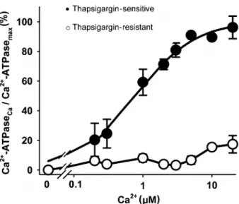
SERCA-associated activity (29), increases with increas-ing Ca2+concentration according to Equation 1.
v=Vmax~ Ca2z
= Ca0:5z Ca2z
(Equation 1)
where Vmaxand Ca0.5have the usual meaning (Figure 1).
The thapsigargin-sensitive activity had a Ca0.5 value of
780 nM and accounted for at least 80% of the total Ca2+
-ATPase activity (compare filled and open symbols). The thapsigargin-resistant activity was very low and, therefore, it was not possible to determine precisely Ca0.5or Vmax.
Vanadate inhibited the thapsigargin-sensitive Ca2+
-ATPase according to Equation 2 and Figure 2.
vi=vo~IC50= IC50z VO43 {
h i
(Equation 2)
where vois the activity in the absence of vanadate, viis
the activity at different inhibitor concentrations, and IC50
(41mM) is the vanadate concentration required to attain 50% of the maximal inhibition.
mono-clonal antibodies anti-SERCA1 or anti-SERCA2 pumps. The SERCA1 isoform was not detected in either the SERCA-enriched membrane or the whole homogenate (Figure 3A), whereas SERCA2 was the isoform present in the same preparations (lanes 1 and 2; Figure 3B). These bands have the same molecular mass as the positive control for SERCA2b (lane 3; chicken cerebellum, 115 kDa), but they also have a molecular mass near the SERCA2a band (lane 4; rat heart, 110 kDa). Figure 3 is a representative blot revealing the same profile that was found in the other two developed with the use of different RVD preparations.
Characterization of CaM and CaMKII from RVD Western blotting analysis indicated a 17-kDa protein in the SERCA-enriched membrane fraction as well as in the crude homogenate of RVD that co-migrated with that found in an enriched CaM preparation (chicken cerebel-lum microsomes; Figure 4A shows a typical assay). Likewise, Figure 4B shows the presence of CaMKII (lanes 1-3) co-migrating with the positive controls (chicken cerebellum, lane 4; electrocyte membrane, lane 5) in the 50-kDa region. Faced with the abundance of these regulatory proteins in RVD, it is possible that both CaM and CaMKII modulate the SERCA pump isoform in RVD smooth muscle, stimulating a regulatory kinase-mediated phosphorylation that has been described in other tissues and species (14,29).
Phosphorylation of the SERCA2 pump from RVD by CaMKII
To investigate whether the SERCA2 pump from RVD is modulated by CaMKII (Figure 4B), phosphorylation assays were used to detect phosphoserine residues by Western blotting. A phosphorylated band of,115 kDa was found in the SERCA-enriched fraction of RVD only after the addition Figure 1. Ca2+ concentration dependence of
thapsigargin-sensitive and -resistant Ca2+-ATPase activity. The assays were
performed in triplicate and data are reported as means ± SE for 3 experiments, using 3 different enzymatic preparations. The smooth curve was fitted to the thapsigargin-sensitive data points (filled circles) using Equation 1 as described in the text. Ca2+
-ATPase at 10mM Ca2+was 1.08 ± 0.11
mmol Pi x mg-1in 2 h. Ordinate legend: Ca2+-ATPase
Caand Ca2+-ATPasemaxrepresent
the activity at each Ca2+concentration and the maximal activity
obtained by fitting the function to the experimental points of thapsigargin-sensitive Ca2+-ATPase values, respectively.
Thapsigargin-resistant activities (open circles) were calculated from the total Ca2+-ATPase activity at each Ca2+concentration in
the same assay.
Figure 2. Inhibition of thapsigargin-sensitive Ca2+-ATPase
activity by vanadate. The assays were performed in triplicate with 10mM Ca2+and the results are reported as means ± SE for
4 experiments, using 3 different preparations. Ca2+-ATPase
activity was measured without vanadate in the control. The smooth curve was fitted to the data points using Equation 2 as described in the text. Ordinate legend: Ca2+-ATPase
iand Ca2+
-ATPaseorepresent activity in the presence and in the absence of
vanadate at each concentration of the inhibitor, respectively.
Figure 3. Expression of sarco(endo)plasmic reticulum Ca2+
-ATPase (SERCA) isoforms.A, Immunoblotting with anti-SERCA1 antibody and B, immunoblotting with anti-SERCA2 antibody.
Lane 1= 24mg SERCA-enriched fraction;lane 2= 32mg of
crude homogenate; lane 3 = 4mg of chicken cerebellum microsomes (positive control for SERCA2b);lane 4= 10mg of rat heart microsomes (positive control for SERCA2a);lane 5= 0.4mg (inA) or 10mg (in B) of heavy microsomes of rat fast
of Ca2+ and CaM (Figure 5), indicating that CaMKII
phosphorylates serine residues of the SERCA2 pump.
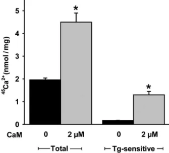
Calmodulin stimulates active Ca2+transport mediated
by SERCA2 in RVD
Since CaM strongly stimulates kinase-mediated phos-phorylation of SERCA2 (Figure 5), 45Ca2+ uptake was
assayed in the absence or presence of CaM to see whether CaMKII-mediated phosphorylation is associated with an increase in Ca2+pumping activity (30). Depletion of CaM by
mild alkaline treatment renders the vesicles leaky and Ca2+
accumulation is barely detectable. Therefore, we tested the influence of exogenous CaM on Ca2+transport. Addition of
CaM stimulates the steady levels of thapsigargin-sensitive
Ca2+ accumulation into the vesicles derived from the
membranes by a factor of 8 (Figure 6).
Discussion
We have demonstrated that SERCA2 is present in subcellular membranes from RVD and that its pumping capacity is stimulated by CaM through a pathway that is probably mediated by CaMKII. In line with the results of our previous study (20), we have now worked with a so called ‘‘nuclear fraction’’ of RVD because i) it has a higher sarcoplasmic reticulum/plasma membrane ratio, and ii) the yield of thapsigargin-sensitive Ca2+-ATPase is 4-fold
higher when compared to the microsomal fraction, often chosen for these studies in several tissues/species (8,31). SERCA pump isoforms have a high degree of sequence identity, but they can be reasonably distin-guished from the type of tissue in which they are expressed by the analysis of biochemical parameters such as Ca2+dependence, vanadate sensitivity, and by
specific antibody recognition (13,32,33). Therefore, these criteria were used to characterize the SERCA isoform expressed in RVD. The Ca2+affinity of SERCA isoforms
reflects a critical functional property of Ca2+-ATPases that
is probably related to specialized Ca2+ environments
(13,34). The SERCA2b isoform, localized in smooth muscle (19) (usually comprising.70% of total SERCA) (25), and non-muscle cells (19) has a 2-fold higher affinity Figure 4.Expression of calmodulin (CaM) and Ca2+
/calmodulin-dependent protein kinase II (CaMKII) in the membranes of the SERCA-enriched fraction and crude homogenate. A, Immunoblotting with anti-calmodulin antibody.Lane 1= 24mg of SERCA-enriched fraction;lane 2= 32mg of whole homogenate;
lane 3= 4mg of chicken cerebellum microsomes (positive control for CaM).B, Immunoblotting with anti-CaMKII antibody.Lanes 1, 2, 3= 40mg of 3 different SERCA-enriched fractions from RVD;lane 4= 4mg of chicken cerebellum microsomes;lane 5= 20mg of innervated fraction of membrane electrocyte of Electrophorus electricus(L.).Lanes 4and5are positive controls for CaMKII. SERCA = sarco(endo)plasmic reticulum Ca2+-ATPase.
Figure 5.Phosphorylation of serine residues in sarco(endo)plas-mic reticulum Ca2+-ATPase (SERCA). Blots were obtained after
incubation of phosphorylated membranes (5mg) with anti-phos-phoserine residue antibody. The phosphorylation assays were performed in the absence (lane 1) or presence (lane 2) of 10mM free Ca2+and 2
mM calmodulin (CaM). Control (lane 3) without
Ca2+, calmodulin and ATP shows no preexisting phosphorylation.
Figure 6. Effect of calmodulin (CaM) on total and thapsigargin (Tg)-sensitive 45Ca2+ uptake by vesicle membranes from the
SERCA-enriched fraction.45Ca2+uptake without (black columns;
controls) or with 2mM exogenous calmodulin (gray columns). The assays were performed in triplicate and the data are reported as means ± SE for 4 experiments using 4 different preparations. SERCA = sarco(endo)plasmic reticulum Ca2+-ATPase. *P
,
for Ca2+ (Ca
0.5 = 0.1-0.3mM) compared to SERCA2a
(Ca0.5= 0.2-0.5mM) or SERCA1 (13,32,34), and 5-fold
higher than SERCA3 (Ca0.5 ,1mM) (34), when expressed in COS or HEK-293 cells. Accordingly, these differences in Ca2+affinity could be due to variations in
the equilibrium between the two main conformations of the enzyme, E1 and E2(12). Experiments in COS cells
using SERCA2b mutants without the putative 11th transmembrane segment indicate that the apparently higher Ca2+ affinity of SERCA2b does not involve
structural differences in Ca2+-binding sites between
SERCA2 isoforms, but instead would be a shift in the E1«E2equilibrium towards the high-affinity E1state (13).
The value of Ca0.5 for the SERCA isoform in RVD
(780 nM) is similar to that of the high-affinity SERCA2 isoform, even though these values are used to compare subcellular preparations similar to ours and preparations of cells transfected with isoforms from other species. The functional properties of the different SERCA isoforms expressed in cells may not be correlated exactly to the values obtained for native enzymes, since positive and negative modulators certainly are also expressed in the native environment (34), such as CaM and/or membrane-anchored CaMKII (27).
SERCA pumps are P-type ion motive ATPases and therefore they are inhibited by vanadate, a transition state analog of phosphate that specifically binds to and stabilizes the E2 conformation (35). The differential
sensitivity to vanadate is useful in distinguishing between the SERCA isoforms (13,34). Our results show that the thapsigargin-sensitive activity in RVD has a very low affinity for vanadate, with an IC50of 41mM compared to
SERCA3 (,10mM) (34). SERCA2b is less sensitive to vanadate when compared to SERCA2a in distinct expression systems, and the IC50 found falls near
100mM for SERCA2b in other systems (25) Again, the decreased sensitivity to vanadate could be attributed to a shift in the equilibrium of the conformations toward E1
(35), as in the case of the higher Ca2+affinity discussed
above. Thus, these data indicate that SERCA2b is possibly the isoform present in RVD.
Translational and post-translational regulation of SERCA2 isoforms has been well documented (17). Although it is well known that phospholamban phosphor-ylation increases the affinity of SERCA2 for Ca2+, direct
CaMKII-dependent phosphorylation of SERCA2 is also an important route to control the enzyme function (17,24,29). Before attempting to characterize this modulatory path-way in RVD, we checked for the presence of CaM and CaMKII in this tissue. Endogenous CaM was found in the SERCA-enriched fraction as well as in the crude homo-genate. The same kind of experiment was performed for CaMKII. The polyclonal antibody employed recognizes all 4 CaMKII isoforms (a,b,c, andd) with molecular weights of 50-65 kDa (36), c and d being the more important isozymes in smooth muscles, with MR of,60 kDa (37).
We demonstrated with this antibody that significant amounts of CaMKII are present in the SERCA-enriched fraction, which suggests a potential regulatory role for CaM/CaMKII in SERCA2 activity. This regulatory machin-ery was previously found in cardiac muscle, another tissue that co-expresses SERCA2, CaM and CaMKII.
In Western blotting assays for phosphorylated SERCA at serine residues, addition of Ca2+and exogenous CaM
promotes an intense phosphorylation at 115 kDa, i.e., at the MR at which the antibody detected the Ca2+ pump.
Therefore, this protein is clearly phosphorylated by CaMKII. However, phosphorylation was not related to the aspartyl-phosphorylated residue that is formed during the catalytic cycle of P-type ATPases, since the latter was unstable in the alkaline conditions used in the electro-phoresis development (38). Even though the recognition of serine-phosphorylated SERCA would imply phosphor-ylation mediated by other kinases, the strong response to Ca2+and CaM as well as the presence of high levels of
CaMKII in the membranes support the idea that this enzyme is responsible for serine phosphorylation.
The addition of CaM in the presence of 10mM free Ca2+
increases active Ca2+accumulation by SERCA2-containing
vesicles by one order of magnitude. This accumulation can only be measured in the presence of ruthenium red used to block the ryanodine receptors (Ca2+ release channels)
activated by CaMKII (24). The huge increase in Ca2+
accumulation is not due to the binding of CaM to SERCA, since only PMCA has a CaM binding domain (39). Therefore, and in line with the strong increase in SERCA2 phosphorylation when Ca2+and CaM are added together,
we conclude that the increase in accumulated Ca2+is due to
CaMKII-mediated phosphorylation. CaMKII also directly or indirectly activates other Ca2+ pumping activities. For
example, CaMKII-mediated phosphorylation of phospho-lamban activates cardiac SERCA2 through an increase in Ca2+affinity (40). A direct CaMKII-mediated
phospholam-ban-independent phosphorylation of SERCA2 results in enhanced maximal velocity of Ca2+transport. However, the
CaMKII dependent-phosphorylation of phospholamban cannot be selectively inhibited under the experimental conditions used (40). Although the presence of phospho-lamban has not been detected in RVD, we recently demonstrated the presence of a functional protein kinase A (PKA) in this tissue (Muzi-Filho H, Bezerra CGP, Souza AM, Boldrini LC, Takiya CM, Oliveira FL, et al. unpublished results). It is possible that PKA phosphorylates phospho-lamban increasing SERCA activity and directly phosphor-ylates PMCA isoforms of the RVD.
The present results show that the Ca2+pump in RVD
smooth muscle is SERCA2, possibly SERCA2b, as demonstrated by its high affinity for Ca2+, low affinity for
phosphorylated at specific serine residues. These obser-vations support the hypothesis that the widely distributed (40) CaMKII-mediated phosphorylation could be a power-ful regulatory mechanism of Ca2+transport in RVD and its
contractile activity.
Acknowledgments
Research supported by CAPES, Fundac¸a˜o Jose´ Bonifa´cio and FAPERJ. J.B.R. Rodriguez was the recipient of a fellowship from CAPES.
References
1. Berridge MJ, Bootman MD, Lipp P. Calcium - a life and death signal. Nature 1998; 395: 645-648, doi: 10.1038/ 27094.
2. Berridge MJ, Bootman MD, Roderick HL. Calcium signal-ling: dynamics, homeostasis and remodelling.Nat Rev Mol Cell Biol2003; 4: 517-529, doi: 10.1038/nrm1155. 3. Cheung WY. Calmodulin plays a pivotal role in cellular
regulation. Science 1980; 207: 19-27, doi: 10.1126/ science.6243188.
4. Hook SS, Means AR. Ca2+/CaM-dependent kinases: from
activation to function.Annu Rev Pharmacol Toxicol 2001; 41: 471-505, doi: 10.1146/annurev.pharmtox.41.1.471. 5. Marin J, Encabo A, Briones A, Garcia-Cohen EC, Alonso
MJ. Mechanisms involved in the cellular calcium home-ostasis in vascular smooth muscle: calcium pumps.Life Sci
1999; 64: 279-303, doi: 10.1016/S0024-3205(98)00393-2. 6. Strehler EE, Treiman M. Calcium pumps of plasma
membrane and cell interior. Curr Mol Med2004; 4: 323-335, doi: 10.2174/1566524043360735.
7. Westfall TD, Westfall DP. Pharmacological techniques for the
in vitrostudy of the vas deferens.J Pharmacol Toxicol Methods
2001; 45: 109-122, doi: 10.1016/S1056-8719(01)00144-7. 8. Darby PJ, Kwan CY, Daniel EE. Selective inhibition of
oxalate-stimulated Ca2+transport by cyclopiazonic acid and
thapsigargin in smooth muscle microsomes.Can J Physiol Pharmacol1996; 74: 182-192.
9. Quintas LE, Lafayette SS, Caricati-Neto A, Jurkiewicz A, Noel F. Role of noradrenaline on the expression of the Na+/
K+-ATPase alpha2 isoform and the contractility of cultured
rat vas deferens.Biochem Pharmacol2002; 64: 1431-1437, doi: 10.1016/S0006-2952(02)01359-X.
10. Quintas LE, Cunha VM, Scaramello CB, da Silva CL, Caricati-Neto A, Lafayette SS, et al. Adaptive expression pattern of different proteins involved in cellular calcium homeostasis in denervated rat vas deferens.Eur J Pharmacol2005; 525: 54-59, doi: 10.1016/j.ejphar.2005.10.006.
11. Periasamy M, Kalyanasundaram A. SERCA pump isoforms: their role in calcium transport and disease.Muscle Nerve
2007; 35: 430-442, doi: 10.1002/mus.20745.
12. Eggermont JA, Wuytack F, Verbist J, Casteels R. Expression of endoplasmic-reticulum Ca2+-pump isoforms
and of phospholamban in pig smooth-muscle tissues.
Biochem J1990; 271: 649-653.
13. Verboomen H, Wuytack F, Van den Bosch L, Mertens L, Casteels R. The functional importance of the extreme C-terminal tail in the gene 2 organellar Ca2+-transport ATPase
(SERCA2a/b).Biochem J1994; 303 (Part 3): 979-984. 14. Toyofuku T, Curotto KK, Narayanan N, MacLennan DH.
Identification of Ser38 as the site in cardiac sarcoplasmic reticulum Ca2+-ATPase that is phosphorylated by Ca2+/
calmodulin-dependent protein kinase. J Biol Chem 1994; 269: 26492-26496.
15. Xu A, Netticadan T, Jones DL, Narayanan N. Serine phosphorylation of the sarcoplasmic reticulum Ca2+
-ATPase in the intact beating rabbit heart. Biochem Biophys Res Commun1999; 264: 241-246, doi: 10.1006/ bbrc.1999.1491.
16. Wegener AD, Simmerman HK, Lindemann JP, Jones LR. Phospholamban phosphorylation in intact ventricles. Phosphorylation of serine 16 and threonine 17 in response to beta-adrenergic stimulation. J Biol Chem 1989; 264: 11468-11474.
17. Vangheluwe P, Raeymaekers L, Dode L, Wuytack F. Modulating sarco(endo)plasmic reticulum Ca2+ ATPase 2
(SERCA2) activity: cell biological implications.Cell Calcium
2005; 38: 291-302, doi: 10.1016/j.ceca.2005.06.033. 18. Witcher DR, Kovacs RJ, Schulman H, Cefali DC, Jones LR.
Unique phosphorylation site on the cardiac ryanodine receptor regulates calcium channel activity. J Biol Chem
1991; 266: 11144-11152.
19. Spencer GG, Yu XH, Khan I, Grover AK. Expression of isoforms of internal Ca2+pump in cardiac, smooth muscle
and non-muscle tissues.Biochim Biophys Acta1991; 1063: 15-20, doi: 10.1016/0005-2736(91)90347-B.
20. Scaramello CB, Cunha VM, Rodriguez JB, Noel F. Characterization of subcellular fractions and distribution profiles of transport components involved in Ca2+
home-ostasis in rat vas deferens.J Pharmacol Toxicol Methods
2002; 47: 93-98, doi: 10.1016/S1056-8719(02)00205-8. 21. Lowry OH, Rosebrough NJ, Farr AL, Randall RJ. Protein
measurement with the Folin phenol reagent.J Biol Chem
1951; 193: 265-275.
22. Fabiato A, Fabiato F. Calculator programs for computing the composition of the solutions containing multiple metals and ligands used for experiments in skinned muscle cells. J Physiol1979; 75: 463-505.
23. Grover AK, Xu A, Samson SE, Narayanan N. Sarcoplasmic reticulum Ca2+pump in pig coronary artery smooth muscle
is regulated by a novel pathway.Am J Physiol1996; 271: C181-C187.
24. Xu A, Narayanan N. Ca2+/calmodulin-dependent
phosphor-ylation of the Ca2+-ATPase, uncoupled from
phospholam-ban, stimulates Ca2+-pumping in native cardiac
sarcoplasmic reticulum. Biochem Biophys Res Commun
1999; 258: 66-72, doi: 10.1006/bbrc.1999.0579.
25. Wu KD, Lee WS, Wey J, Bungard D, Lytton J. Localization and quantification of endoplasmic reticulum Ca2+-ATPase
isoform transcripts.Am J Physiol1995; 269: C775-C784. 26. Campbell AM, Wuytack F, Fambrough DM. Differential
distribution of the alternative forms of the sarcoplasmic/ endoplasmic reticulum Ca2+-ATPase, SERCA2b and
SERCA2a, in the avian brain.Brain Res1993; 605: 67-76, doi: 10.1016/0006-8993(93)91357-X.
calmodulin-dependent protein kinase II is an essential mediator in the coordinated regulation of electrocyte Ca2+
-ATPase by calmodulin and protein kinase A. J Biol Chem
2005; 280: 30611-30618, doi: 10.1074/jbc.M501880200. 28. Lutsenko S, Kaplan JH. Organization of P-type ATPases:
significance of structural diversity. Biochemistry1995; 34: 15607-15613, doi: 10.1021/bi00048a001.
29. Inesi G, Sagara Y. Specific inhibitors of intracellular Ca2+
transport ATPases.J Membr Biol1994; 141: 1-6.
30. Hawkins C, Xu A, Narayanan N. Sarcoplasmic reticulum calcium pump in cardiac and slow twitch skeletal muscle but not fast twitch skeletal muscle undergoes phosphorylation by endogenous and exogenous Ca2+/calmodulin-dependent
protein kinase. Characterization of optimal conditions for calcium pump phosphorylation. J Biol Chem 1994; 269: 31198-31206.
31. Landeira-Fernandez A. Ca2+transport by the sarcoplasmic
reticulum Ca2+-ATPase in sea cucumber (Ludwigothurea
grisea) muscle.J Exp Biol2001; 204: 909-921.
32. Verboomen H, Wuytack F, De Smedet H, Himpens B, Casteels R. Functional difference between SERCA2a and SERCA2b Ca2+pumps and their modulation by
phospho-lamban.Biochem J1992; 286 (Part 2): 591-595.
33. Greene AL, Lalli MJ, Ji Y, Babu GJ, Grupp I, Sussman M, et al. Overexpression of SERCA2b in the heart leads to an increase in sarcoplasmic reticulum calcium transport func-tion and increased cardiac contractility.J Biol Chem2000; 275: 24722-24727, doi: 10.1074/jbc.M001783200.
34. Lytton J, Westlin M, Burk SE, Shull GE, MacLennan DH. Functional comparisons between isoforms of the sarcoplas-mic or endoplassarcoplas-mic reticulum family of calcium pumps.J Biol Chem1992; 267: 14483-14489.
35. Yamasaki K, Yamamoto T. Existence of high- and low-affinity vanadate-binding sites on Ca2+-ATPase of the
sarcoplasmic reticulum.J Biochem1991; 110: 915-921. 36. Gaertner TR, Kolodziej SJ, Wang D, Kobayashi R, Koomen
JM, Stoops JK, et al. Comparative analyses of the three-dimensional structures and enzymatic properties of alpha, beta, gamma and delta isoforms of Ca2+
-calmodulin-dependent protein kinase II. J Biol Chem 2004; 279: 12484-12494, doi: 10.1074/jbc.M313597200.
37. Schworer CM, Rothblum LI, Thekkumkara TJ, Singer HA. Identification of novel isoforms of the delta subunit of Ca2+/
calmodulin-dependent protein kinase II. Differential expres-sion in rat brain and aorta.J Biol Chem1993; 268: 14443-14449.
38. Smith KE, Hammes GG. Studies of the phosphoenzyme intermediate of the yeast plasma membrane proton-translo-cating ATPase.J Biol Chem1988; 263: 13774-13778. 39. Carafoli E. Calcium pump of the plasma membrane.Physiol
Rev1991; 71: 129-153.
40. Odermatt A, Kurzydlowski K, MacLennan DH. The vmax of the Ca2+-ATPase of cardiac sarcoplasmic reticulum (SERCA2a) is
not altered by Ca2+/calmodulin-dependent phosphorylation or