characterization and biological role
Ana Catarina Maurício Brito Ataíde Montes
Dissertation presented to obtain the Doutoramento (Ph.D.) degree in
Biochemistry at the Instituto de Tecnologia Química e Biológica,
Universidade Nova de Lisboa
Supervisor: Dr. Júlia Costa
Opponents: Dr. Thierry Galli, Dr. Leonor David
Supervisor: Dr. Júlia Costa.
President of the Jury: Dr. Claudina Rodrigues-Pousada.
Members of the Jury: Dr. Thierry Galli; Dr. Harald S. Conradt; Dr. Leonor David; Dr. Paula M. Alves; Dr. Ana Luísa Simplício.
Cover: By author. Immunofluorescence microscopy of NT2N neurons with labeled low molecular weight neurofilament protein.
© 2007
Ana Catarina Maurício Brito Ataíde Montes
Ph.D. Thesis
Laboratório de Glicobiologia
Instituto de Tecnologia Química e Biológica, Universidade Nova de Lisboa
Av. da República, Estação Agronómica Nacional
2780-157 Oeiras, Portugal
Acknowledgements
This PhD thesis would not come to print without the help of many people to whom I would like to express my sincere gratitude.
When I finished my final report to obtain the Biochemistry degree, six years ago, I took the resolution that, one day, when writing the PhD thesis, I would not leave the acknowledgements for last. By that time, I felt that I was too much in a hurry, too tired and too emotive to do it adequately. Life runs so fast that only now I realize that the PhD thesis is finished and I was not able to keep up with my resolution. Once again, I feel I am not expressing properly the gratitude to the people whose help and support was so important to me throughout these years. I must apologize for such weak words to describe so strong feelings.
First, I have to thank to my supervisor, Dr. Júlia Costa, for introducing me to the Glycobiology and Membrane Trafficking fields, opening me the doors of the fascinating universe of Science. From the beginning, when she accepted me as an undergraduate student in her Laboratory, her guidance was invaluable. Julia was always available, transmitting knowledge, experience, ideas and also listening, discussing. Her commitment, intelligence and rigor shaped me as an investigator and, over the years, she gave me the freedom to explore my own ideas. I am grateful for the confidence she has placed in me.
I would like to thank Dr. Harald Conradt for his great contribution to this thesis work, namely the clarification of fucosyltransferase IX specificity towards glycoproteins. Most importantly, I thank him for his availability for sharing his vast and deep knowledge in Glycoscience and for transmitting me his profound scientific rigor and critical sense. Our fruitful meetings were always challenging and enlightening.
There are many memorable days in one’s life, but some are crucial as they open surprising perspectives and envision unsuspected new paths. I had the revelation that “Lex was in
the TI-VAMP compartment” during Dr. Thierry Galli presentation, the first time he came to ITQB. I thank him for his enthusiasm and valuable suggestions, since that first hour, and for welcoming me in his Laboratory and providing me the best working conditions. In Paris, Dr. Lydia Danglot embraced my project with her amazing energy and competence and I am very grateful for that. I also have to thank her for teaching me the tricky business of hippocampal primary cultures and for sharing her knowledge about the hippocampus, the “real” neurons, the mechanisms. Specially, thank you Lydia, for the exchange of experience, for all the discussions, ideas, thoughts, that made my short stay in Paris one of the most challenging periods of my PhD.
suggestions, protocols and tips he gave me over the years.
I thank to Dr. Paula Alves for the encouragement and advice. To Cristina Peixoto, reliable colleague and friend, always available for support and wise suggestions. To Telmo Graça, for the HPAEC-PAD, for his commitment with the work, and also for the nice neighborhood. To Vítor Hugo, Luís Maria Fonseca, Ana Barbas, Sandra Viegas, José Andrade, Guida Serra, Isabel Marcelino, Marcos Sousa, Elisabete Nascimento, Sofia Leite and other corridor colleagues, for the help whenever needed, and for creating a friendly environment. To Susana Lopes, for having lightened the last difficult months. To Sofia, for the friendship from early breakfast to late snacks.
A special thanks to all the Glico colleagues, the present and the former, for the help and support. To Ricardo Gouveia, who volunteered to be the proofreader of this thesis; to Eda Machado, more than a bench partner, a close companion; to Catarina Gomes and Cristina Escrevente. Thank you all for sharing the work, the worries, the stressful times and also the joys, the fun, the relaxing moments. To Angelina Palma, the ideal friend and co-worker, for her unconditional and absolute companionship, for listening so much, for being with me at all times and for much more this page is too small to contain. To Vítor Sousa, for being a good honest friend, in laugh and annoy, for what he taught me, never oversimplifying things or patronizing me, discussing and making me grow scientifically. To both Angie and Vítor for the unforgettable year. To Vanessa Morais, the challenging lab partner, for all the experimental discussion, for a friendship against all odds. To Nuno Barata, always a patient and pleasant friend, despite I often was not in such good mood.
I thank my friends, the ones who carried me during the last years and the ones that patiently await me without giving up. Thank you Gonçalo, Natacha, Miguel & Luísa, Irina & Alexandre, Ana & Domingos, Ana, Susana & Luís, Vasco, Susana, Patrícia, Jun, Pedro. In the context of this thesis, I thank to Rodrigo Almeida and Nuno Raimundo for the careful readings. Thank you Nuno for the honest criticism that only a true friend is capable.
Agradeço à minha família pelo apoio incondicional, compreensão, carinho e paciência infinda. Mãe, obrigada pelo encorajamento constante, pela fé inabalável em mim, pela ajuda indispensável e insubstituível até à última hora. Pai, obrigada por me teres ensinado a sonhar. Avó, obrigada por cuidares de mim, sempre. Cristina e Nuno, obrigada pela irmandade perfeita, a capacidade de saberem quando mais vos preciso. Glória e Osvaldo, obrigada pelo carinho. Às mães, Círia e Glória, obrigada pela lição de vida de força e resistência.
Por fim, a ti Pedro, a quem dedico esta tese, obrigada pelo nós, motor da Vida.
victory is the art of persevering
Summary
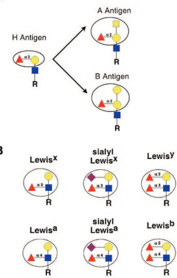
α3/4-Fucosyltransferases (α3/4-FUTs) are glycosyltransferases (GTs)
that catalyze the transfer of fucose in an α3/4-linkage onto the N-acetylglucosamine residue from acceptors containing the type II or type I
(Galβ4/3GlcNAc, respectively) structures, thus synthesizing the fucosylated Lewis (Le) carbohydrate determinants. Fucosyltransferase IX (FUT9), the most
recently identified member of the family, presents the higher divergence from the other FUTs and its sequence is the only highly conserved among species. FUT9
synthesizes the Lewisx (Lex) epitope (Galβ4(Fucα3)GlcNAc). Recent evidence has suggested that it is the enzyme responsible for the synthesis of Lex in the mouse brain. Lex expression has been described in glycoproteins, proteoglycans and glycolipids from the central nervous system (CNS) of diverse species, including rodents and humans.
The major goal of the work presented in this thesis has been to characterize human FUT9 and elucidate the functional role of Lex in differentiating neurons in vitro. In Chapter 2, FUT9 has been cloned from human NT2N neurons and overexpressed in HeLa cells (FUT9wt), where it presented in
vivo activity with de novo synthesis of the Lex determinant. In vitro specificity studies towards small synthetic oligosaccharide acceptors linked to a hydrophobic moiety showed that FUT9wt synthesized preferentially Lex and Ley, whereas Lea was produced with low efficiency, and only in the presence of the activating manganese cation. The corresponding oligosaccharides substituted
with α2,3-linked neuraminic acid were not acceptors of FUT9wt. FUT9wt has been found to efficiently fucosylate desialylated N-glycans from glycoproteins,
such as human asialoerythropoietin, but not native sialylated erythropoietin. Detailed analysis of the modified glycan structures from asialoerythropoietin by
synthesizing the corresponding peripherally monofucosylated structures. Difucosylated glycans were also detected, although in minor amounts and only in
tetrantennary structures with repeats. These results demonstrated the high specificity of FUT9 for the synthesis of the Lex determinant in N-linked glycans from glycoproteins. Furthermore, FUT9 is an adequate enzyme for in vitro synthesis of Lex-carrier glycoproteins.
FUT9wt was localized in the trans-Golgi and trans-Golgi network (TGN) of
HeLa cells, colocalizing with the trans-Golgi and TGN resident enzyme
β4-galactosyltransferase and with the TGN marker TGN-46. Deletion of the cytoplasmic tail of FUT9, or just the Ser/Thr cluster contained in it, caused a shift to earlier Golgi sub-compartments, as indicated by higher colocalization with the
cis-Golgi marker GM130 and redistribution into an endoplasmic reticulum-like pattern after incubation with brefeldin A. These results indicated that the
information for intra-Golgi localization is contained in the cytosolic tail of FUT9. With the purpose of obtaining high amounts of recombinant FUT9, in
Chapter 3, a soluble form of the enzyme, sFUT9, was overexpressed in the Spodoptera frugiperda Sf9 insect cell line. sFUT9 was efficiently secreted and it presented higher efficiency for fucosylating glycoproteins, both sialylated and
non-sialylated, than full-length FUT9wt from HeLa cells. Thus, sFUT9 is suitable for synthetic purposes. Moreover, it can also be used for further functional and
structural studies of FUT9, such as the determination of its three-dimensional structure.
membrane protein-1 (LAMP-1) and the v-SNARE tetanus neurotoxin-insensitive
vesicle-associated membrane protein (TI-VAMP). Furthermore, Lex was found in the cell bodies and neurites of NT2N neurons. These results associated for the
first time the Lex epitope with the TI-VAMP mediated exocytic pathway, known to be involved in neurite outgrowth. Moreover, antibody incubation assays with an
anti-Lex monoclonal impaired adhesion of NT2N neurons to the surface matrix and inhibited neurite initiation of outgrowth.
The levels of the Lex epitope during neuronal development in rat primary hippocampal cultures were also studied, as addressed in Chapter 5. Lex was detected in a subset of GABAergic interneurons, where it was also found to
colocalize with the TI-VAMP marker similarly to that found in NT2N human neurons. On the plasma membrane of mature hippocampus GABAergic neurons,
Lex was detected in spine-like protrusions or phillopodia, dendritic domains adjacent to synaptic regions.
In conclusion, the work described in this thesis showed that FUT9 synthesized the Lex epitope in human NT2N neurons, underlying neuronal adhesion and initiation of neurite outgrowth. Localization of Lex in the TI-VAMP exocytic pathway in both human and rat neurons suggested common functions
Sumário
Os α3/4-fucosiltransferases (α3/4-FUTs) são glicosiltransferases (GTs) que catalizam a transferência de fucose, numa ligação α3/4, para resíduos de N-acetilglucosamina de aceitadores que contenham estruturas do tipo II ou do
tipo I (Galβ4/3GlcNAc, respectivamente), sintetizando assim epítopos fucosilados designados determinantes Lewis (Le). O fucosiltransferase IX (FUT9) foi o último
elemento da família a ser identificado, sendo o que apresenta maior divergência em relação aos outros FUTs e o único com elevada homologia entre espécies. O
FUT9 sintetiza o epítopo Lewisx (Lex) (Galβ4(Fucα3)GlcNAc). Estudos recentes sugeriram que o FUT9 é responsável pela síntese do Lex no cérebro de murganho. A expressão do Lex foi descrita em glicoproteínas, proteoglicanos e glicolípidos do sistema nervoso central (SNC) de várias espécies, incluindo roedores e humanos.
O objectivo principal do trabalho apresentado nesta tese foi a caracterização do FUT9 humano e a elucidação da função do Lex na diferenciação neuronal in vitro. No Capítulo 2, o FUT9 humano foi clonado de neurónios NT2N e sobrexpresso em células HeLa (FUT9wt), tendo apresentado
actividade in vivo, com síntese de novo do determinante Lex. In vitro, estudos de especificidade usando aceitadores sintéticos, constituídos por pequenos oligossacáridos ligados a uma porção hidrófoba, revelaram que o FUT9wt
sintetizou preferencialmente Lex e Ley, enquanto o Lea foi produzido com baixa eficiência e apenas na presença do catião manganês como activador. Os
oligossacáridos correspondentes substituídos com ácido neuramínico, numa
ligação α2,3, não foram aceitadores do FUT9wt. Foi observado que o FUT9wt fucosilou eficientemente N-glícidos não sialilados de glicoproteínas, como a asialoeritropoietina, mas não a eritropoietina nativa sialilada. A análise por
próxima, com ou sem repetições, sintetizando as estruturas correspondentes com monofucosilação periférica. Foram ainda detectados glícidos bifucosilados,
embora em menores quantidades, e apenas em estruturas tetrarramificadas com repetições. Estes resultados demonstraram a elevada especificidade do FUT9
para sintetizar o determinante Lex em N-glícidos de glicoproteínas. Indicaram ainda que o FUT9 é um enzima adequado para a síntese in vitro de
glicoproteínas contendo Lex.
O FUT9wt foi detectado no trans-Golgi e no trans-Golgi reticular (TGN) de
células HeLa, colocalizando com o enzima β4-galactosiltransferase, residente no trans-Golgi e no TGN, e com o TGN-46, marcador do TGN. A remoção total da cauda citoplasmática do FUT9, ou apenas do cluster de Ser e Thr nela contido,
provocou uma deslocalização do enzima para sub-compartimentos anteriores, revelada pela colocalização aumentada com a proteína GM130, marcadora do
cis-Golgi, e pela redistribuição por compartimentos com uma marcação típica de retículo endoplasmático após incubação com brefeldina A. Estes resultados
indicaram que a informação que determina a localização intra-Golgi do FUT9 está contida na cauda citoplasmática do enzima.
Com o objectivo de obter quantidades elevadas de FUT9 recombinante,
no Capítulo 3, foi sobrexpressa uma forma solúvel do enzima - sFUT9 - na linha celular de insecto Spodoptera frugiperda Sf9. O sFUT9 foi secretado
eficientemente e apresentou uma eficácia superior na fucosilação de glicoproteínas, tanto sialiladas como não sialiladas, à apresentada pela forma
total do enzima, FUT9wt, expressa em células HeLa. Assim, o sFUT9 é adequado para fins de síntese. Adicionalmente, poderá também ser utilizado em
estudos funcionais e estruturais do FUT9, incluindo a determinação da estrutura tridimensional do enzima.
blot e na síntese eficiente de Lex e Ley em ensaios de actividade in vitro. Estudos de microscopia confocal de imunofluorescência revelaram que o determinante Lex se encontrava na membrana plasmática e em vesículas intracelulares positivas para o marcador de endossomas secundários/lisossomas LAMP1 (do inglês lysosomal-associated membrane protein 1) e para a v-SNARE TI-VAMP
(do inglês tetanus neurotoxin-insensitive vesicle-associated membrane protein). Foi ainda observado que o Lex se encontrava nos corpos celulares e nos prolongamentos neuronais dos neurónios NT2N. Estes resultados relacionaram pela primeira vez o epítopo Lex com a via de exocitose mediada pela TI-VAMP, que se sabe estar envolvida no crescimento dos prolongamentos neuronais.
Para além disso, a incubação com um anticorpo monoclonal anti-Lex, reduziu a adesão dos neurónios NT2N à superfície da matriz e inibiu a iniciação do
crescimento dos prolongamentos neuronais.
Os níveis do epítopo Lex foram também estudados durante o desenvolvimento de culturas primárias do hipocampo de rato, como descrito no Capítulo 5. O Lex foi detectado num subgrupo de interneurónios GABAérgicos, tendo-se observado que colocalizava com o marcador TI-VAMP, à semelhança do verificado em neurónios humanos NT2N. Na membrana plasmática de
neurónios GABAérgicos maturos, o Lex foi detectado em protuberâncias semelhantes a espículas ou filopodia, domínios dendríticos adjacentes a regiões
sinápticas.
Em conclusão, o trabalho descrito nesta tese mostrou que o FUT9 sintetizou o epítopo Lex em neurónios humanos NT2N, estando na base dos processos de adesão neuronal e de iniciação do crescimento dos prolongamentos neuronais. A localização do Lex no compartimento exocítico definido pela TI-VAMP, tanto em neurónios humanos como de rato, sugeriu funções comuns para o determinante glicídico no SNC de ambos os organismos.
Ademais, a descoberta de síntese eficiente de Lex em N-glícidos de glicoproteínas pelo FUT9 indicou que os efectores dessas funções biológicas
Table of contents
Thesis Outline……...…..……….…………... xxiii
List of Figures………..……….………... xxv
List of Tables…………..……….………… xxvi
Abbreviations ………….………. xxvii
Chapter 1 – General introduction……….……… 1
1.1. Membrane transport in mammalian cells……….… 3
1.1.1. The secretory pathway……….... 3
1.1.1.1. The endoplasmic reticulum……… 3
1.1.1.2. The Golgi apparatus……...……… 6
1.1.1.2.1. COPI transporters……… 8
1.1.1.2.2. Membrane targeting……… 9
1.1.1.3. The trans-Golgi network.………….……..……… 10
1.1.2. The endocytic pathway………...……….... 11
1.1.3. Non-conventional endosomal secretion……...……….... 13
1.2. Protein glycosylation……….………..……… 14
1.2.1. N-linked oligosaccharides…….………... 15
1.2.2. O-linked oligosaccharides ……….………. 17
1.2.3. Glycosaminoglycans……..………..…..………. 19
1.2.4. Glycosyltransferases………....………..………. 20
1.2.4.1. Glycosyltransferase localization ………... 21
1.2.4.2. Glycosyltransferase structure and mechanism….……... 23
1.3. Fucosylation……….…… 25
1.3.1. The α2- and α6-fucosyltransferases.……… 27
1.3.2. The α3/4-fucosyltransferase family.………..……… 28
1.4. NTera2 cell line as model system for human neuronal differentiation… 34
1.5. Aims of this thesis work.……….……… 38
Chapter 2 – Human fucosyltransferase IX efficiently fucosylates N-linked asialoglycoproteins with the synthesis of the adhesion Lewisx determinant……….. 41
2.1. Summary.……….…… 43
2.2. Introduction.……….. 45
2.3. Materials and methods.……….. 47
2.3.1. Cell culture……….... 47
2.3.2. Cloning of human FUT9 from NT2N neurons……….….... 47
2.3.3. Plasmid construction……….... 48
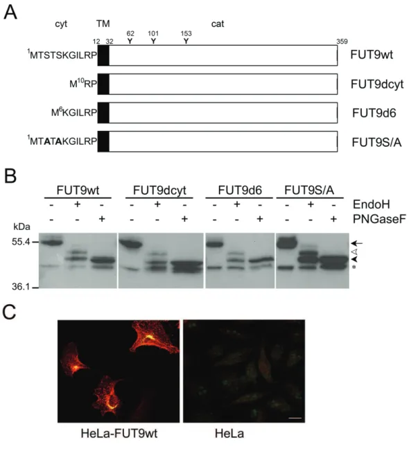
2.3.4. Western blot analysis of FUT9wt and mutants overexpressed in HeLa cells………. 49
2.3.5. Fucosyltransferase activity assays ………..…….... 50
2.3.6. Native and desialylated (asialoEPO) EPO…………..………….... 51
2.3.7. Immunoaffinity isolation of desialylated EPO from FUT9 incubations for N-glycan analysis………. 51
2.3.8. Release of N-glycans from EPO protein, purification and desalting……… 52
2.3.9. Analysis of neutral oligosaccharides via HPAEC-PAD………….. 53
2.3.10. Analysis of N-glycans by Matrix-Assisted Laser Desorption Ionization/Time-of-Flight Mass Spectrometry (MALDI/TOF-MS)... 53
2.3.11. Immunofluorescence microscopy and image quantification…... 54
2.4. Results.………. 55
2.4.1. Overexpression of human FUT9wt in HeLa cells and Lex biosynthesis……….…. 55
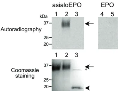
2.4.3. FUT9wt efficiently fucosylates asialoglycoproteins...….. 60
2.4.4. FUT9wt efficiently fucosylates di-, tri- and tetrantennary N-glycans from asialoEPO………..……….. 62
2.4.5. TGN localization of FUT9 is determined by the cytosolic tail…… 67
2.5. Discussion.………... 72
2.6. Acknowledgements.……… 75
Chapter 3 – Stable expression of an active soluble recombinant form of human fucosyltransferase IX in Spodoptera frugiperda Sf9 Cells….………. 77
3.1. Summary……….………. 79
3.2. Introduction……….……….. 80
3.3. Materials and methods……….……….. 81
3.3.1. Plasmid construction ……….. 81
3.3.2. Cell culture……….……… 82
3.3.3. Stable expression of human FUT9 in Sf9 cells……… 83
3.3.4. Fucosyltransferase activity………. 83
3.3.5. Protein analysis……….………... 84
3.4. Results and discussion…………..………..….. 85
3.4.1. Expression of soluble human FUT9 (sFUT9) in Sf9 cells……….. 85
3.4.2. Characterization of sFUT9……….…….……… 91
3.4.2.1. N-glycosylation of sFUT9………... 91
3.4.2.2. Acceptor specificity of sFUT9………... 93
3.5. Conclusions……….…. 97
3.6. Acknowledgements ……… 97
Chapter 4 – Increased levels of fucosyltransferase IX and carbohydrate Lewisx adhesion determinant in human NT2N neurons …………..…………. 99
4.1. Summary ………. 101
4.3.1. Neuronal culture……….…….. 104
4.3.2. Fucosyltransferase assays……….……… 104
4.3.3. SDS-PAGE and Western blot analysis ……….... 105
4.3.4. Glycosidase hydrolysis….………...……… 106
4.3.5. Cell-surface biotinylation.…….………...……… 107
4.3.6. RNA extraction and reverse transcription….……… 107
4.3.7. Immunofluorescence microscopy…………...………... 108
4.3.8. Recycling assay……….…………...………... 110
4.3.5. Anti-Lex incubation assays………...………... 111
4.4. Results ……….………..….. 111
4.4.1. Level of the Lex determinant is increased during NT2N neurons differentiation……… 111
4.4.2. Lex from NT2N neurons is synthesized by FUT9……….………... 114
4.4.3. Identification of a Lex-carrier glycoprotein associated specifically with NT2N neurons………... 119
4.4.4. Lex determinant is localized at the plasma membrane and in the TI-VAMP compartment of NT2N neurons………... 123
4.4.5. Lex is recycled to the plasma membrane of NT2N neurons..…… 128
4.4.6. Anti-Lex antibody impairs initiation of neurite outgrowth and adhesion of NT2N neurons……….…..……..………... 130
4.5. Discussion………...…. 132
4.6. Acknowledgements ……… 135
Chapter 5 – Levels of Lewisx during differentiation of rat primary hippocampal cultures ………...…………. 137
5.1. Summary ………. 139
5.2. Introduction ……….. 140
5.3.1. Neuronal culture……….…….. 141
5.3.2. Immunofluorescence microscopy…………...………... 142 5.3.3. SDS-PAGE and Western blot analysis ……….... 143
5.4. Results ……….………..….. 144
5.4.1. Lex expression is induced during development of rat
hippocampal cultures……….. 144
5.4.2. GABAergic neurons from rat hippocampus express Lex-carriers. 146 5.4.3. The Lex determinant is localized at the plasma membrane and
in the TI-VAMP compartment of rat hippocampus neurons...………… 148 5.4.4. Lex-carrier glycoproteins are expressed by neurons in rat
hippocampal cultures………...…….. 150
5.5. Discussion………..……….…. 153
5.6. Acknowledgements ……… 156
Chapter 6 – General discussion and conclusions ……… 157
6.1. General discussion and perspectives ………. 159
6.1.1. Characterization of recombinant full-length and soluble forms of
human FUT9………....…… 160
6.1.2. Localization of human FUT9………...…… 164 6.1.3. Expression of FUT9 and Lex during neuronal development…….. 168 6.2. General conclusions………...…… 172
Thesis Outline
The work described herein concerns the study of fucosyltransferase IX in
the central nervous system. Two lines of research were followed. One of them envisaged the characterization of human fucosyltransferase IX, while the other
focused on the investigation of the biosynthetic product of the enzyme in neurons, the Lewisx determinant, and its biological role during neuronal differentiation.
This dissertation starts with a general introduction to the endomembranous system of mammalian cells, where post-translational
modifications, including glycosylation, occur. Particular relevance is given to the Golgi apparatus, as fucosyltransferase IX is a Golgi-resident protein. This is
followed by an overview about protein glycosylation, including a summary of current knowledge of localization, structure and mechanism of
glycosyltransferases. Fucosylation is focused in detail, specially the
α3/4-fucosyltransferase family and the Lewisx determinant, one of its products. Finally, a brief characterization of the NTera 2 cell line as a model system for human neuronal differentiation is presented.
Chapter 2 describes the cloning, overexpression and characterization of
human fucosyltransferase IX. Studies concerning the substrate specificity of the enzyme, in particular towards glycoproteins, and its subcellular localization are
presented.
In Chapter 3, the production and characterization of a recombinant soluble
form of human fucosyltransferase IX from Spodoptera frugiperda Sf9 cells is described.
Chapter 4 is concerned with the levels of Lewisx determinant during differentiation of human NT2N neurons and with the assignment of
neurons in culture are studied and the subpopulation of neurons that express the fucosylated determinant investigated.
List of Figures
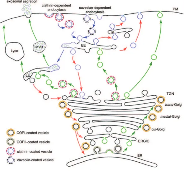
Figure 1 Page 5 Schematic representation of the secretory and endocytic pathways.
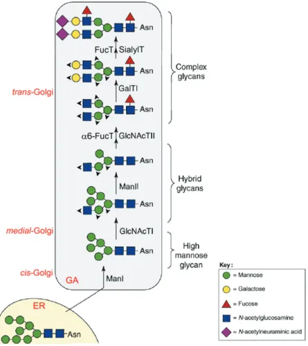
Figure 2 Page 16 Schematic representation of N-linked oligosaccharide biosynthetic pathways in the Golgi apparatus.
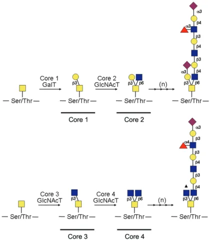
Figure 3 Page 18 Schematic representation of core 1, core 2, core 3 and core 4
O-linked oligosaccharide subtype biosynthetic pathways.
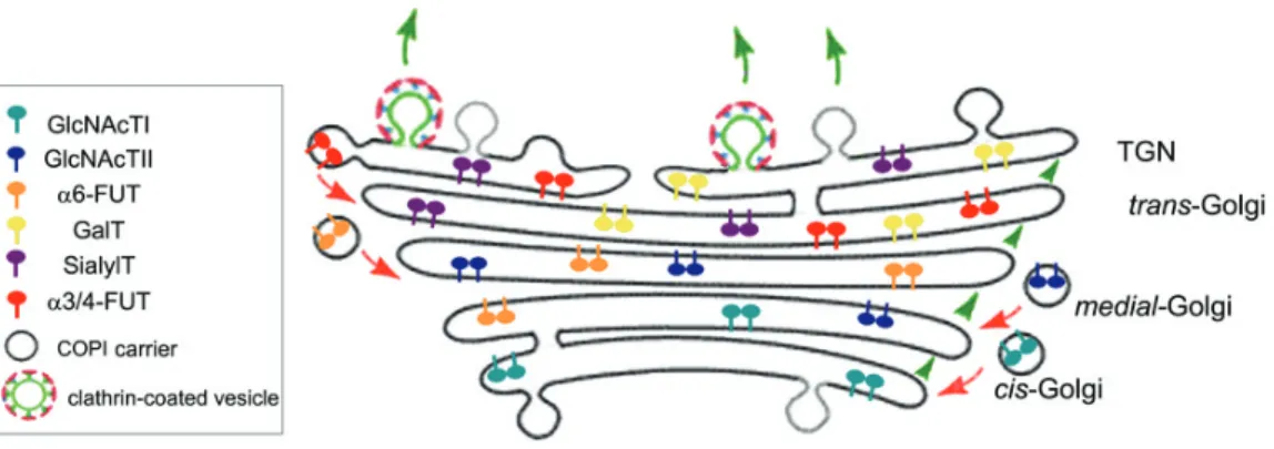
Figure 4 Page 21 Schematic representation of the distribution of glycosyltransferases in the Golgi apparatus.
Figure 5 Page 26 Structures of common fucosylated epitopes.
Figure 6 Page 29 Schematic representation of human α3/4-fucosyltransferases.
Figure 7 Page 31 Schematic representation of human α3-fucosyltransferase IX (FUT9) reaction.
Figure 8 Page 56 Overexpression of FUT9wt and mutants in HeLa cells.
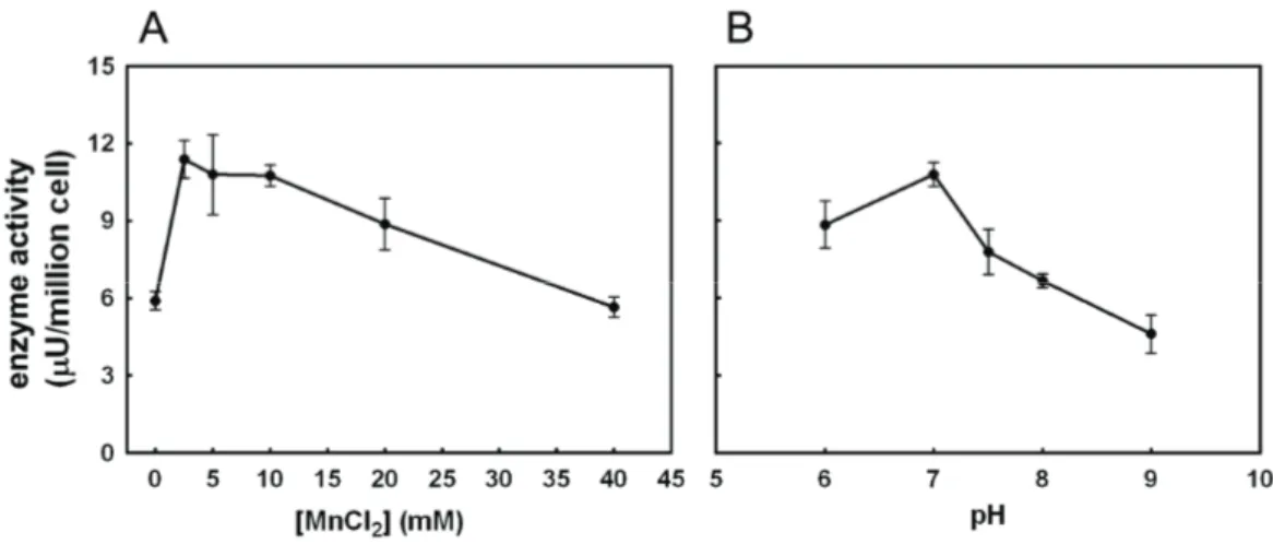
Figure 9 Page 59 Effect of Mn2+ (A) and pH (B) on FUT9wt activity.
Figure 10 Page 62 Analysis of asialoerythropoietin and erythropoietin after incubation with FUT9wt.
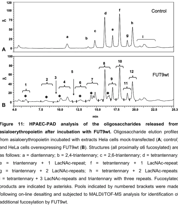
Figure 11 Page 63 HPAEC-PAD analysis of the oligosaccharides released from asialoerythropoietin after incubation with FUT9wt.
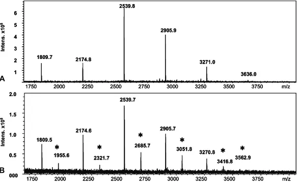
Figure 12 Page 64 MALDI-TOF mass-spectrometric analysis of the oligosaccharides obtained from asialoerythropoietin after FUT9wt incubation.
Figure 13 Page 69 Subcellular localization analysis of FUT9wt and mutants.
Figure 14 Page 71 Effect of Brefeldin A on the localization of FUT9wt and cytosolic tail mutants.
Figure 15 Page 86 Schematic representation of human FUT9 constructs.
Figure 16 Page 87 Production of FUT9wt and sFUT9 from SF9 cells grown in shake flasks.
Figure 17 Page 88 α3-Fucosyltransferase activity of Sf9-FUT9wt and Sf9-sFUT9 cells.
Figure 18 Page 89 Western blot analysis of secreted (A) and intracellular (B) sFUT9.
Figure 19 Page 91 Western blot analysis of FUT9 transfection assays in HeLa cells.
Figure 20 Page 92 Deglycosylation and Western blot analysis of secreted sFUT9.
Figure 21 Page 112 Immunodetection of Lewis determinants in NT2- cells and NT2N neurons by immunofluorescence confocal microscopy.
Figure 22 Page 115 RT-PCR analysis of fucosyltransferases along NT2N neurons differentiation induced by retinoic acid.
Figure 25 Page 120 Immunodetection by Western blot of Lex-carrier proteins along NT2N neurons differentiation induced by retinoic acid and of LAMP-1 in NT2N neurons.
Figure 26 Page 122 Characterization of Lex-carrier protein of NT2N neurons.
Figure 27 Page 124 Localization of Lex in NT2N neurons by immunofluorescence confocal microscopy.
Figure 28 Page 127 Western blot analysis of biotinylated plasma membrane proteins containing the Lex determinant.
Figure 29 Page 129 Plasma membrane distribution of recycled Lex in NT2N neurons by immunofluorescence confocal microscopy.
Figure 30 Page 131 Effect of anti-Lex antibody L5 on the adhesion and initiation of neurite outgrowth from NT2N neurons.
Figure 31 Page 145 Detection of the Lex determinant along rat hippocampal cultures development by immunofluorescence confocal microscopy.
Figure 32 Page 147 Detection of the Lex determinant in 14 DIV rat hippocampus neurons by immunofluorescence microscopy.
Figure 33 Page 149 Colocalization analysis of Lex in hippocampus neurons by immunofluorescence confocal microscopy.
Figure 34 Page 151 Immunodetection by Western blot of Lex-carrier proteins from hippocampal cultures.
List of Tables
Table 1 Page 60 Substrate specificity of recombinant FUT9wt from HeLa cells.
Table 2 Page 66 Observed mass signals (m/z) by MALDI/TOF-MS analysis and predicted composition of the N-glycans from asialoerythropoietin.
Table 3 Page 94 Substrate specificity of sFUT9 from Sf9 cells.
Abbreviations
Abbreviation Full form
A absorbance AP adaptor protein ApoE apolipoprotein E ara-C cytosine arabinoside ARF1 ADP ribosylation factor 1
ARFGAP GTPase activating protein for ARF1 BFA brefeldin A BHK baby hamster kidney
BSA bovine serum albumin CCD conserved oligomeric Golgi complex-dependent ChABC chondroitinase ABC
CHO chinese hamster ovary cells CHX cycloheximide CNS central nervous system
CNX calnexin COG conserved oligomeric Golgi COP coat protein complex
CSPG chondroitin sulfate proteoglycan C-type lectin calcium-dependent type lectin
DC-SIGN dendritic cell-specific ICAM3 grabbing nonintegrin dHex deoxyhexose
DIV days in vitro
DMEM-HG Dulbecco’s modified Eagle’s medium- high glucose DOC sodium deoxycholate
EC embryonic carcinoma ECM extracellular matrix
EDTA ethylenediaminetetra-acetic acid EE early endosome
EEA-1 early endosome antigen-1 EGF epidermal growth factor
ES embryonic stem Fab antigen binding fragment
FBS foetal bovine serum Fuc fucose
FUT fucosyltransferase GA Golgi apparatus GABA γ-aminobutyric acid
GAD glutamate decarboxylase GAG glycosaminoglycan Gal galactose GalNAc N-acetylgalactosamine
GalNAcT N-acetylgalactosaminyltransferase GalT galactosyltransferase GDP guanoside-5’-diphosphate GEF guanine-nucleotide exchange factor
Glc glucose GlcA glucoronic acid GlcNAc N-acetylglucosamine
GlcNAcT N-acetylglucosaminyltransferase GP glycoprotein GT glycosyltransferase GTP guanoside-5’-triphosphate HBBS Hank’s balanced salt solution
HeLa Henrietta Lack’s HEK human embryonic kidney
HEPES N-(2-hydroxyethyl)-piperazine-N'-2-ethanesulfonic acid Hex hexose
HexNAc N-acetylhexosamine
HNK-1 human natural killer-1 cells epitope
HPAEC-PAD high pH anion exchange chromatography with pulsed amperometric detection
Abbreviation Full form
IL interleukin LacNAc N-acetylactosamine
LAMP-1 lysosomal associated membrane protein - 1 LE late endosome Lea Lewisa
Leb Lewisb Lex Lewisx Ley Lewisy
Lyso lysosome
MALDI/TOF-MS matrix-assisted laser desorption ionization/time-of-flight mass spectrometry
Man mannose Man6P mannose 6-phosphate group MAP microtubule-associated protein MEM minimum essential medium
MI mitosis inhibitors
MOPs 4-morpholinepropanesulfonic acid MVBs multivesicular bodies
NCAM neuronal cell adhesion molecule NeuAc N-acetylneuraminic acid
NF-L low molecular weight neurofilament protein
NF-M P- hypophosphorylated medium molecular weight neurofilament protein NF-M P+ hyperphosphorylated medium molecular weight neurofilament protein NP-40 Nonidet-P40
NT2 cell line NTera2/cl.D1 cell line
NT2- cells NTera2/cl.D1 undifferentiated cells
NT2N neurons post-mitotic neurons derived from NT2- cells NT2A astrocytes astrocytes derived from NT2- cells
OD optical density PAGE polyacrylamide gel electrophoresis PBS phosphate buffered saline
PBSCM phosphate buffered saline supplemented with CaCl2 and MgCl2
PCR polymerase chain reaction
PNGaseF peptide-N-glycosidase F
PO poly-DL-ornithine RA retinoic acid RE recycling endosome
RPTPβ receptor-type protein-tyrosine phosphatase β RT-PCR reverse transcription polymerase chain reaction
SCGs sympathetic cervical ganglia
SD standard deviation SDS sodium dodecyl sulphate
sFUT9 soluble fucosyltransferase IX SialylT sialyltransferase sLacNAc sialyl-N-acetylactosamine sLea sialyl-Lewisa
sLex sialyl-Lewisx
SNAP synaptosomal associated protein
SNARE soluble N-ethylmaleimide-sensitive factor attachment protein receptor SSEA-1 stage specific embryonic antigen-1
suc sucrose SYT7 synaptotagmin VII TFA trifluoracetic acid TGF-β transforming growth factor-β TGN trans-Golgi Network
TI-VAMP tetanus neurotoxin-insensitive vesicle associated membrane protein TM transmembrane domain
Tris tris(hydroxymethyl)aminomethane TX-100 Triton X-100
UV ultra-violet
Amino Acid nomenclature:
Abbreviations Amino acid name
Ala A Alanine Arg R Arginine Asn N Asparagine
Asp D Aspartate (Aspartic Acid) Cys C Cysteine
Gln Q Glutamine
Glu E Glutamate (Glutamic Acid) Gly G Glycine
Chapter 1
1. General introduction
1.1. Membrane transport in mammalian cells
A hallmark of eukaryotic cells is the presence of an elaborate
endomembrane system that is responsible for the exchange of macromolecules between cells and their environment. The secretory pathway delivers newly
synthesized proteins, carbohydrates and lipids to the plasma membrane and extracellular matrix, modifying them during the trafficking process (Figure 1, green arrows and membranes), whereas the endocytic pathway takes up
macromolecules into the cell (Figure 1, blue arrows and membranes). Transport along these pathways occurs through anterograde or retrograde translocation of
material between different membrane-enclosed organelles, via small vesicles and large transport containers. Despite this transfer, each organelle maintains its
characteristic set of resident macromolecules via several mechanisms, of which retrieval (Figure 1, red arrows) plays a fundamental role (Bonifacino and
Lippincott-Schwartz, 2003).
1.1.1.The secretory pathway
1.1.1.1. The endoplasmic reticulum
The nascent proteins are targeted to the membrane of the endoplasmic reticulum (ER) and translocated into or across the ER via a cotranslational
process. Once in the ER, proteins are processed for trafficking through the secretory pathway. One of the initial events in this process is the transfer of a
polypeptides into their correct three-dimensional conformation is also started
co-translationally in the ER. The quality control machinery in the lumen of the ER assures that only correctly folded proteins exit this compartment. Misfolded
proteins are retained, triggering the unfolded protein response (UPR) pathway, which will regulate a battery of genes involved in ER-associated protein
degradation (ERAD) but also in protein translocation, folding, glycosylation, and trafficking (reviewed by Ruddock and Molinari, 2006).
Following ER processing, proteins are concentrated in ER export sites (ERES), specialized domains of the ER membrane, also known as transitional ER (tER), responsible for the export of secretory cargo. In mammalian cells,
ERES are distributed throughout the cytoplasm but concentrated in the juxtanuclear region adjacent to the Golgi apparatus (GA) (reviewed by Watson
and Stephens, 2005). These sites are characterized by the presence of the coat protein complex II (COPII) (Figure 1), formed by the assembly of a set of soluble
proteins, which mediates the biogenesis of transport carriers. The COPII mediates the selection of cargo from within the ER lumen, either directly
(transmembrane cargo molecules) or indirectly via specific cargo receptors (soluble cargo and some transmembrane cargo molecules) (reviewed by
Tang et al., 2005). After detaching from the membrane of the ER, COPII carriers (vesicles and/or tubules) loose the COPII coat and undergo fusion to form
pleiotropic membrane structures termed VTCs (vesicular-tubular clusters), that constitute the ER-to-Golgi-intermediate compartment (ERGIC, Figure 1). VTCs are then delivered to the cis-Golgi, fusing with the Golgi membrane and being
4
secretory compartments, fusing with the PM. Multivesicular bodies (MVBs) derived from the LE can fuse with the PM, releasing the internal vesicles as exosomes. Retrieval is observed in the exocytic and endocytic pathways (red arrows): recycling of ER-resident proteins from the GA and intra-Golgi recycling are mediated by COPI-coated vesicles; internalized material can be recycled from the EE back to the PM directly or via recycling endosomes (RE). Adapted from Kartberg et al., 2005; Keller et al., 2006; van der Goot and Gruenberg, 2006; van Niel et al., 2006.
1.1.1.2.The Golgi apparatus
The GA comprises a stack of distinct compartments (cisternae) arranged
from cis to trans. It acts as the central organelle of the secretory pathway, receiving newly synthesized proteins and lipids from the ER in the cis-side,
covalently modifying them while they move through the stack to the trans-side, until the trans-Golgi network (TGN), where they are targeted to the final cellular
destinations (anterograde transport) (Munro, 2005). Concomitantly, recycling of proteins and lipids to previous compartments (retrograde transport) occurs.
A century after the GA discovery, fundamental molecular mechanisms
underlying the maintenance of Golgi’s morphological and functional integrity, despite the constant flow of lipids and proteins along the exocytic pathway,
remain unclear. These have been extensively addressed in the last decades, although they are still far from being fully understood.
The observation of numerous, small sized (70-90 nm), peri-Golgi vesicles, which bud from the Golgi cisternae in a COPI coat dependent manner (Orci et al.,
1986; Malhotra et al., 1989), contributed to the proposal of several models. For many years, the predominant model defended vesicular transport and stable
compartments, stating that newly synthesized cargo was transported in the anterograde direction by default. Transport would occur first via COPII vesicles
sequentially with Golgi cisternae. On the other hand, Golgi-resident proteins
would be confined to the stable cisternae via retention signals excluding them from the traffickingvesicles (reviewed by Rothman and Wieland, 1996).
However, the vesicular transport/stable compartments model could not explain the transport through the GA of molecules like 300 nm procollagen fibers
(Bonfanti et al., 1998), far too large to fit in the vesicular transporters. Furthermore, it was observed that glycosyltransferases (GTs) localized in the
cis-Golgi were processed by late-Golgi acting enzymes (Hoe et al., 1995; Harris and Waters, 1996) and that retrograde COPI vesicles recycled membrane-bound proteins containing the cytoplasmic ER recycling motif K(X)KXX (Cosson and
Letourneur, 1994; Letourneur et al., 1994).
Despite some divergent data (Orci et al., 2000; Cosson et al., 2002), in
vitro and in situ studies showed that COPI vesicles were enriched in several Golgi-resident proteins, in a process dependent of GTP hydrolysis by the small
GTPase ADP ribosylation factor 1 (ARF1) (Lanoix et al., 1999), and depleted of anterograde cargo that remained in the cisternae (Lanoix et al., 2001;
Martinez-Menarguez et al., 2001; Mironov et al., 2001; Malsam et al., 2005). In previous work we have observed that COPI vesicles, formed in vitro from Golgi
membranes of Baby Hamster Kidney (BHK) cells, were enriched in the trans-Golgi and TGN resident enzyme fucosyltransferase III (FUT3), contrary to
anterograde cargo, such as transferrin receptor (plasma membrane resident) or ApoE (soluble secretory protein), which were not detected in the same vesicles (Sousa et al., 2003). Lately, evidence suggested trafficking via transient tubular
connections that may mediate several processes (reviewed by Mironov et al., 2005), such as the recycling of GA resident enzymes (Kweon et al., 2004).
All this data pointed to cisternal maturation/progression models, which stated that retrograde traffic via COPI transporters and/or tubular connections
Although this is still a theme subject to intense debate, the cisternal
maturation model collects nowadays more substantial support from experimental evidence. Two very recent studies in yeast have shown that markers of the early
Golgi were consistently replaced with late Golgi markers (but never vice versa) (Losev et al., 2006; Matsuura-Tokita et al., 2006), confirming by direct
observation the cisternal maturation hypothesis. So, throughout this thesis localization in the GA will be discussed in the context of the cisternal maturation
model.
Assuming the cisternal maturation model, Golgi-resident proteins would be concentrated in retrograde COPI transporters and retrieved back to previous
compartments. Therefore, Golgi localization would depend on sorting into COPI transporters and targeting to the acceptor membrane.
1.1.1.2.1COPI transporters
COPI coat formation involves the interaction of the soluble coatomer protein complex to the membrane, via specific motifs present in the cytoplasmic
domains of resident proteins (as, for example, the di-Lys K(X)KXX ER-retrieval motif) and via the small GTPase ARF1 in its GTP bound form. The several
components polymerize in a lattice on the membrane, inducing sorting, budding and fission. Subsequent disassembly of the coat requires GTP hydrolysis on
ARF1 by the membrane-associated GTPase activating protein for ARF1 (ARFGAP) (reviewed by Lippincott-Schwartz and Liu, 2006). A role for ARFGAP in cargo sorting into COPI vesicles has also been suggested (reviewed by
Kartberg et al., 2005) in a mechanism that would involve direct interactions with cytoplasmic domains of cargo proteins (Lanoix et al., 2001).
Recently, new insights into the dynamics of COPI coat components, given by live-cell imaging techniques and photobleaching studies, suggested that all
components and regulators involved in the process cycle on and off membranes, in a mechanism uncoupled from vesicle formation and independently of vesicle
self-assemble, acting as a molecular machine that drives membrane sorting and
trafficking processes (Lippincott-Schwartz and Liu, 2006).
1.1.1.2.2. Membrane targeting
After COPI vesicle formation, the processes of directioning, targeting,
docking and finally fusion will depend on conserved proteins that act in a regulated cascade: SNAREs (soluble N-ethylmaleimide-sensitive factor
attachment protein receptors) on vesicle and target membranes, tethering factors and regulatory Rab small GTPases (Lupashin and Sztul, 2005).
SNARES are a family of short coiled-coil membrane associated proteins
that act in docking and fusion events, via the formation of a trans-SNARE complex (or SNAREpin) between one v-SNARE in the vesicle membrane and
three t-SNAREs chains associated with the target compartment (reviewed by Hong, 2005). The combinatory use of different SNAREs gives rise to a wide
variety of complexes. For example, the SNARE complexes consisting of
GS15−Ykt6−GS-28−syntaxin-5 and membrin−Bet1−Sec22b−Syntaxin-5 are both involved in Golgi transport. After fusion, the trans-SNARE complex is converted into a cis-SNARE complex and then disassembles in a process driven by ATP hydrolysis (reviewed by Hong, 2005).
Direct interactions between SNAREs and COPI coat have been observed (Rein et al., 2002). Formation of trans-SNARE complexes involved in intra-Golgi
transport has been shown to require tethering factors, like the golgin p115 (Shorter et al., 2002; Puthenveedu and Linstedt, 2004) and the multisubunit
conserved oligomeric Golgi (COG) complex (Suvorova et al., 2002). Tethering factors, recruited by small GTPases, act as physical links, maintaining the
vesicles and the acceptor membrane in close proximity (reviewed by Lupashin and Sztul, 2005). The number of known compartment-specific tethering factors
2007). This suggests a critical role for tethering factors in the fusion process,
imposing membrane selectivity to vesicle-docking events.
The identification of different subpopulations of COPI vesicles enriched in
distinct cargo molecules, defined by different tethering factors and different SNAREs (Malsam et al., 2005), confirmed the importance of vesicle targeting
events in the regulation of intra-Golgi transport. In summary, the concerted action of the large variety of SNAREs and tethering complexes, together with their
effectors and regulators, impose membrane selectivity to vesicle-docking events and contributes to the spatial and temporal regulation of COPI transporters fusion with target Golgi membranes.
1.1.1.3.The trans-Golgi network
At the TGN, cargo is sorted into carriers moving to different destinations in the cell, including the endosomal compartments and the plasma membrane. In
some cell types there is also regulated secretion from specific storage compartments. Evidence from live-imaging studies suggested that proteins
travelling along the exocytic pathway leave the GA predominantly via tubular carriers emerging from specific cargo domains of the TGN (reviewed by Gleeson
et al., 2004).
Cargo targeted to the endosomal compartments is incorporated into clathrin-coated vesicles (Figure 1). The machinery needed for the formation of
clathrin-coated vesicles is complex and involves the recruitment of clathrin adaptors which simultaneously bind to clathrin and to other machinery
components including specific receptors (Owen et al., 2004). Clathrin adaptors, the heterotetrameric adaptor protein (AP) complexes AP-1, AP-2, AP-3, and
A classical example is the mannose 6-phosphate group (Man6P) present
in lysosomal hydrolases. This glycan motif is recognized by the Man6P-receptor that recycles between the TGN and the endosomal compartment (reviewed by
Gu et al., 2001). Other sorting signals that mediate endosomal and lysosomal targeting include the Tyr motifs, based on the amino acid sequence YXXØ
(where X and Ø represents any amino acid residue and one with a bulky hydrophobic side chain, respectively), and the dileucine-based motifs
(D/EXXXLL/I) (reviewed by Traub, 2005).
Recent models propose that sorting continues at post-TGN locations. Evidence accumulated in the last years consolidates the hypothesis that
endosomal intermediates are part of the secretory pathway (reviewed by Rodriguez-Boulan and Musch, 2005), as described below in Section 1.2.3.
1.1.2.The endocytic pathway
The endocytic pathway starts with the invagination of specific areas of the plasma membrane and internalization of the resulting vesicles (Figure 1).
Endocytosis can occur via three main mechanisms: clathrin-coated vesicles, caveolae or non-caveolar non-clathrin entry pathways.
With the exception of caveolae, whose destination can be regulated, all endocytic routes are thought to converge into endosomes. Endocytosed molecules first appearing in early endosomes (EE, Figure 1) where sorting takes
place. Plasma membrane receptors are recycled directly or through recycling endosomes (RE, Figure 1), whereas other molecules are routed towards the
TGN (Figure 1). Concomitantly, specific regions of the EE limiting membrane suffer local invaginations, which generate intraluminal vesicles. These contain
cytoplasmic material and also membrane proteins with the extracellular domains exposed at the surface. EE regions enriched in intraluminal vesicles detach and
derived from the TGN fuse with the LE releasing digestive enzymes (Figure 1)
(reviewed by van der Goot and Gruenberg, 2006; Perret et al., 2005).
From the LE, some molecules are recycled to the TGN (Figure 1), like the
Man6P-receptor, others integrated in intraluminal vesicles by reverse budding. These can be targeted to lysosomes (Lyso, Figure 1) for degradation or be
integrated in multivesicular bodies (MVBs, Figure 1) that are targeted to the plasma membrane for exocytosis (Section 1.1.3) (reviewed by Keller et al., 2006;
van Niel et al., 2006).
Caveolae-mediated endocytosis (Figure 1) represents a low capacity but highly regulated pathway, restricted to certain cell types, contrary to the
ubiquitous clathrin-mediated endocytosis. It occurs via caveolae, first described as smooth-surfaced flask-shaped pits covering the surface of many mammalian
cell types (reviewed by Perret et al., 2005). The major protein constituent of calveolae is caveolin-1/caveolin-2 (or caveolin-3 in the muscle), associated with
lipid raft domains (reviewed by Kirkham and Parton, 2005). Caveolar budding is induced by specific ligands such as viruses and toxins, like the simian virus 40
and the cholera toxin. From caveolae, molecules can be sorted into EE or be transported to the ER or to the GA. In polarized cells, caveolae are also involved
in basolateral-to-apical transcytosis (reviewed by Perret et al., 2005).
Concerning the non-caveolar non-clathrin endocytosis, although its
mechanisms and components are not well understood, it has been suggested to be a specialized high capacity pathway for lipids. Lipid-based sorting appears to be an important feature, as shown by sensitivity to cholesterol depleting agents
1.1.3.Non-conventional endosomal secretion
Recently, novel findings suggested that endosomal compartments are key
players not only in the endocytic pathway but also in the sorting of proteins in the biosynthetic routes, and that non-conventional secretion associated with
endosomal compartments can also occur (reviewed by Rodriguez-Boulan and Musch, 2005).
A mechanism of endosomal secretion occurs via MVBs, which can fuse with the plasma membrane and release their cargo into the extracellular space (Figure 1). The released intraluminal vesicles are called exosomes and many cell
types secrete them via this mechanism, including haematopoietic cells, B- and T-lymphocytes, dendritic cells, mast cells, platelets, intestinal epithelial cells,
astrocytes, tumor cells and neurons (reviewed by Keller et al., 2006; van Niel et al., 2006).
Additionally, lysosomes also function as secretory organelles undergoing Ca2+-triggered exocytosis in non-specialized secretory cells. This pathway is involved in the membrane repair mechanism in several cell types, such as fibroblasts (Rodriguez et al., 1997; Reddy et al., 2001), and is subverted by the
trypanosome parasite to invade the host cell (reviewed by Andrews, 2002). Melanosomes are also a form of secretory lysosome in melanocytes that store
and release melanin (reviewed by Stinchcombe et al., 2004).
Although the set of proteins required for effective lysosomal secretion varies in each cell type, lysosomal exocytosis involved in membrane repair has
been shown to be regulated by the lysosomal Ca2+-sensor synaptotagmin VII (SYT7) (Martinez et al., 2000; Reddy et al., 2001). Membrane fusion is facilitated
by the interaction of SYT7 with the plasma membrane t-SNARE, composed of synaptosomal associated protein of 23 kDa (SNAP-23) and syntaxin 4, which
Secretory lysosomes seem to share many of the intricate molecular
machinery required for neurite outgrowth: expression of TI-VAMP mutants (Martinez-Arca et al., 2000; Martinez-Arca et al., 2001) and TI-VAMP silencing in
neuronal cells (Alberts et al., 2003) impaired neurite outgrowth; TI-VAMP forms trans-SNARE complexes with the plasma membrane syntaxin 4 and SNAP-25,
the neuronal homologue of SNAP-23 (reviewed by Proux-Gillardeaux et al., 2005). Furthermore, the involvement of SYT7 in neurite outgrowth was recently
demonstrated (Arantes and Andrews, 2006), suggesting that Ca2+-regulated exocytosis from TI-VAMP-containing lysosomal compartments are the membrane source for neurite outgrowth.
The non-conventional secretion events associated with endosomal compartments suggest the existence of cross talk between the secretory and
endocytic pathways. A new perception of the endomembrane system and intracellular traffic emerges where the classical boundaries separating secretory
and endocytic compartments become blurred.
1.2. Protein glycosylation
Glycosylation is the major post-translational modification of lipids and
proteins that occurs along the secretory pathway of eukaryotic systems. Most secreted and membrane-bound proteins are glycosylated, from immunoglobulins to proteins of the extracellular matrix such as fibronectin and laminin (reviewed by
Jones et al., 2005). The oligosaccharide components of glycoproteins and glycolipids were often thought of as elaborate sugar ornaments, but their
importance in cell-cell communication, cell adhesion, cell protection and quality control has begun to be appreciated.
group of an Asn residue and the O-glycosidic bond via the oxygen of any residue
containing an hydroxyl group, like Ser or Thr (reviewed by Spiro, 2002).
1.2.1.N-linked oligosaccharides
In N-linked glycosylation, a preformed oligosaccharyl moiety
(Glc3Man9GlcNAc2) is transferred from a dolichol-linked pyrophosphate donor to
an Asn residue from a nascent polypeptide within the consensus sequence Asn-X-Ser/Thr, where X can be any amino acid residue except Pro. This initial
step in the N-glycosylation pathway is catalyzed by the oligosaccharyltransferase (reviewed by Yan and Lennarz, 2005) and is the most ubiquitous co-translational
protein modification occurring in the lumen of the ER. Initial processing of the oligosaccharide occurs in the ER, including removal of the three glucose (Glc)
and one mannose (Man) units by glucosidases I and II, and α-mannosidase, respectively. Correct folding of the nascent glycoprotein is accompanied by the
removal of the third Glc residue by glucosidase II, and subsequent trimming of a
Man residue by α2-mannosidase marks the exit from the ER (reviewed by Herscovics, 1999).
Processing continues in the GA with further trimming and addition of monosaccharides at the non-reducing end of the oligosaccharide by several GTs.
Three different categories of N-linked oligosaccharides may be formed (Figure 2): high-mannose type, when the oligosaccharides remain unaltered after the initial
trimming reactions, maintaining five to nine Man residues; complex-type, when there is further processing in the GA and the final structure presents a core of
Man3GlcNAc2 and branches defined by N-acetylglucosamine (GlcNAc) residues
after the addition of the first GlcNAc residue, resulting in complex structures on
one arm and Man residues on the other (reviewed by Dennis et al., 2001).
Figure 2: Schematic representation of N-linked oligosaccharide biosynthetic pathways in the Golgi apparatus. The high-mannoseMan8GlcNAc2 core structure exits
the ER to the Golgi apparatus (GA) where hybrid and complex N-glycans are formed by the sequential action of glycosidases and glycosyltransferases: mannosidase I (ManI);
N-acetylglucosaminyltransferase I (GlcNAcTI); mannosidase II (ManII); N-acetylglucosaminyltransferase II (GlcNAcTII); α6-fucosyltransferase (α6-FucT);
β4-galactosyltransferase I (GalTI), α2,3-sialyltransferase (SialylT) and α3/4-fucosyltransferase (FucT). Arrowheads indicate locations of possible elongation and
N-glycosylation presents enormous variation: macroheterogeneity arises from the fact that it does not occur at every potential glycosylation site, even among different molecules of the same protein, and microheterogeneity is caused
by the different structures presented by the oligosaccharide moiety occupying each glycosylation site (reviewed by Jones et al., 2005). The glycosylation profile
depends upon the sequential action of Golgi-resident glycosidases and especially of GTs. Factors such as differential tissue-specific expression of the enzymes,
order of distribution of the enzymes along the secretory pathway, enzyme specificity and site accessibility of the substrate, donor availability, among others, affect the final structure of the oligosaccharide (reviewed by Czlapinski and
Bertozzi, 2006).
1.2.2.O-linked oligosaccharides
In O-linked glycosylation, monosaccharide residues are sequentially
added directly onto the protein, in contrast to the pre-assembled oligosaccharyl precursor that initiates N-linked glycosylation. There is not a consensus
sequence and there are several possible O-glycosidic linkages, the mucin-type
(GalNAc-α-Ser/Thr) being the best characterized. Mucin-type glycosylation is based on a set of 8 core O-linked oligosaccharide structures, which are further
elongated by the addition of Gal, Fuc and NeuAc residues, with core 1 to core 4-types being the most common (Figure 3). Core 1-type structures are
formed by the addition of a β3 linked Gal residue to N-acetylgalactosamine (GalNAc) and further substitution of GalNAc with a GlcNAc residue in a β6 linkage gives core 2-type structures. Core 3-type structures are formed by the
structures can be further elongated with GlcNAc, Gal, Fuc and NeuAc as
exemplified in Figure 4 (reviewed by Peter-Katalinic, 2005).
Figure 3: Schematic representation of core 1, core 2, core 3 and core 4 O-linked oligosaccharide subtype biosynthetic pathways. Core 1 and core 2 O-glycans
subtype formation is regulated by β3-galactosyltransferase (Core 1 GalT) and β6-N-acetylglucosaminyltransferase (Core 2 GlcNAcT) activities, whereas core 3 and
core 4 subtypes are controlled by β3-GlcNAcT (Core 3 GlcNAcT) and β6-GlcNAcT (Core 4 GlcNAcT). Branching by sequential action of several glycosyltransferases is represented by (n). Arrowhead indicates location of possible additional linkage. Symbols
Although less investigated, O-GlcNAc-type glycosylation, O-fucosylation,
O-mannosylation, and O-glucosylation are also observed (reviewed by Spiro, 2002). O-linked attachment of GlcNAc to Ser and Thr residues of nuclear and
cytoplasmic proteins is not elongated nor further modified. It has gained considerable attention in the last years, not only because of its unusual cellular
localization but also because it is rapidly reversible. Cycling of O-GlcNAc is
controlled by soluble N-acetylglucosaminyltransferase (GlcNAcT) and
N-acetylglucosaminidase in response to various cellular stimuli. This is a highly dynamic post-translational modification that plays a key role in signal transduction pathways, acting as a nutrient and stress sensor (reviewed by Hart et al., 2007).
Numerous proteins have been identified as targets of O-GlcNAc, and modulation of O-GlcNAc levels has been shown to modify DNA binding, enzyme activity,
protein–protein interactions, protein half-life and subcellular localization (reviewed by Zachara and Hart, 2004). Deregulation of the O-GlcNAc glycosylation cycle
has been associated with several pathologies, including diabetes, cardiomyopathy and neurological disorders (reviewed by Hart et al., 2007).
1.2.3. Glycosaminoglycans
Historically, proteoglycans (PG) have been considered a distinct class of glycoproteins due to their unique structural characteristics. They are composed of a protein core that has one or more long linear glycosaminoglycan (GAG) chains,
made up of repeating disaccharide units. Depending upon the composition of the GAG chains, PGs can be classified as heparin, chondroitin sulfate, dermatan
sulfate, heparan sulfate or keratan sulfate PGs. GAGs are characterized by their high number of negatively charged functional groups, like carboxylate and sulfate
(reviewed by Cattaruzza and Perris, 2006).
Usually PGs are heavily O-glycosylated, via an O-glycosaminoglycan
(reviewed by Peter-Katalinic, 2005). The sequential addition of two β-Gal residues and a β3-linked glucoronic acid (GlcA) form the common tetrasaccharide from which the polymerization reactions occur. In the case of keratan sulfates, the GAG chains can be N- or O-linked to the protein core
(Bulow and Hobert, 2006).
PGs are components of the extracellular matrix (ECM) of several tissues,
contributing to configuration and integrity, especially in structural tissues like cartilage, blood vessels and brain (reviewed by Cattaruzza and Perris, 2006).
1.2.4. Glycosyltransferases
Glycosyltransferases are a large family of enzymes, involved in the biosynthesis of glycoconjugates that catalyze the transfer of a monosaccharide
residue from an activated nucleotide sugar donor to the acceptor glycoconjugate, forming a glycosidic bond. GTs are characterized by its exclusivity for a nucleotide-sugar donor, being named according to it, and by a very precise
specificity for the acceptor substrates. The monosaccharide residues are transferred individually to the acceptor, allowing the stepwise building of the
oligosaccharide chain by the sequential action of different GTs. These are distributed along the exocytic pathway, mainly in the GA. Golgi-resident GTs are
type II membrane proteins with a short N-terminal cytosolic tail, a single transmembrane domain (TM), a flexible region adjacent to the TM called stem
1.2.4.1. Glycosyltransferase localization
Much of the GTs that catalyze the glycosylation of proteins and lipids in
mammalian cells are localized in the GA. As referred in Section 1.2.1, relative localization of GTs is a factor of extreme importance in the determination of the
oligosaccharide structures. Early-acting enzymes are concentrated in cis cisternae while late acting enzymes are enriched in the medial- and trans-Golgi,
in an organization that has often been compared to an assembly line. Nevertheless, GTs are not strictly confined to particular cisternae but distributed in gradients along the Golgi stack, with consequent overlap of several enzymes
(Nilsson et al., 1993). In Figure 4, this is exemplified for GTs from the N-linked oligosaccharide biosynthetic pathway.
Figure 4: Schematic representation of the distribution of glycosyltransferases in
the Golgi apparatus. Golgi-resident GTs are constantly recycled within the stack by retrograde transport via COPI carriers (red arrows) maintaining their distribution gradients. N-acetylglucosaminyltransferase I and II (GlcNAcTI and GlcNAcTII, respectively); α6-fucosyltransferase (α6-FucT); β4-galactosyltransferase (GalT); α2,3-sialyltransferase (SialylT); α3/4-fucosyltransferase (α3-FucT). Anterograde cargo is
Newly synthesized GTs are themselves cargo molecules of the exocytic
pathway, transported from the ER to their specific destination in the GA. Thus, the mechanisms involved in their localization will be dependent on the
mechanism of transport through the GA and also on the mechanisms underlying the maintenance of steady-state composition of the organelle referred in Section
1.1.1.2.
Common sorting motifs have never been found on the peptide sequences
of the cytosolic tails of GTs. Studies with deletion mutants of resident GTs or fusion proteins with non-Golgi proteins have been carried out and revealed that the TM was essential for Golgi localization. Its presence was sufficient to target
proteins from other subcellular compartments to the GA, as shown for
β4-galactosyltransferase (GalT) (Teasdale et al., 1992; Teasdale et al., 1994), although the cytosolic tail of this trans-Golgi enzyme has also been shown to be involved in its localization (Nilsson et al., 1991). It was observed for other GTs
that TM flanking regions were important for Golgi localization, for example,
α2,6-sialyltransferase (SialylT) cytosolic tail and stem spaced by a TM of the same length as that of SialylT were sufficient for Golgi retention (Munro, 1991). In
the last years, special attention has been given to the cytosolic tail proposed to play an accessory role to TM regions, conferring the specific sub-Golgi
localization. Actually, for α2-FUT and α3-GalT the cytosolic tails have been shown to be sufficient for Golgi localization (Milland et al., 2001; Milland et al.,
2002). Furthermore, removal of the cytosolic tail of FUT3, a trans-Golgi/TGN resident enzyme, caused a localization shift to previous Golgi cisternae (Sousa
et al., 2004). For the glycolipid SialylTII and N-acetylgalactosaminyltransferase (GalNAcT), proximal and distal Golgi enzymes respectively, swapping of the cytosolic domains did not affect Golgi localization but was sufficient to invert
sub-Golgi location of chimeric proteins (Uliana et al., 2006a). These data substantiates the role of the cytosolic tail of Golgi-resident GTs on the fine