Infre que nt p5 3 ge ne alte ratio ns
in ulce rative co litis
Laboratório de Provas Funcionais do Aparelho Digestivo, Departamento de Gastroenterologia, Hospital das Clínicas, Faculdade de Medicina, Universidade de São Paulo, São Paulo, SP, Brasil
R. Mattar, A.M. Alexandrino and A.A. Laudanna
Abstract
The purpose of this study was to determine whether point mutations and loss of the p53 gene take place in ulcerative colitis which is histologically negative for dysplasia. DNA was extracted from 13 frozen rectal or colon biopsies and blood samples. Ulcerative colitis was classified histologically as active (10 cases) and inactive (3 cases). Exons 5-8 were amplified by PCR, treated with exonuclease and shrimp alkaline phosphatase and sequenced by the dideoxy chain termination method with the Sequenase Version 2.0 DNA sequencing kit. PCR products of intron 6 and exon 4 were digested with MspI and AccII, respectively, for RFLP analysis. No p53 gene mutation was detected in these cases. The number of informative patients for loss of heterozygosity (LOH) at the p53 intron 6 was high, 11 out of 12 (92%), whereas no LOH was observed. LOH affecting p53 exon 4 was not detected in lesions from 5 of 12 patients (42%). In ulcerative colitis, tumor progression is similar to that in sporadic colon cancer, and other oncogenes and tumor suppressor genes are likely to be mutated before the p53 gene.
Co rre spo nde nce R. Mattar
Laboratório de Provas Funcionais do Aparelho Digestivo Hospital das Clínicas, FM, USP Av. Dr. Enéas C. de Aguiar, 255 2º andar, Bloco 6
05403-000 São Paulo, SP Brasil
Fax: + 55-11-282-7599 E-mail: shiroineko@ uol.com.br
Presented at the World Congress of Gastroenterology, Vienna, September 6-11, 1998.
Research supported by CAPES. Publication supported by FAPESP.
Received November 6, 1998 Accepted June 1, 1999
Ke y wo rds
·p53 gene ·Ulcerative colitis ·LO H
·Mutation
Intro ductio n
When Lane and Crawford (1) and Linzer et al. (2) first described p53 protein as a tumor antigen in 1979, they probably never imagined that its gene would become the most important tumor suppressor gene ever studied. It took some time to realize that the p53 gene was a tumor suppressor (3-6), not an oncogene, as was thought at the beginning (7,8). Frequent deletion of the 17p13.1 re-gion (p53 locus) associated with mutation of the remaining allele in a variety of tumors (9), including sporadic colon cancer (6,10) and colon cancer associated with ulcerative colitis (11-14), indicated that wild type p53
was a tumor suppressor gene.
p53 protein functions as a tumor suppres-sor, arresting the cell cycle in the G1 phase when DNA is damaged (15), inducing the expression of the p21 protein, an inhibitor of Cdk kinase and proliferating-cell nuclear antigen (PCNA) (16,17). The amino-termi-nal domain of p21 inhibited cyclin-Cdk ki-nases and the carboxy-terminal domain in-hibited PCNA, two different targets essential for cell-cycle progression and DNA replica-tion (16). Thus, damaged DNA would not replicate, providing time for the repair sys-tem to act (15). If this syssys-tem failed, p53 would induce apoptosis (18,19).
genes by deletion and mutation of the re-maining allele is considered to play an im-portant role in carcinogenesis (20). The mul-tiple step process in colon tumorigenesis requires the mutational activation of an on-cogene and the loss of several tumor sup-pressor genes. According to the genetic mo-del of sporadic colon cancer progression, carcinoma is preceded by precancerous le-sions, i.e., small and large adenomas (21). The loss of the p53 gene by mutation and deletion seems to be a marker for the conver-sion of adenoma to carcinoma (22), since p53 gene alterations occur near the transi-tion from benign to malignant growth (6). In dysplastic and cancerous ulcerative colitis lesions, p53 gene mutations are also fre-quent, i.e., they occurred in 35 and 90% of cases, respectively, suggesting that p53 gene inactivation in ulcerative colitis-associated neoplasm progression may be an early event, different from sporadic colon cancer (12).
The purpose of this study, therefore, was to determine whether point mutations and loss of the p53 gene take place in ulcerative colitis which is histologically negative for dysplasia.
Mate rial and Me tho ds
Tissue sam ple s
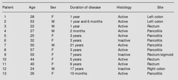
The study was approved by the Scientific Ethics Committee and the Council of the Department. Rectal or colon biopsy speci-mens and blood samples were obtained from 13 patients with ulcerative colitis documented by endoscopic and histologic findings who entered the study after giving written in-formed consent (one patient refused venous blood puncture). The histological lesions were classified according to Riddell et al. (23). These patients were symptomatic; ten patients had active disease but were negative for dysplasia, and three patients had inactive colitis (Table 1). ACD (citric acid/sodium citrate/glucose) was used as anticoagulant.
Tissue and blood samples were stored at -70oC prior to DNA extraction.
D NA e xtractio n
DNA was extracted from frozen blood and tissues by the phenol-chloroform method according to Sambrook et al. (24). Up to 43 µg genomic DNA was obtained from a small endoscopic biopsy specimen which was suf-ficient for the study.
Po lym e rase chain re actio n
One microgram or 500 ng of genomic DNA was used as a template in a reaction volume of 50 µl containing 50 pmol of each primer (Table 2), 200 µM of deoxynucleotide triphosphate (dNTP) and 2.5 U of Taq DNA polymerase (Perkin Elmer, Branchburg, NJ, USA). PCR was performed in a 2400 GeneAmp PCR system (Perkin Elmer). Ex-ons 5, 6, 7 and 8 of p53 gene were amplified in 40 cycles according to the following sched-ule: 93o
C for 1 min, 57o
C for 2 min and 70o
C for 2 min. Intron 6 and exon 4 were ampli-fied in 35 cycles according to the following schedule: 94o
C for 1 min, 62o
C for 50 s and 72o
C for 1 min.
Re strictio n fragme nt le ngth po lymo rphism
analysis o f the p53 ge ne
PCR products of intron 6 were digested with 60 U of MspI at 37o
C overnight (25); moreover, PCR products of exon 4 were digested with 12 U of AccII at 37oC
over-night (26). The DNA fragments were sepa-rated by electrophoresis on 4% low melting point agarose gel.
Chain-te rminatio n D NA se que ncing
PCR products were sequenced by the dideoxy chain termination method with the T7 Sequenase version 2.0 DNA sequencing kit (United States Biochemical). Radioactive label was incorporated with a-35SdATP
(Amersham, Cleveland, OH, USA). Samples were run on 6% gels; dried gels were ex-posed to Kodak TMG/RA-1 film at room temperature for 2-5 days.
Re sults
D NA se que ncing
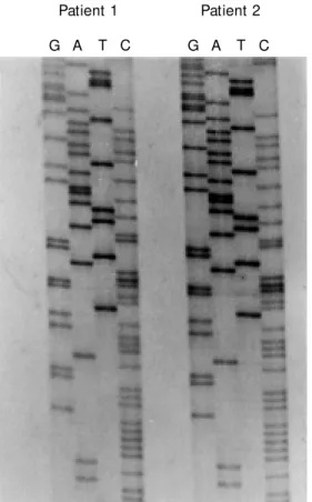
Thirteen ulcerative colitis biopsies were analyzed for mutation in the p53 gene. Ex-ons 5-8 were amplified with primers
con-taining the EcoRI restriction site for cloning in the pBluescript plasmid. However, direct sequencing of treated PCR products with exonuclease I and shrimp alkaline phos-phatase was performed. Direct sequencing was fast, easy and gave excellent results (Figure 1). No p53 gene mutation was de-tected in any of the cases studied.
PCR-RFLP analysis o f the p53 ge ne
DNA from tissue samples of twelve pa-tients with ulcerative colitis paired with DNA from leukocytes were studied for the detec-tion of p53 gene loss of heterozigosity (LOH). Two polymorphic sites were chosen, AccII in exon 4 and MspI in intron 6. Primers for
Table 1 - Clinical and pathological data.
Patient Age Sex Duration of disease Histology Site
1 28 F 1 year Active Left colon
2 53 M 1 year and 6 months Active Left colon
3 22 M 1 year Active Rectum
4 27 M 2 months Active Pancolitis
5 25 F 3 years Active Pancolitis
6 32 F 3 years Inactive Pancolitis
7 50 M 21 years Active Pancolitis
8 57 F 3 years Active Pancolitis
9 50 F 7 years Inactive Rectum/sigmoid
10 44 F 5 years Active Rectum
11 40 F 6 years Active Rectum
12 24 M 17 years Inactive Right colon
13 26 F 10 months Active Pancolitis
Table 2 - Primer sets used in PCR, sequencing and PCR-RFLP.
Primer set/ Primer sequences Priming region Assays References
1 5’-GGGCCCAGGGGTCAGCGGCA-3 ' Exon 7 antisense Sequencing
2 (27) 5’-GGAATTCTCCTAGGTTGGCTCTGAC-3 ' Exons 7-8 PCR/Sequencing 5’-GGAATTCCTGCTTGCTTACCTCGCT-3 ' Exons 7-8 PCR/Sequencing
3 (27) 5’-GGAATTCCTCTTCCTACAGTACTCC-3' Exons 5-6 PCR/Sequencing 5’-GGAATTCAGTTGCAAACCAGACCTCA-3' Exons 5-6 PCR/Sequencing
4 (25) 5’-AGGTCTGGTTTGCAACTGGG-3' Intron 6 PCR/RFLP 5’-GAGGTCAAATAAGCAGCAGG-3' Intron 6 PCR/RFLP
exon 4 span a region of 259 bp, AccII diges-tion of the amplified fragments identifies a second allele of 160 and 99 bp. LOH affect-ing the p53 exon 4 locus was not detected in lesions from five of twelve informative pa-tients (42%). MspI identifies a 2-allele poly-morphism with bands of 63 + 44 bp and 107 bp. The number of informative patients for LOH at the p53 intron 6 locus was high, eleven of twelve patients (92%), whereas no LOH was observed.
D iscussio n
There is convincing evidence that pa-tients with ulcerative colitis have a higher incidence of colorectal cancer than the gen-eral population. The risk increases with the duration of the disease (23,28), extensive ulcerative colitis (29) and older age at symp-tom onset (28). Dysplasia has been a histo-logic marker of malignant transformation and an indicator for colectomy in these
pa-tients (23). It is, thus, of great interest to identify markers for those cases that are likely to progress to high-grade dysplasia or adenocarcinoma.
We examined p53 gene alterations, al-lelic deletion and mutation in thirteen symp-tomatic ulcerative colitis patients negative for dysplasia. Exons 5-8 were amplified and sequenced from DNA of all cases in order to eliminate the possibility of missing a muta-tion. No p53 gene mutation was detected in these specimens.
The frequency of the loss of heterozygos-ity in the p53 gene was analyzed by PCR-RFLP. The regions examined were exon 4 and intron 6. Even though the number of informative cases was high, 42% and 92% in exon 4 and intron 6, respectively, LOH was not found in these cases. Our purpose was to observe the inflammatory process itself, since other genetic alterations have been observed at this stage.
We did not observe any p53 gene altera-tion in ulcerative colitis negative for dyspla-sia, in agreement with others (30) who did not detect p53 protein overexpression in sec-tions of indefinite dysplasia and no LOH of the p53 gene in epithelium associated with chronic inflammation, except in one case which had dysplasia elsewhere in the speci-men (31).
In a recent study on gastric cancer (Mattar R, Alexandrino A and Laudanna AA, un-published results), we also failed to observe allelic loss of the p53 gene in any of the cases analyzed. Thus, perhaps in our environment the p53 gene is not a frequent target of genetic alterations but other tumor suppres-sor genes are. This possibility should be considered and verified in a large number of sporadic colon cancer specimens.
Geographical differences in the p53 gene spectrum of mutations were previously re-ported in hepatocellular carcinoma from sub-Saharan Africa and China (Qidong). In these areas with risk for aflatoxins, a specific mu-tation of codon 249 of the p53 gene was
Patient 1 Patient 2
G A T C G A T C
found, whereas in other countries, mutations were scattered over four of the five evolu-tionarily conserved domains, which include codon 249 (32).
Rates of mutation or allelic deletion of the p53 gene were also slightly lower in dysplastic and cancerous ulcerative colitis than in other human cancers (11,13), or they were frequent (12,14) depending on the study referenced. In ulcerative colitis, p53 gene mutation was present in mucosa adjacent to the tumor that was histologically negative for dysplasia or cancer (14), quite different from sporadic colon cancer, where p53 gene mutation is a late event (6).
In conclusion, p53 is not a useful marker of malignant progression in our routine clini-cal practice since its alterations do not pre-cede dysplasia; other markers should be fur-ther evaluated for this purpose.
The activation of the src proto-oncogene was reported as being an early event in the genesis of ulcerative colitis colon cancer since src tyrosine kinase activity and protein abundance were elevated in neoplastic ul-cerative colitis epithelia. However, src ac-tivity measured in active colitis was remark-ably similar to that in normal mucosa or mild quiescent colitis (33).
Another promising marker of malignant progression in ulcerative colitis is the mu-cin-associated carbohydrate antigen sialosyl-Tn (ssialosyl-Tn) that correlates with malignant trans-formation in sporadic colonic neoplasms. It was expressed in 86% of ulcerative colitis
patients who developed cancer in at least one prior nondysplastic surveillance biopsy specimen from the same site (34).
Microsatellites are short nucleotide re-peat sequences present throughout the hu-man genome. In a subset of colorectal tu-mors and other tutu-mors, errors in DNA repair occur, resulting in altered DNA lengths at these regions, a phenomenon which has been termed microsatellite instability. This abnor-mality occurred at the same frequency in approximately one-fourth of patients with ulcerative colitis-associated dysplasia and carcinoma (35). Recently, microsatellite in-stability has been detected in 50% of ulcer-ative colitis patients whose colonic mucosa was negative for dysplasia (36). The authors suggested that the inability of DNA repair mechanisms to compensate for the stress of chronic inflammation might be one mech-anism for heightened neoplastic risk in ul-cerative colitis.
In ulcerative colitis tumor progression, other oncogenes and tumor suppressor genes are likely to be mutated, preceding the p53 gene as in sporadic colon cancer, or another process may occur in the genetic pathway. The observation that the mismatch repair function is lost due to the inflammatory pro-cess in patients with short-term ulcerative colitis (36) and the expression of the antigen sTn in biopsy specimen (34) open a new field of investigation in ulcerative colitis surveillance programs.
Re fe re nce s
1. Lane DP & Craw ford LV (1979). T antigen is bound to a host protein in SV 40-trans-formed cells. Nature, 278: 261-263. 2. Linzer DIH, M altzman W & Levine AJ
(1979). The SV 40 A gene product is re-quired for the production of a 54,000 M W cellular tumor antigen. Virology, 998: 308-318.
3. Finlay CA, Hinds PW, Tan T-H, Eliyahu D, Oren M & Levine AJ (1988). Activating mutations for transformation by p53
pro-duce a gene product that forms a hsc-70-p53 complex w ith an altered half-life. M o-lecular and Cellular Biology,8: 531-539. 4. Nigro JM , Baker SJ, Preisinger AC,
Jessup JM , Hostetter R, Cleary K, Bigner SH, Davidson N, Baylin S, Devilee P, Glover T, Collins FS, Weston A, M odali R, Harris CC & Vogelstein B (1989). M uta-tions in the p53 gene occur in diverse human tumour types. Nature, 342: 705-708.
5. Hinds PW, Finlay CA, Quartin RS, Baker SJ, Fearon ER, Vogelstein B & Levine AJ (1990). M utant p53 DNA clones from hu-man colon carcinomas cooperate w ith ras in transforming primary rat cells: a com-parison of the “ hot spot” mutant pheno-types. Cell Grow th and Differentiation, 1: 571-580.
gene mutations occur in combination w ith 17p allelic deletions as late events in colo-rectal tumorigenesis. Cancer Research, 50: 7717-7722.
7. Parada LF, Land H, Weinberg RA, Wolf D & Rotter V (1984). Cooperation betw een gene encoding p53 tumour antigen and ras in cellular transformation. Nature, 312: 649-651.
8. Eliyahu D, Raz A, Gruss P, Givol D & Oren M (1984). Participation of p53 cellular tu-mour antigen in transformation of normal embryonic cells. Nature, 312: 646-649. 9. Levine AJ, M omand J & Finlay CA (1991).
The p53 tumour suppressor gene. Nature, 351: 453-456.
10. Rodrigues NR, Row an A, Smith M EF, Kerr IB, Bodmer WF, Gannon JV & Lane DP (1990). p53 mutations in colorectal can-cer. Proceedings of the National Acade-my of Sciences, USA, 87: 7555-7559. 11. Greenw ald BD, Harpaz N, Yin J, Huang Y,
Tong Y, Brow n VL, M cDaniel T, New kirk C, Resau JH & M eltzer SJ (1992). Loss of heterozygosity affecting the p53, Rb, and mcc/apc tumor suppressor gene loci in dysplastic and cancerous ulcerative coli-tis. Cancer Research, 52: 741-745. 12. Yin J, Harpaz N, Tong Y, Huang Y, Laurin
J, Greenw ald BD, Hontanosas M , New kirk C & M eltzer SJ (1993). p53 point muta-tions in dysplastic and cancerous ulcer-ative colitis lesions. Gastroenterology, 104: 1633-1639.
13. Chaubert P, Benhattar J, Saraga E & Costa J (1994). K-ras mutations and p53 alter-ations in neoplastic and nonneoplastic le-sions associated w ith longstanding ulcer-ative colitis. American Journal of Patholo-gy, 144: 767-775.
14. Brentnall TA, Crispin DA, Rabinovitch PS, Haggitt RC, Rubin CE, Stevens AC & Burmer GC (1994). M utations in the p53 gene: an early marker of neoplastic pro-gression in ulcerative colitis. Gastroenter-ology, 107: 369-378.
15. Kastan M B, Onyekw ere O, Sidransky D, Vogelstein B & Craig RW (1991). Partici-pation of p53 protein in the cellular re-sponse to DNA dam age. Cancer Re-search, 51: 6304-6311.
16. Chen J, Jackson PK, Kirschner M W & Dutta A (1995). Separate domains of p21 involved in the inhibition of Cdk kinase and PCNA. Nature, 374: 386-388. 17. Waga S, Hannon GJ, Beach D & Stillman
B (1994). The p21 inhibitor of
cyclin-de-pendent kinases controls DNA replication by interaction w ith PCNA. Nature, 369: 574-578.
18. Shaw P, Bovey R, Tardy S, Sahli R, Sordat B & Costa J (1992). Induction of apoptosis by w ild-type p53 in a human colon tumor-derived cell line. Proceedings of the Na-tional Academy of Sciences, USA, 89: 4495-4499.
19. El-Deiry WS, Harper JW, O’Connor PM , Velculescu VE, Canman CE, Jackman J, Pietenpol JA, Burrell M , Hill DE, Wang Y, Wiman KG, M ercer WE, Kastan M B, Kohn KW, Elledge SJ, Kinzler KW & Vogelstein B (1994). WAF1/CIP1 is induced in p53-mediated G1 arrest and apoptosis. Cancer
Research, 54: 1169-1174.
20. Knudson Jr AG (1985). Hereditary cancer oncogenes and antioncogenes. Cancer Research, 45: 1437-1443.
21. Vogelstein B, Fearon ER, Hamilton SR, Kern SE, Preisinger AC, Leppert M , Nakamura Y, White R, Smits AM M & Bos JL (1988). Genetic alterations during colo-rectal-tumor development. New England Journal of M edicine, 319: 525-532. 22. Yokota J & Sugimura T (1993). M ultiple
steps in carcinogenesis involving alter-at ions of m ult iple t um or suppressor genes. FASEB Journal, 7: 920-925. 23. Riddell RH, Goldman H, Ransohoff DF,
Appelman HD, Fenoglio CM , Haggitt RC, Ahren C, Correa P, Hamilton SR, M orson BC, Sommers SC & Yardley JH (1993). Dysplasia in inflammatory bow el disease: standardized classification w ith provi-sional clinical applications. Human Pathol-ogy, 14: 931-968.
24. Sambrook J, Fritsch EF & M aniatis T (1989). M olecular Cloning - A Laboratory M anual. 2nd edn. Cold Spring Harbor Laboratory, New York.
25. M cDaniel T, Carbone D, Takahashi T, Chumakov P, Chang EH, Pirollo KF, Yin J, Huang Y & M eltzer SJ (1991). The M spI polymorphism in intron 6 of p53 (TP53) detected by digestion of PCR products. NucleicAcids Research, 19: 4796. 26. De la Calle-M artin O, Fabregat V, Romero
M , Soler J, Vives J & Yägue J (1990). AccII polymorphism of the p53 gene. Nucleic Acids Research, 18: 4963.
27. Tohdo H, Yokozaki H, Haruma K, Kajiyama G & Tahara E (1993). p53 gene mutations in gastric adenomas. Virchow s Archiv. B, Cell Pathology, 63: 191-195.
28. Lashner BA, Silverstein M D & Hanauer
SB (1989). Hazard rates for dysplasia and cancer in ulcerative colitis. Digestive Dis-eases and Sciences, 34: 1536-1541. 29. Gyde SN, Prior P, Allan RN, Stevens A,
Jew ell DP, Truelove SC, Lof berg R, Brostrom O & Hellers G (1998). Colorec-tal cancer in ulcerative colitis: a cohort study of primary referrals from three cen-tres. Gut, 29: 206-217.
30. Papatheodoridis GV, Zizi A-S, Xourgias V, Elemenoglou I & Karamanolis DG (1998). Indefinite dysplasia in ulcerative colitis has little clinical significance and is not associ-at ed w it h p53 prot ein accum ulassoci-at ion. American Journal of Gastroenterology, 93: 285-286.
31. Fogt F, Vort m eyer AO, Goldm an H, Giordano TJ, M erino M J & Zhuang Z (1998). Comparison of genetic alterations in colonic adenoma and ulcerative colitis-associated dysplasia and carcinoma. Hu-man Pathology, 29: 131-136.
32. Ozturk M , Bressac B, Puisieux A, Kew M , Volkmann M , Bozcall S, M ura JB, De la M ont S, Carlson R, Blum H, Wands J, Takahashi H, Von Weizsäcker F, Galun E, Kar S, Car BI, Schröder CH, Erken E, Varinli S, Rustgi VK, Prat J, Toda G, Koch HK, Liang XH, Tang Z-Y, Shouval D, Lee H-S, Vyas GN & Sarosi I (1991). p53 mutation in hepatocellular carcinoma after aflatoxin exposure. Lancet, 338: 1356-1359. 33. Cartw right CA, Coad CA & Egbert BM
(1994). Elevated c-src tyrosine kinase ac-tivity in premalignant epithelia of ulcer-ative colitis. Journal of Clinical Investiga-tions, 93: 509-515.
34. Itzkow itz SH, M arshall A, Kornbluth A, Harpaz N, M cHugh JBD, Ahnen D & Sachar DB (1995). Sialosyl-Tn antigen: ini-tial report of a new marker of malignant progression in long-standing ulcerative colitis. Gastroenterology, 109: 490-497. 35. Suzuki H, Harpaz N, Tarmin L, Yin J, Jiang