a Department of Pathology, Orthopaedics and Traumatology Institute, São Paulo University Medical School – São Paulo/SP, Brazil.
b Endocrinology Division and Molecular Genetics Laboratory, São Paulo University Medical School – São Paulo/SP, Brazil.
c Statistics Department, São Paulo University School of Public Health - São Paulo/SP, Brazil.
Email: [email protected] Received for publication on August 28, 2006. Accepted for publication on October 11, 2006.
CLINICAL SCIENCES
CLASSICAL OSTEOBLASTOMA, ATYPICAL
OSTEOBLASTOMA, AND OSTEOSARCOMA. A
COMPARATIVE STUDY BASED ON CLINICAL,
HISTOLOGICAL, AND BIOLOGICAL PARAMETERS
Cláudia Regina Gomes Cardim Mendes de Oliveira,a Berenice Bilharino
Mendonça,b Olavo Pires de Camargo,a Emilia Modolo Pintob,Sérgio Antonio
Barbosa Nascimentoa, Maria do Rosario D. O. Latorre,c Maria Claúdia Nogueira
Zerbinia
Oliveira CRGCM de, Mendonça BB, Camargo OP de, Pinto EM,Nascimento SAB, Latorre M do RDO,Zerbini MCN. Classical osteoblastoma, atypical osteoblastoma, and osteosarcoma. a comparative study based on clinical, histological, and biological parameters. Clinics. 2007;62(2):167-74.
OBJECTIVE: To investigate the biological behavior of classical and atypical osteoblastomas in comparison to osteosarcomas.
METHODS: Based on histological parameters, 30 osteoblastomas were subclassified as classical osteoblastomas (23/30) or atypical osteoblastoma (high cellularity, prominent blue osteoid, and epithelioid osteoblasts–7/30). Comparative immunohistochemical and clinical analysis was performed in 17 cases of patients with high-grade osteosarcoma. Formalin-fixed, paraffin-embedded archival tissue was immunostained for p53 and proliferation cell nuclear antigen. Tumors with positive p53 stain underwent molecular analyses for fragments of exon 10.
RESULTS: The mean proliferating cell nuclear antigen indexes for classical osteoblastoma, atypical osteoblastoma, and osteosarcoma were 33%, 61%, and 79%, respectively. The atypical subgroup showed similar results to those of the osteosarcoma group (P < 0.001). p53 protein was detected in 4 (13%) osteoblastomas (3 of these were atypical osteoblastoma), and 4 osteosarcomas (23%) also showed p53 positivity. DNA mutation performed in p53-positive cases was confirmed in exon 10 in 2 atypical osteoblastomas (2/3), 1 classical osteoblastoma (1/1), and 1 osteosarcoma (1/4). Disease recurrence was correlated with p53 expression (P = 0.009), atypical subtype (P = 0.031), spiculated blue bone on histology (P = 0.018), and proliferatingcell nuclear antigen labeling index ≥ 40 (P = 0.015).
CONCLUSION: These results validate atypical osteoblastoma as an entity, with p53 and proliferation cell nuclear antigen immunoexpression closer to that of osteosarcoma than of classical osteoblastoma. Proliferating cell nuclear antigen labeling index and p53 may be useful predictors of recurrence.
KEYWORDS: Bone neoplasms. Cell proliferation. Proliferating cell nuclear antigen. Immunohistochemistry. Osteoblastoma. p53 gene. p53 protein.
INTRODUCTION
Osteoblastoma is an uncommon bone tumor that is
considered the benign counterpart of osteosarcoma. Locally aggressive behavior has been noted in some osteoblastomas; however, malignant transformation of the tumors ia rare.1–5
Malignant osteoblastoma or aggressive osteoblastoma,6,7 are
terms used to describe some lesions that show atypical cytologic features and may be correlated with recurrence. Osteoblastoma accounts for approximately 1% of all bone tumors.8,9 The most commonly affected sites are the spine,
of life.1 Approximately 25% of patients have radiographic
and histologic aspects that may suggest malignancy,10 such
as large epithelioid osteoblasts, bizarre cells, mitosis, and lace- or sheet-like osteoid production. Several reports6,7,11
have proposed a designation of a borderline osteoblastic tumor, whose radiographic and histologic features vary be-tween the osteoblastoma and osteosarcoma. The term ag-gressive osteoblastoma was considered preferable because it designates a tumor with local destructive behavior, but without metastatic potential. According to several studies, osteoblastomas may primarily undergo malignant transfor-mation.1,2,4,5 Moreover, cases of osteoblastoma-like
osteosa-rcoma are well-recognized.12,13 Further reports of large
se-ries of osteoblastomas have not confirmed the predictive value of the microscopic appearances,1,14 raising the
ques-tion of whether atypical osteoblastoma really does exist. The assessment of the proliferative antigen expression yields predictive indicators in soft tissues and bone tumors, with several studies demonstrating the clinical value of anti-PCNA (proliferating cell nuclear antigen) and anti-p53 monoclonal antibodies in these tumors.15–19
Immunohisto-chemical expression of the p53 protein and the analysis of p53 gene mutations have also been described in 10% to 35% of soft tissue tumors.18 However, only few studies have
reported the importance of p53 changes in borderline or low-grade osteoblastic tumors, with controversial results.20– 22 Rossner et al23 reported mutation of the p53 gene at exon
5 in a patient with malignant osteoblastoma. Radig et al20
described another mutation at exon 7 in a patient with osteoblastoma-like osteosarcoma. Luca et al24 have
previ-ously reported abnormalities in exon 10 of the p53 gene of osteosarcomas, suggesting that exon 10, which is prob-ably involved in the protein oligomerization, is essential for protein function.
The aim of this study was to contribute to understand-ing this rare bone neoplasm based on morphology, prolif-eration rate, and p53 mutation analysis. In addition, high-grade osteosarcomas were evaluated to determine discrimi-nating features.
MATERIALS AND METHODS
Patients
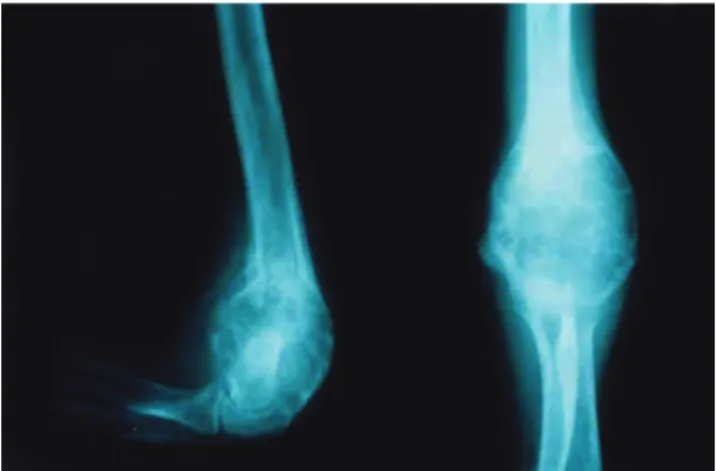
A retrospective review of 30 patients with osteoblastoma who were treated at the Instituto de Ortopedia e Traumatologia of the São Paulo University Medical School Hospital was performed. The group of pa-tients evaluated consisted of 19 men and 11 women, and the median age was 18.5 years (range, 4-41 years). The most commonly affected sites were the spine (33%) and
the long bones of the appendicular skeleton (23%). Typi-cally, a radiolucent and well-circumscribed expansive le-sion with calcifications was observed (Figure 1). Seventeen patients with high-grade osteosarcomas were evaluated in order to compare results from immunohistochemical and molecular analyses. In all patients, hematoxylin-eosin– stained sections confirmed the diagnosis. Tumors were clas-sified as atypical osteoblastomas (n = 7) according to the histologic features described by Schajowicz and Lemos7
and Dorfman.6 Schajowicz and Lemos7 proposed the term
malignant osteoblastoma for lesions that present with high cellularity, nuclear atypia, and immature tumor bone (spicu-lated blue bone). Dorfman and Weiss6 proposed the term
aggressive osteoblastoma for lesions that present with cel-lular zones of epithelioid osteoblasts. Radiographic find-ings and follow-up were documented based on the clinical records.
Immunohistochemistry (IHQ)
Deparaffinized formalin-fixed sections were immunostained using monoclonal antibodies to the p53 pro-tein (DO7, 1:2000, DAKO Corporation) and PCNA (PC10, 1:8000, DAKO Corporation). Sections underwent heat-in-duced epitope retrieval using a pressure-cooker. Antigens were identified using the strepta-avidin-biotin method. For p53, positivity was defined as ≥ 10% of immunostained tumor cells.25 The percentage of tumoral cells positive for
PCNA was determined from 500 tumor cells, and results were expressed as PCNA labeling index (PCNA-LI).
Molecular Analyses
Preparing DNA (deoxyribonucleic acid)
Genomic DNA was isolated from the archival paraffin-embedded material from the 4 cases of osteoblastoma and
the 4 cases of osteosarcoma with positive immunohisto-chemistry for the p53 protein. Paraffin-block sections from tumor tissues were stained by hematoxylin-eosin to select areas of neoplastic and normal tissues. According to pre-viously published guidelines,26 DNA extraction was
per-formed using paraffin-embedded tissue with the standard phenol chloroform extraction method.
Polymerase chain reaction (PCR)
PCR was used to amplify DNA fragments. In all cases, genomic DNA was amplified using a 100-µL PCR mixture of oxynucleotide triphosphate (100 µmol/L), MgCl2 (50 mmol/L), 10 pmol of each of the primers, and Taq polymer-ase (2.5 U) (Pharmacia Biotech, Uppsala, Sweden). Ther-mal cycling was performed using a Gene Amp PCR Sys-tem (Perkin Elmer Cetus, Norwalk, CT). Polymerase chain reaction was done at 94°C for 5 minutes followed by a 30-cycle denaturation at 94°C for 5 minutes, and a 30-30-cycle denaturation at 94°C for 30 seconds. In all PCR experi-ments, reactions without DNA were considered negative controls. All PCR products were visualized using 2% ethidium bromide-stained agarose gels. Fragments of exon 10, exon 5, and exon 7 of the p53 with 195 bp were am-plified using 2 pairs of oligonucleotide.23
Exon 5: 5’ A T C T G T T C A C T T G T G C C C T G A C T T T C 3’ and 5’ A C C C T G G G C A A C C A G C C C T G T 3’
Exon 10: 5’ G C T G TA T A G G T A C T T G A A G T G C 3’ and 5’ C A T A G G A T T C C A T T C T C A T C 3’ Exon 7: C T C C T A G G T T G G C T C T G 3’ and 5’ G A G G C T G G G G C A C A G C A G G C C A G T G 3’
Direct Sequencing
Polymerase chain reaction products were prepared for DNA sequencing using exonuclease enzyme (Amersham, Arlington Heights, IL) and shrimp alkaline phosphatase (Amersham) at 37°C for 15 minutes and at 80°C for 15 minutes. Automatic direct DNA sequencing of exon 10 was done using the AB I Prism Dye Terminator Sequencing Kit (Perkin Elmer Cetus for PCR products).
Statistical analysis
Analyses of correlations between immunohistochemi-cal expression of p53 protein, histologic features, and out-comes were carried out using the Fisher exact test. Analy-ses of the PCNA proliferation index, expression of the p53 protein, and outcomes were done using the Mann-Whitney test. A P value < 0.05 was considered statistically signifi-cant. Recurrence was fixed as event of interest, which was evaluated by the Kaplan-Meier test, correlated with p53 immunoexpression, PCNA index ≥ 40 and < 40, and
sub-group of osteoblastoma (atypical osteoblastoma x classi-cal osteoblastoma).
RESULTS
Pathologic findings
Histologically, the major components of the osteoblastoma were osteoblast-like cells, stromal cells, osteoclasts, osteoid tissue deposition, and blood vessels. These cases were classified as classical osteoblastomas (n = 23) (Figure 2). Five patients had destructive infiltration and cortical invasiveness (interruption). In these patients, the most commonly affected sites were the spine, flat bones, and long bones. Atypical osteoblastomas were di-agnosed by the following features: epithelioid osteoblasts were identified in 5 patients (Figure 3) and a zone of spicu-lated blue bone,5 was observed in 6 patients (Figure 4).
Figure 2 - Classical histologic aspects of osteoblastoma with long inter anastomosing trabeculae of woven bone and osteoid rimmed by a single row of osteoblasts (Case 21, hematoxylin-eosin stain, X 200)
Occasionally, osteoblasts showed a focal clustering pattern and hyperchromatic nuclear staining (Figure 5). Cases with features described above were classified as atypical osteoblastomas (n = 7), and the distinction from osteosar-coma was based on the absence of atypical mitotic figures, cellular pleomorphism, neoplastic cartilage, and permeative growth into adjacent bone tissue.13
p53 immunohistochemistry / gene mutation
Positive immunoexpression of p53 was identified in 4 cases of osteoblastomas (13.3%), with 3 of these classified as atypical osteoblastoma (Figure 6). Significantly more cases were p53-positive in atypical tumors (P = 0.03). In the osteosarcoma group (n = 17), 4 cases were p53-posi-tive (23.5%). When the 3 groups were compared, immunoexpression of p53 in atypical osteoblastoma was similar to that of osteosarcoma, and both were different from classical osteoblastoma (P < 0.005) (Figure 7). Es-sentially, expression of the p53 protein was statistically
cor-related with recurrence (P = 0.009), atypical osteoblastoma classification (P = 0.031), and with the presence of spicu-lated blue bone (P = 0.018). Mutational changes of the p53 at exon 10 (codon 337) were identified in 3 of 4 patients with osteoblastoma (2 patients with atypical osteoblastoma, and 1 patient with classical osteoblastoma) (Figure 8) and 1of 4 patients with osteosarcoma. Exons 5 and 7 could not
Figure 4- Atypical osteoblastoma. Focal areas of fine, lace-like osteoid (Case 29, hematoxylin-eosin stain, X 400)
Figure 5 - Atypical osteoblastoma. High cellularity and atypical cells. No evidence of osteoid production (Case 27, hematoxylin-eosin stain, X 400)
Figure 6- Atypical osteoblastoma. Immunohistochemical detection of the p53 protein with DO7. A distinct nuclear reaction is seen the majority of the tumor cells (Case 26, immunohistochemistry, X 400)
Figure 8 - Direct genomic sequence analysis of the p53 gene in patient no.30. Heterozygous G to A substitution in exon 10
be evaluated in the samples because of the limited amount of adequately sized DNA fragments obtained from the ar-chival paraffin embedded tissues, which failed to amplify by PCR.
PCNA labelling index (PCNA-LI)
The atypical osteoblastoma and osteosarcoma groups had higher mean PCNA-LI values than those of classical osteoblastoma (P < 0.001) (Figures 9 and 10). The mean PCNA indexes for classical osteoblastoma, atypical osteoblastoma, and osteosarcoma were 33.7%, 61.7%, and 79.3%, respectively. Results were divided into 2 groups: low PCNA index (< 40%) and high PCNA index (≥ 40%) (30 patients)
Correlation between clinical outcome, osteoblastoma classification, p53 expression, morphological
parameters, and PCNA labelling index
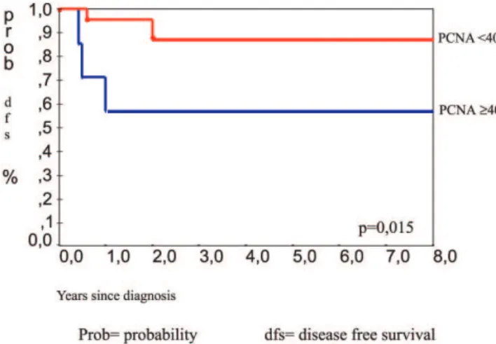
The clinical data of 30 patients were available with a 48-month period of follow-up. All patients were treated by total surgical ressection. Five patients (16.6%) had recur-rence of the tumor. Three of the patients were in the group with atypical osteoblastoma. Analysis by the Kaplan-Meyer test showed that recurrence was statistically correlated with the atypical osteoblastoma subgroup (P = 0.031), (Figure 11), p53 protein expression (P = 0.009), (Figure 12), pres-ence of spiculated blue bone (P = 0.041), and PCNA la-belling index stratified by a 40% cut-off value (P = 0.015) (Figure 13).
DISCUSSION
The accurate detection and differentiation and treatment of benign and malignant bone tumors can be a serious chal-lenge. 27,28 The histologic appearance of osteoblastoma
var-ies widely. At one end of the spectrum are the lesions char-acterized by trabecular organization of the newly formed
Figure 12 - Kaplan-Meier test for event recurrence, comparing p53 immunoexpression positive and negative
Figure 10 - PCNA% in classical osteoblastomas (1.0), atypical osteoblastomas (2.0), and osteosarcomas (3.0)
Figure 9 - Mean values of PCNA labeling indexes in classical osteoblastoma, (33.7%) atypical osteoblastoma (61.7%), and osteosarcoma. (79.3%)
osteoid, highly vascular background, and low cellularity. At the other end of this spectrum are the lesions charac-terized by high cellularity, prominent blue osteoid, mito-sis, and epithelioid osteoblasts. These cases may be desig-nated as atypical or aggressive osteoblastomas. In our study, the presence of a lace-like or a sheet-like osteoid deposi-tion was correlated with recurrence (P = 0.041) and ag-gressive behavior. As with other series, the agag-gressive pat-tern of this tumor entity was not correlated with other his-tological features, such as the evidence of epithelioid osteoblasts and infiltrative histologic growth.1,14
The recurrence rate presented in this series of patients (16.6%) was similar to that of earlier observations.14 The
PCNA-LI was correlated with recurrence and the highest in-dices were associated with atypical groups, as observed in the osteosarcoma group. A PCNA-LI < 40% was rarely found in atypical osteoblastoma (1/7) or osteosarcoma (0/17).
The frequency of p53 immunostaining varied among the different subtypes of osteoblastoma and osteosarcoma, be-ing observed predominantly in osteosarcoma and in atypi-cal osteoblastoma. Interestingly, out of the 2 classiatypi-cal osteoblastomas associated with recurrence, 1 was positive for p53 immunostaining and showed a DNA mutation in exon 10. This is the first study that shows DNA mutation at exon 10 in osteoblastomas. Other studies have also re-ported p53 mutations and the immunohistochemical pro-tein detection.22,29-31 Immunopositivity for p53 was detected
in cases that did not have evidence of p53 gene mutation. This may have occurred for several reasons, including that
Figure 13 - Kaplan-Meier test for event recurrence, comparing PCNA labeling index ³40 and < 40
mutational changes of p53 gene may occur outside the re-gion of the gene analyzed. In this study, Exon 5 and exon 7 were not evaluated in all the samples because the size of the amplified fragments is restricted in archival paraffin-embedded tissues. The quality of DNA extracted from this sort of sample is highly variable, and it depends on decal-cification, fixing, and storing of the tissues samples.32
The sample size of the current study was too small to draw definitive conclusions regarding the relationship be-tween clinicopathologic features and p53 changes. How-ever, the results indicating that atypical osteoblastoma, as described by its morphological features, seems to be a real entity. When evaluated by means of proliferation rate and p53 mutations, atypical osteoblastoma exhibits a behavior that is in between classical osteoblastomas and osteossarcomas. The major diagnostic challenge is in dif-ferentiating malignant or aggressive osteoblastoma from osteosarcoma on the basis of the pathologic features,since the diagnosis markedly changes treatment in patients.33,34
None of our cases presented metastases. These findings raise the possibility that new genetic events may occur in the group of osteosarcomas that are responsible for its abil-ity to acquire metastatic potential. Our results suggest that the detection of p53 immunopositivity, evidence of a high PCNA labeling index (³ 40), and mutations of the p53 gene (exon 10) may have a role in the pathogenesis of osteoblastomas and may reflect the tumoral progression with recurrences, but not exert a sufficient deleterious ef-fect to produce malignant behavior, as is observed in oste-osarcoma.
Thus, further investigation, including that regarding functional analysis of mutations, is essential to demonstrate the role of genetic changes in the clinical and histopatho-logic features of osteoblastoma.
ACKNOWLEDGMENTS
RESUMO
Oliveira CRGCM de, Mendonça BB, Camargo OP de, Pinto EM,Nascimento SAB, Latorre M do RDO,Zerbini MCN. Osteoblastoma clássico, osteoblastoma atípico e osteos-sarcoma. Um estudo comparativo baseado em parâmetros clínicos, histológicos e biológicos. Clinics. 2007;62(2): 167-74.
OBJETIVOS: Investigar o comportamento biológico de
osteoblastomas clássicos e atípicos comparados com osteossarcomas.
MÉTODOS: Com base em parâmetros histológicos
clas-sificamos um grupo de 30 osteoblastomas nos subgrupos de osteoblastomas clássicos (23/30) e de osteoblastomas atípicos (que apresentam como característica grande celularidade, osteóide azul proeminente e osteoblastos epitelióide—7/30). Como efeito de comparação dos resul-tados imunohistoquímicos e análise clínica, avaliamos 17 pacientes com osteosarcoma de grau avançado. Os cortes histológicos com bloco de parafina fixado em formalina foram imunocorados para p53 e antígeno nuclear de célu-la em proliferação. Tumores com coloração positiva para p53 tiveram análise molecular para fragmentos do exon 10.
RESULTADOS: O índice médio de antígeno nuclear de
cé-lula em proliferação para osteoblastoma clássico,
osteoblastoma atípico e osteosarcoma foram de 33%, 61% e 79%, respectivamente. O subgrupo atípico demonstrou resul-tados similares aos dos osteosarcomas (p<0,001). Foram de-tectadas proteína p53 em 4 (13%) osteoblastomas; 3 desses foram osteoblastomas atípicos, sendo que 4 osteosarcomas (23%) também demonstraram p53 positivo. A mutação do DNA nos casos positivos de p53 foi confirmada no exon 10 em dois osteoblastomas atípicos (2/3), um osteoblastoma clás-sico (1/1) e um osteosarcoma (1/4). A recorrência da doença foi correlacionada com a expressão do p53 (p=0,009), subtipo atípico (p=0,031), osso azul espiculado no resultado da histologia (p=0,018), e índice de marcação pelo antígeno nu-clear de célula em proliferação ≥ 40 (p=0,015).
CONCLUSÃO: Esses resultados validam os
osteoblas-tomas atípicos como entidade real, com imunoexpressão das proteínas p53 e antígeno nuclear de célula em prolife-ração mais perto do osteosarcoma do que do osteoblastoma clássico. O índice de marcação pelo antígeno nuclear de célula em proliferação e o p53 podem ser úteis fatores de prognóstico da recorrência.
UNITERMOS: Neoplasias ósseas. Proliferação celular.
PCNA. Imunohistoquímica. Osteoblastoma. Proteína p53. Gene p53.
REFERENCES
1 Lucas DR, Unni KK, Mcleod RA, O’Connor MJ, Sim FH. Osteoblastoma: clinicopathologic study of 306 cases.Hum Pathol. 1994;25:117-34.
2 Mitchell ML, Ackerman LV. Metastatic and pseudomalignant osteoblastoma. Skeletal Radiol. 1986;15:213-8.
3 Moon KS, Jung S, Lee TY, Kim SH, Kang SS. Benign osteoblastoma of the occipital bone. Case report and literature review. Neuropathology. 2006;26:141-6.
4 Morton KS, Quenville NF, Beauchamp CP. Aggressive osteoblastoma. J Bone Joint Surg. 1989;71:428-31.
5 Seki T, Fukuda H, Hanaoka H, Yatabe S, Takano M, Koide O. Malignant transformation of benign osteoblastoma. J Bone Joint Surg. 1975;57:424-6.
6 Dorfman HD, Weiss SW. Boderline osteoblastic tumors: problems in the differential diagnosis of aggressive osteoblastoma and low grade osteosarcoma. Semin Diagn Pathol. 1984;1:215-34.
7 Schajowicz F, Lemos C. Malignant osteoblastoma. J Bone Joint Surg. 1976; 58:202-11.
8 Dorfman HD, Czerniak B. Benign osteoblastic tumors. In: Bone tumors. St Louis: Mosby; 1998. p. 103-25.
9 Saghieh S, Rameh C, Birjawi G, Lakki S. Sacral osteoblastoma presenting as a L5-S1 disc herniation. Int Surg. 2005;90:289-92. 10 Mcleod RA, Dahlin DC, Beabout, JW. The spectrum of osteoblastoma.
Am J Roentgenol. 1976;126:321-35.
11 Scully RE, Galdabini JJ, Mc Neely BU. Case records of the Massachusetts General Hospital. New Engl J Med. 1980;303:866-73. 12 Bertoni F, Unni KK, Mcleod RA, Dahlin DC. Osteosarcoma resembling
osteoblastoma. Cancer. 1985;55:416-26.
13 O’ Connor E, Stacy G. Osteoblastoma. emedicine [ serial on line]. 2004, June 16. Avaiable from: http.www.emedicine.com/radio/topic494.htm 14 Della Rocca C, Huvos AG. Osteoblastoma: varied histological presentations with a benign clinical course. Am J Surg Pathol. 1996;20:841-50.
15 Dreinhofer KE, Akerman M, Willen H, Anderson C, Gustafson P. Proliferating cell nuclear antigen (PCNA) in high-grade malignant fibrous histiocytoma: prognostic value in 48 patients. Int J Cancer. 1994;59:379-82.
17 Kroese MOS, Rutgers DH, Wils IS, Van Unnik JAM, Roholl PJM. The relevance of the DNA index and proliferation rate in the grading of benign and malignant soft tissue tumors. Cancer. 1990;65:1782-8. 18 Lonardo F, Ueda T, Huvos AG, Healey J, Ladanyi M. p53 and mdm2
alterations in osteosarcomas. Cancer. 1997;79:1541-47.
19 Niezabitowski A, Rys J, Roessner A, Lackowska B, Schneider-Stock R, Gruchala A, et al. Assessment of proliferative activity, DNA values and some clinicopathologic parameters in mesenchymal tumors. Gen Diagn Pathol. 1997;142:327-33.
20 Radig K, Schneider-Stoch R, Mittler V, Neumann HW, Roessner A. Genetic instability in osteoblastic tumors of the skeletal system. Pathol Res Pract. 1998;194:664-7.
21 Toguchida J, Yamaguchi T, Ritchi B, Beauchamp RL, Dayton SH, Herrera GE, et al. Mutation spectrum of p53 gene in bone and soft tissue sarcomas. Cancer Res. 1992;52:6194-9.
22 Ueda Y, Dockhorn-Dwurniczak B, Blasius S. Analysis of mutant p53 protein in osteosarcomas and other malignant and benign lesions of bone. J Cancer Res Clin Oncol. 1993;119:172-8.
23 Roessner A, Schneider-Stoch R, Radig K, Neumann W, Mittler U. Alterations of p53gene in soft tissue and bone tumors. Gen Diagn Pathol. 1997;143:1-13.
24 Luca JW, Strong LC, Hansen MF. A germline missense mutation R 337c in exon10 of the human P53 gene.Hum Mutat. 1998; Suppl 1: S58-61. 25 Park YB, Kim HS, Oh JH, Lee SH. The co-expression of p53 protein and p-glycoprotein is correlated with a poor prognosis in osteosarcoma. Int Orthop (SICOT). 2001;24:307-10.
26 Wright DK, Manos MM. Sample preparation from paraffin-embedded tissues. In: Innis M, editor. PCR protocols: a guide to methods and applications.San Diego: Academic Press; 1990. p153.
27 Camargo OP de, Croci AT, Oliveira CRGMC de, Baptista AM, Caiero MT. Functional and radiographic evaluation of 214 aggressive benign bone lesions treated with curettage, cauterization, and cementation: 24 years of follow-up. Clinics. 2005;60(6):439-444.
28 Guerra RB, Tostes MD, Miranda L da C, Camargo OP de, Baptista AM, Caiero MT. Comparative analysis between osteosarcoma and Ewing’s sarcoma: evaluation of the time from onset of signs and symptoms until diagnosis. Clinics. 2006;61:99-106.
29 Cordon-Cardo C, Latres E, Drobnjak M, Oliva MR, Pollack D, Woodruff JM, et al. Molecular abnormalities of mdm2 and p53 in adult soft tissue sarcomas. Cancer Res. 1994; 54:794-9.
30 Moússes S, Mcauley L, Bell RS, Kandel R, Andrulis IL. Molecular and Immunohistochemical identifications of p53 alterations in bone and soft tissue sarcomas.Mod Pathol. 1996;9:1-6.
31 Yokoyama R, Schneidner-Stock R, Radig K, Wex T, Roessner A. Clinicopathologic implications of mdm2, p53 and K-ras gene alterations in osteosarcomas: mdm2 amplification and p53 mutations found in progressive tumors. Pathol Res Pract. 1998;194:615-21.
32 Jackson DP, Lewis FA, Taylor GR, Boylsto AW, Quirke NP. Tissue extraction of DNA and RNA and analysis by the polymerase chain reaction. J Clin Pathol 1990; 43:499-504.
33 White LM, Kandel R. Osteoid-producing tumors of bone. Semin Musculoskelet Radiol. 2000;4:25-43.