Re ce nt advance s in angio te nsin II
signaling
1Multidisciplinary Research Group on Hypertension, Clinical Research Institute of
Montreal, University of Montreal, Q uebec, Canada
2Department of Medicine and Therapeutics, Western Infirmary, University of Glasgow,
Glasgow, Scotland, UK R.M. Touyz1
and C. Berry2
Abstract
Angiotensin II (Ang II)* is a multifunctional hormone that influences
the function of cardiovascular cells through a complex series of intracellular signaling events initiated by the interaction of Ang II with AT1 and AT2 receptors. AT1 receptor activation leads to cell growth, vascular contraction, inflammatory responses and salt and water reten-tion, whereas AT2 receptors induce apoptosis, vasodilation and natri-uresis. These effects are mediated via complex, interacting signaling pathways involving stimulation of PLC and Ca2+ mobilization;
activa-tion of PLD, PLA2, PKC, MAP kinases and NAD(P)H oxidase, and stimulation of gene transcription. In addition, Ang II activates many intracellular tyrosine kinases that play a role in growth signaling and inflammation, such as Src, Pyk2, p130Cas, FAK and JAK/STAT. These events may be direct or indirect via transactivation of tyrosine kinase receptors, including PDGFR, EGFR and IGFR. Ang II induces a multitude of actions in various tissues, and the signaling events following occupancy and activation of Ang receptors are tightly controlled and extremely complex. Alterations of these highly regu-lated signaling pathways may be pivotal in structural and functional abnormalities that underlie pathological processes in cardiovascular diseases such as cardiac hypertrophy, hypertension and atherosclero-sis.
Co rre spo nde nce
R.M. Touyz
Clinical Research Institute of Montreal
110 Pine Ave. West Montreal, Q uebec, H2W 1R7 Canada
Fax: + 2-514-987-5523 E-mail: touyzr@ ircm.qc.ca
Presented at the IV International Symposium on Vasoactive Peptides, Belo Horizonte, MG, Brazil, O ctober 19-21, 2001.
Research supported by funds from the Canadian Institutes of Health Research (CIHR), Heart and Stroke Foundation of Canada, Canadian Hypertension Society (CHS) and the Fonds de la Recherche en Santé du Q uebec. R.M. Touyz is supported by a scholarship from the CIHR/CHS. C. Berry is supported by a Medical Research Council Clinical Training fellowship.
Received December 6, 2001 Accepted January 29, 2002
Ke y wo rds
·Angiotensin receptors
·Second messengers
·Phospholipase
·Protein kinase
·Reactive oxygen species
Akt/PKB, protein Ser/Thr kinase/protein kinase B
Ang II, angiotensin II
EGFR, epidermal growth factor receptor
ERK, extracellular signal-regulated kinase
FAK, focal adhesion kinase
IGFR, insulin growth factor receptor
IP3, inositol triphosphate
JAK, Janus kinase
JNK, c-Jun N-terminal protein kinase
MAPK, mitogen-activated protein kinase
MEK, MAPK/ERK kinase
MKP-1, MAPK phosphatase 1
PD GF(R), platelet-derived growth factor (receptor)
PG, prostaglandin
PI3K, phosphatidylinositol 3-kinase
PKC, protein kinase C
PLA, phospholipase A
PLC, phospholipase C
PLD, phospholipase D
RTK, receptor tyrosine kinase
SAPK, stress-activated protein kinases
STAT, signal transducers and activators of transcription
VSMC, vascular smooth muscle cells
Intro ductio n
Angiotensin II (Ang II) regulates blood pressure, plasma volume, sympathetic ner-vous activity and thirst responses. It also plays an important pathophysiological role in car-diovascular disease, including cardiac hyper-trophy, myocardial infarction, hypertension and atherosclerosis. Ang II is produced sys-temically via the classical renin-angiotensin system and locally via the tissue renin-angio-tensin system (1). Ang II was initially de-scribed as being primarily a vasoconstrictor peptide. However, recent studies demonstrate that Ang II has growth factor and cytokine-like properties as well. At the cellular level Ang II modulates contraction, it regulates cell growth, apoptosis and differentiation, it influences cell migration and extracellular matrix deposition, it is proinflammatory, it stimulates production of other growth factors (e.g., platelet-derived growth factor, PDGF) and vasoconstrictors (e.g., ET-1), and it transactivates growth factor receptors (e.g., PDGFR, epidermal growth fac-tor recepfac-tor (EGFR), and insulin-like growth factor receptor (IGFR)) (2). The multiple ac-tions of Ang II are mediated via specific, highly complex intracellular signaling path-ways that are stimulated following initial bind-ing of the peptide to its specific receptors (3). In mammalian cells Ang II binds to two dis-tinct high-affinity plasma membrane recep-tors, AT1 and AT2. The term intracellular
signaling pathway includes the complex in-terrelated molecular cascades that transmit information from the membrane receptor to the intracellular proteins that regulate cell ac-tivities. The present review focuses on recent advances in Ang II signal transduction in car-diovascular cells. The growth factor and cy-tokine-like properties of this vasoconstrictor peptide will be highlighted and implications in cardiovascular disease will be discussed.
Angio te nsin re ce pto rs
AT1 and AT2 receptors, which are both
seven transmembrane spanning G protein-coupled receptors, have been cloned and phar-macologically characterized. Pharmacologi-cally the receptors can be distinguished ac-cording to inhibition by specific antagonists. AT1 receptors are selectively antagonized by
biphenylimidazoles such as losartan, whereas tetrahydroimidazopyridines such as PD123319 specifically inhibit AT2 receptors (3,4). Two
other Ang receptors have been described, AT3
and AT4 (5). However, the pharmacology of
these receptors has not been fully character-ized, and therefore these receptors are not yet included in a definitive classification of mam-malian Ang receptors (6).
AT1 re ce pto r
In humans there is only a single AT1
recep-tor type, whereas in rodents, two subtypes of the AT1 receptor have been identified (AT1a
and AT1b). To date, AT1 receptors have been
shown to mediate most of the physiological actions of Ang II and this subtype is predomi-nant in the control of Ang II-induced vascular functions (1). In the vasculature, AT1
recep-tors are expressed mainly in smooth muscle cells (7). In the heart, AT1 receptors are
pres-ent in cardiomyocytes and fibroblasts (7). Ligand-receptor binding leads to activation of G proteins through exchange of GTP for GDP, resulting in the release of a and ßg complexes, which mediate downstream actions. AT1
re-ceptors interact with various heterotrimeric G proteins including Gq/11, Gi, Ga12 and Ga13. The different G protein isoforms couple to distinct signaling cascades. For example, Gq activation results in activation of phospholi-pase C (PLC), whereas GaI leads to cGMP formation. Although G protein-coupled re-ceptors do not contain intrinsic kinase activity, they are phosphorylated on serine and threo-nine residues by members of the G protein receptor kinase family. AT1 receptors are
family kinases, and focal adhesion kinase (FAK) can tyrosine phosphorylate AT1
recep-tors (8).
AT2 re ce pto r
The second major isoform of the Ang re-ceptor, AT2, is normally expressed at high
levels in fetal tissues, and decreases rapidly after birth (9). In adults, AT2 receptor
expres-sion is detectable in the pancreas, heart, kid-ney, adrenals, myometrium, ovary, brain and vasculature (9). In blood vessels, AT2
recep-tors predominate in the adventitia and are detectable in the media. The AT2 receptor is
re-expressed in adults after vascular and car-diac injury and during wound healing and renal obstruction, suggesting a role for this receptor type in tissue remodeling, growth and/or development. The functional roles of AT2 receptors are unclear, but these receptors
may antagonize, under physiological condi-tions, AT1-mediated effects by inhibiting cell
growth, and by inducing apoptosis and vasodi-lation (10,11). Recently, in vivo studies of forearm blood flow in healthy human subjects found that the AT2 receptor was not involved
in modulating vascular tone in these subjects (12). Other studies suggest that AT2 receptors
also contribute to pathological processes asso-ciated with cardiac hypertrophy and inflam-mation (13). Signaling pathways through which AT2 receptors mediate cardiovascular actions
have recently been elucidated. Four major cascades are involved including 1) activation of protein phosphatases and protein dephos-phorylation, 2) regulation of the nitric oxide-cGMP system, 3) stimulation of PLA2 and
release of arachidonic acid, and 4) sphingolipid-derived ceramide. These pathways have been reviewed elsewhere (14) and will not be dis-cussed further here.
Intrace llular signals induce d by AT1
re ce pto rs
Ang II promotes its effects by acting
directly through Ang II receptors, indirectly through the release of other factors, and via cross-talk with intracellular signaling path-ways of other vasoactive agents, growth fac-tors and cytokines. AT1 receptors are coupled
to multiple, specific signaling cascades, lead-ing to diverse biological actions. The signal-ing processes are multiphasic with distinct temporal characteristics.
AT1 signaling thro ugh pho spho lipids
(Figure 1)
Phospholipase C. One of the earliest de-tectable events resulting from Ang II stimu-lation is a rapid, PLC-dependent hydrolysis of phosphatidylinositol-4,5-bisphosphate (15). The PLC family includes three related enzymes: PLC-ß, PLC-g, and PLC-d which are regulated by either G proteins a and ßg ß), by tyrosine phosphorylation (PLC-g) or by Ca2+ (PLC-d). Classically, AT
1
re-ceptor activation results in a rapid produc-tion of 1,4,5-inositol triphosphate (IP3),and
a more sustained release of diacylglycerol, which are involved in Ca2+ mobilization from
Figure 1. Phospholipid-derived second messengers resulting from Ang II-activated phos-pholipases. Show n are the major phospholipid-derived products from reactions catalyzed by phospholipase A (PLA), phospholipase C (PLC) and phospholipase D (PLD). AT1R =
angio-tensin 1 receptor, IP3 = inositol triphosphate, DAG = diacylglycerol, PKC = protein kinase C.
AT1R
PLA2 PLC PLD
Phosphatidylcholine
Lysophospholipid Arachidonic
acid IP3 DAG
PKC
Phosphatidic acid
the sarcoplasmic reticulum and stimulation of protein kinase C (PKC) (16), respectively. Ang II-stimulated IP3 generation may also be
mediated, in part, via tyrosine kinase-depen-dent pathways. Increased intracellular Ca2+
results in vascular smooth muscle cell (VSMC) contraction, whereas PKC activa-tion regulates intracellular pH through the Na+/H+ exchanger (15,16). PLC activation
correlates temporally with initiation of con-traction in isolated VSMC, as well as in intact small resistance arteries, and most likely constitutes the early signaling path-way for initiation of the Ca2+-dependent,
calmodulin-activated phosphorylation of the myosin light chain which leads to cellular contraction. The cellular processes underly-ing Ang II-induced rise in [Ca2+]
i and pHi
following AT1 receptor stimulation have been
extensively reviewed elsewhere (15,17). Phospholipase D. Unlike PLC, which preferentially acts on phospholipids contain-ing phosphoinositol, PLD hydrolyzes phos-phatidylcholine. The sustained activation of PLD is a major source of prolonged second messenger generation in VSMC and cardio-myocytes (18). Hydrolysis of phosphatidyl-choline by PLD leads to the production of phosphatidic acid and subsequent genera-tion of diacylglycerol by phosphatidic acid phosphohydrolase (18). Diacylglycerol is the physiological activator of PKC and is also a source of arachidonic acid. A number of PKC isoforms have been described includ-ing a, ß, d, e, µ, V, and l. Molecular mechan-isms coupling AT1 receptors to PLD involve
Gßg and their associated Ga12subunits, Src and RhoA (19). The downstream pathways associated with Ang II-induced activation of PLD in VSMC are PKC independent, but involve intracellular Ca2+ mobilization and
Ca2+ influx that is tyrosine kinase dependent.
Ang II-induced PLD signaling has been im-plicated in cardiac hypertrophy, VSMC pro-liferation, and vascular contractility (20,21). These actions are mediated via phosphatidic acid and other PLD metabolites, that
influ-ence vascular generation of superoxide an-ions by stimulating NAD(P)H oxidase, which activate tyrosine kinases and Raf and modu-late intracellular Ca2+ signaling (18). The
long-term signaling events associated with Ang II-stimulated growth and remodeling in the cardiovascular system are dependent, in large part, on PLD-mediated responses.
Phospholipase A2. Ang II induces activa-tion of PLA2, which is responsible for
re-lease of arachidonic acid from cell mem-brane phospholipids (22). Released arachi-donic acid is metabolized by cyclooxygena-ses, lipoxygenases or cytochrome P450 oxygenases to many different eicosanoids in vascular and renal tissues. Cyclooxygenases catalyze the formation of prostaglandin (PG) PGH2, subsequently converted to
thrombox-ane by thromboxthrombox-ane synthase, to PGI2 (or
prostacyclin) by prostacyclin synthase, or to PGE2, PGD2 or PGF2a, by different enzymes
(22). Lipoxygenases catalyze the formation of 5-, 12-, or 15-HPETEs, that then undergo spontaneous or peroxidase-catalyzed reduc-tion to the corresponding HETEs and, in the case of 5-HPETE, to leukotrienes (22). Cy-tochrome P450 oxygenases catalyze arachi-donic acid epoxidation to epoxyeicosa-trieenoic acids, w and w-1 hydroxylation to 20- and 19-HETE, and allylic oxidation to other HETEs.
PLA2-derived eicosanoids influence
vas-cular and renal mechanisms important in blood pressure regulation. In VSMC and endothelial cells, these effects are mediated via AT1 receptors, whereas in neonatal rat
cardiac myocytes, neuronal cells and renal proximal tubule epithelial cells, Ang II-in-duced activation of PLA2 occurs via AT2
receptors (23). Ang II-elicited activation of vascular PLA2 is dependent on [Ca2+]i, Ca2+
-calmodulin-dependent protein kinase II and mitogen-activated protein kinases (MAPK) (22,23). Activated PLA2 and its metabolites
in turn activate Ras/MAPK-dependent sig-naling pathways, amplifying PLA2 activity
a positive feedback mechanism. Ang II-gen-erated eicosanoids regulate vascular con-traction and growth, possibly by activating MAPK and redox-sensitive pathways. Thromboxanes are involved in Ang II-stimu-lated contraction, whereas vasorelaxant PGs such as PGE2 and PGI2 attenuate Ang
II-mediated vasoconstriction in some vascular beds. Lipoxygenase-derived eicosanoids also influence Ang II-elicited actions in VSMC. 12-HETE facilitates the stimulatory actions of Ang II on Ca2+ transients in cultured cells.
Lipoxygenase inhibitors attenuate the vaso-constrictor action of Ang II and decrease blood pressure in SHR (24).
AT1-m e diate d tyro sine pho spho rylatio n
A recent development in the field of Ang II signaling is the demonstration that AT1
receptor activation is associated with in-creased protein tyrosine phosphorylation and activation of MAPK. These processes are characteristically associated with growth fac-tors and cytokines. Accordingly, it is becom-ing increasbecom-ingly evident that in addition to its potent vasoconstrictor properties, Ang II has mitogenic- and inflammatory-like character-istics. Ang II stimulates phosphorylation of many non-receptor tyrosine kinases includ-ing PLC-g, Src family kinases, JAK (JAK and TYK), FAK, Ca2+-dependent tyrosine
kinases (e.g., Pyk2), p130Cas and phospha-tidylinositol 3-kinase (PI3K). In addition Ang II influences activity of receptor tyrosine kinases (RTK), such as EGFR, PDGFR and IGFR. The role of tyrosine kinases in Ang II-mediated signaling has been extensively re-viewed (1,2,8). Only recent developments are discussed here.
No n-re ce pto r tyro sine kinase activatio n
(Figure 2)
Src family kinases. To date at least 14 Src-related kinases have been identified, of which the 60-kDa c-Src is the prototype.
c-Src is abundantly expressed in vascular smooth muscle and is rapidly activated by Ang II in VSMC (25). Src plays an important role in Ang II-induced phosphorylation of PLC-g and IP3 formation. Src, intracellular
Ca2+ and PKC regulate Ang II-induced
phos-phorylation of p130Cas, a signaling mole-cule involved in integrin-mediated cell ad-hesion. Src has also been associated with Ang II-induced activation of Pyk2 and extra-cellular signal-regulated kinases (ERKs) as well as activation of other downstream pro-teins including pp120, p125Fak, paxillin, JAK2, signal transducers and activators of transcription 1 (STAT1), Ga, caveolin, and the adapter protein, Shc (26). Studies in VSMC isolated from human resistance ar-teries suggest that c-Src may also be impor-tant in the regulation of Ang II-stimulated
Figure 2. Tyrosine kinase pathw ays stimulated by angiotensin II (Ang II) in vascular smooth muscle cells. Ang II activates Src, w hich regulates phospholipase C-g (PLC-g)- and extracel-lular signal-regulated kinase (ERK)-dependent signaling pathw ays. Ang II binding to the angiotensin 1 receptor (AT1R) induces the physical association and activation of JAK2/TYK2
(Janus kinase/s) as indicated by the dashed line. JAK2/TYK2 phosphorylates STAT proteins (signal transducers and activators of transcription) that are translocated to the nucleus w here they activate gene transcription. Ang II also activates focal adhesion kinase (FAK) w hich possesses sites favored for phosphorylation by Src. FAK associates w ith paxillin and talin that associate w ith actin. The link betw een AT1 receptor and FAK is unknow n, but the
Rho family of GTPases are potential candidates. Pyk2 and CADTK (Ca2+-dependent tyrosine
kinase) are activated by Ang II through Ca2+-dependent pathw ays. Activated Pyk2 regulates
Src and ERK-dependent signaling cascades. p130Cas is transiently activated by Ang II, possibly via a Ca2+-dependent pathw ay. Phosphorylated p130Cas may be important in the
regulation of a-actin expression. PI3K activation by Ang II leads to Akt/protein kinase B activation, w hich in turn stimulates cell survival pathw ays and activation of p70 S6-kinase.
Ang II
AT1R
p130Cas
Gq JAK2/TYK2
Src FAK
PLC-g
Pyk2/ CADTK
Shc-Grb2-Sos
Ras Raf
M EK
ERK Ca2+
DNA synthesis
STATs
IP3
p
RhoA GTP?
Ca2+ mobilization (27). Furthermore, c-Src
mediates Ang II regulation of plasminogen activity in bovine aortic endothelial cells. Activation of c-Src is required for cytoskel-etal reorganization, focal adhesion forma-tion, cell migration and cell growth (28). Increased activation of c-Src by Ang II may be an important mediator of altered VSMC function in hypertension.
Janus family kinases, tyrosine kinase and STAT activation. Similar to classical cytokine receptors, the AT1 receptor stimulates JAK2
and TYK2, members of the JAK family (29). AT1 receptor-induced activation of JAK leads
to phosphorylation of the STAT proteins p91/84 (STAT1a/ß), p113 (STAT2) and p92 (STAT3), which are transcription factors. Ang II-induced tyrosine phosphorylation and nuclear translocation of STAT1 require JAK2 and p59 Fyn kinase (a member of the Src family of kinases). p59 Fyn appears to act as a docking protein for both JAK2 and STAT1, which facilitates JAK2-mediated phospho-rylation of STAT1. JAK proteins are key mediators of mRNA expression and are char-acterized as early growth response genes. JAK phosphorylates STAT proteins that are translocated to the nucleus, where they acti-vate gene transcription (30). Electroporation of antibodies against STAT1 and STAT3 abolished VSMC proliferative responses to Ang II, but not to other growth factors, impli-cating an essential role of STAT proteins in Ang II-induced cell proliferation (30). The JAK-STAT signaling pathway activates early growth response genes, and may be a mecha-nism whereby Ang II influences vascular and cardiac growth, remodeling and repair.
Focal adhesion kinase and proline-rich tyrosine kinase 2. Ang II promotes cell mi-gration and induces changes in cell shape and volume by activating FAK-dependent signaling pathways (31). Focal adhesion com-plexes, specialized sites of cell adhesion, act as supramolecular structures for the assem-bly of signal transduction mediators. The best-characterized tyrosine kinase localized
to focal adhesion complexes is a 125-kDa protein, FAK. FAK exhibits extracellular matrix-dependent tyrosine autophosphory-lation and physically associates with two non-RTK, c-Src and p59 Fyn (pp59), via their SH2 domains (32). FAK autophospho-rylation may also result in physical associa-tions with PI3K, which is a downstream tyrosine kinase involved in trophic cellular responses. As a consequence of its associa-tion with c-Src, FAK undergoes further ty-rosine phosphorylation, which results in FAK binding to Grb2, an association with the GDP-GTP exchange protein, Sos, and Ras. This in turn leads to ERK1/2 activation. FAK is abundant in developing blood ves-sels, and elevation of its phosphotyrosine content in VSMC is a rapid response to Ang II. Ang II-induced activation of FAK causes its translocation to sites of focal adhesion with the extracellular matrix and phosphory-lation of paxillin and talin, which may be involved in the regulation of cell morphol-ogy and movement (32). AT1-induced FAK
activation also plays an important role in Ang II-mediated hypertrophic responses in VSMC. The link between the AT1 receptor
and FAK is unknown, but the Rho family of GTPases may be important.
Another FAK family member, Pyk2, also called cell adhesion kinase-ß, related adhe-sion focal tyrosine kinase and calcium-de-pendent tyrosine kinase (the rat homologue of Pyk2), is activated by AT1 receptors and is
dependent on increased intracellular Ca2+
(32). Since Pyk2 is a candidate to regulate c-Src and to link G protein-coupled vaso-constrictor receptors with protein tyrosine kinase-mediated contractile, migratory and growth responses, it may be a potential point of convergence between Ca2+-dependent
p130Cas. p130Cas is an Ang II-activated tyrosine kinase that plays a role in cytoskel-etal rearrangement (34). This protein serves as an adapter molecule because it contains proline-rich domains, an SH3 domain, and binding motifs for the SH2 domains of Crk and Src. p130Cas is important for integrin-mediated cell adhesion, by recruitment of cytoskeletal signaling molecules such as FAK, paxillin and tensin to the focal adhe-sions. In cultured VSMC, Ang II induces a transient increase in p130Cas tyrosine phos-phorylation (28). Some investigators have found this phosphorylation to be dependent on Ca2+, c-Src and PKC, and to require an
intact cytoskeletal network (28). Other stud-ies reported that Ang II-induced activation of p130Cas is Ca2+ and PKC independent
(35). Although the exact functional signifi-cance of Ang II-induced activation of p130Cas is unclear, it might regulate a-actin expression, cellular proliferation, migration and cell adhesion. p130Cas also plays a criti-cal role in cardiovascular development and actin filament assembly.
Phosphatidylinositol 3-kinase. PI3K is a heterodimeric enzyme composed of a p85 adapter and a p110 catalytic subunit. PI3K catalyzes the synthesis of 3-phosphorylated phosphoinositides. The major products of PI3K influence cell survival, metabolism, cytoskeletal reorganization and membrane trafficking and have recently been identified to play an important role in the regulation of VSMC growth (36). PI3K, characteristically associated with tyrosine kinase receptors, is also activated by AT1 receptors (37,38). In
VSMC Ang II stimulates activity, phospho-rylation and migration of PI3K, and induces translocation of the p85 subunit from the perinuclear area to foci throughout the cyto-plasm and the cytoskeletal apparatus (37). PI3K inhibition by wortmannin and LY294002 blocks Ang II-stimulated hyper-plasia in cultured rat cells, suggesting the important regulatory role of this non-RTK in VSMC growth (37). Several molecular
tar-gets for PI3K have been identified, including centaurin, the actin-binding-protein profilin, phosphoinositide-dependent kinases, the atypical PKCs, PLC-g, Rac1, c-Jun N-termi-nal protein kinase (JNK) and the protein Ser/ Thr kinase (Akt)/protein kinase B (PKB) (38). Akt/PKB has recently been identified as an important PI3K downstream target in Ang II-activated VSMC. It regulates protein synthesis by activating p70 S6-kinase and it modulates Ang II-mediated Ca2+ responses
in aortic cells by stimulating Ca2+ channel
currents. Akt/PKB has also been implicated to protect VSMC from apoptosis and to pro-mote cell survival by influencing Bcl-2 and c-Myc expression and by inhibiting caspases (38). Mechanisms whereby the AT1 receptor
mediates activation of PI3K-dependent Akt/ PKB are unclear, but redox-sensitive path-ways and c-Src may be important. Although the exact role of PI3K in Ang II signaling in VSMC has not yet been established, it is possible that this complex pathway may con-trol the balance between mitogenesis and apoptosis.
Re ce pto r tyro sine kinase s
Increasing evidence suggests that mito-genic responses to AT1 receptor activation
may be mediated by activation of RTK. Ang II can activate RTK, even though it does not directly bind to RTK. This process of trans-activation has been demonstrated for EGFR, PDGFR and IGFR and has recently been reviewed (39). Mechanisms underlying Ang II-induced transactivation of RTKs include activation of tyrosine kinases (Pyk2 and Src) and redox-sensitive processes (39). EGFR transactivation seems to be a Ca2+
-depend-ent process, whereas PDGFR transactiva-tion is Ca2+ independent. Recently, a novel
tetradecanoyl-phorbol-13-acetate), heparin binding-EGF is generated by cleavage of pro-heparin-bind-ing-EGF by metalloproteinase (40). Free hep-arin-binding-EGF then binds to EGFR re-sulting in EGFR homodimerization and au-tophosphorylation. Similar processes have been demonstrated for IGFR transactivation. The role of these mechanisms in RTK trans-activation by Ang II is unclear as some stud-ies failed to demonstrate a role for metallo-proteinase in AT1-mediated EGFR
transacti-vation in VSMC and cardiac fibroblasts (39). In vitro evidence suggests that AT1
re-ceptor-induced EGFR transactivation is im-portant for some of the trophic effects of Ang II. For example, AT1 receptor-elicited
tyrosine phosphorylation and activation of EGFR stimulated downstream activation of ERK1/2 and VSMC hyperplasia (41). In rat
VSMC, both Ang II-induced nuclear proto-oncogene expression and increase in c-Fos protein were prevented by treatment with EGFR kinase inhibitor. Ang II-mediated EGFR transactivation also plays a role in p70 ribosomal protein S6-kinase-induced protein synthesis (41). Furthermore, recent studies have demonstrated that EGFR acti-vation is involved in Ang II-induced vascu-lar contraction.
Mito ge n-activate d pro te in kinase s (Figure 3)
Mitogen-activated protein kinases are a family of serine/threonine protein kinases that mediate nuclear transduction of extra-cellular signals by intraextra-cellular protein phos-phorylation, leading to a cascade of tran-scription factor activation, enhanced gene expression and trophic cellular responses. Mammalian MAPKs are grouped into six major subfamilies: a) ERK1/2 (also known as p42-kDa MAPK and p44-kDa MAPK, respectively), b) JNK/stress-activated pro-tein kinases (JNK/SAPK), c) p38 MAPK, d) ERK6, p38-like MAPK, e) ERK3, and f) ERK5 (also called Big MAPK 1) (42). MAPK-dependent signaling pathways have been associated with cellular growth and apoptosis, cellular differentiation and trans-formation and vascular contraction. ERK1/2 is activated in response to growth and differ-entiation factors, whereas JNKs and p38 MAPK are usually activated in response to inflammatory cytokines and cellular stress (42). Ang II differentially activates the three major members of the MAPK family, ERK1/2, JNKs and p38 MAPK (43,44). In-duction of MAPK activation typically in-volves phosphorylation by a MAPK kinase, also known as MEK. MEK is, in turn, regu-lated by other MEK kinases, including Raf-1. Although activated by similar stimuli, the signaling processes leading to JNK and p38 MAPK activation are quite different. The best characterized MAPK cascade is the
Raf-AT1R
Src Shc
Grb2 Sos
Guanine nucleotide exchange
Ras
GDP Ras GTP
GTP
Ras
Raf-p
M EK1/2-p
ERK1/2 ERK1/2-p
Transcription factors M EK1/2
PLA2
S6-kinase Ca2+ channels
Na+/H+ exchanger
M APKAPK-2
M KP-1
Figure 3. Ang II-stimulated extracellular signal-regulated kinase (ERK)-dependent signaling pathw ays in vascular smooth muscle cells. The ERK phosphorylation cascade is initiated by Ang II binding to angiotensin 1 receptors (AT1R) that induce Shc-Grb2-Sos formation (tyrosine
Ras-MEK-ERK1/2 pathway.
Events downstream to MAPK activation are numerous and heterogeneous and in-clude PLA2, cytoskeletal proteins, the
MAPK-activated protein kinase 2, and the pp90rsk protein kinase, which can translocate
to the nucleus and activate transcription fac-tors (42). Once phosphorylated, ERKs locate to the nucleus to phosphorylate trans-cription factors and thereby regulate gene expression of cell cycle-related proteins. In VSMC, another downstream target of ERK is the serine/threonine protein kinase pp90rsk,
which phosphorylates the S6 ribosomal protein and stimulates protein synthesis. ERK1/2 activation ultimately results in en-hanced proto-oncogene expression, and ac-tivation of the activator protein 1 complex transcription factor and probably regulates cell cycle progression as well as protein synthesis in VSMC. Ang II may also induce protein synthesis by an ERK-independent pathway in part via activation of the 70-kDa S6-kinase. Other downstream targets of MAPK include cyclooxygenase-2, the con-tractile regulatory protein h-caldesmon, the high-molecular weight form of caldesmon, myelin basic protein, microtubule-associated protein, Ca2+ channels and the Na+/H+
ex-changer (42). The functional outcome of MAPK activation probably depends in part on the availability of downstream substrates. Ang II activates the MAPK signaling cas-cade at various intracellular levels. It in-duces tyrosine and threonine phosphoryla-tion of ERK1/2, JNK/SAPK and p38 MAPK in cultured VSMC, as well as in intact arter-ies. It stimulates phosphorylation of Ras, Raf and Shc and it increases activity of MEK kinase and MEK. In addition, Ang II in-creases activation of vascular Src and Pyk2, potential links between the Ang receptor and ERK. Ang II-stimulated ERK1/2 is associ-ated with increased expression of the early response genes c-fos, c-myc and c-jun, DNA synthesis, cell growth and differentiation and cytoskeletal organization (42).
In addition to ERKs, Ang II activates JNK/SAPKs, which regulate VSMC growth by promoting apoptosis or by inhibiting growth (45). Ang II induces phosphorylation of JNK/SAPK via p21-activated kinase (aPAK) which is dependent on intracellular Ca2+ mobilization and on PKC activation.
Following phosphorylation, the isoforms JNK-1 and JNK-2 translocate to the nucleus to activate transcription factors, such as c -Jun, ATF-2 and Elk-1 (46). Ang II appears to activate VSMC ERK1/2 and JNK/SAPK via different signaling pathways. ERK phospho-rylation occurs via a Ca2+-dependent or
-independent pathway that involves c-Src and the atypical PKC isoform PKC-x, whereas JNK/SAPK activation occurs via a Ca2+-dependent pathway that involves a
ty-rosine kinase other than Src and a novel PKC isoform (47). Furthermore, whereas Ang II-induced phosphorylation peaks within 5 min, kinase activation is maximal at about 30 min. The functional effects of Ang II-in-duced signaling of ERK1/2 and JNK/SAPK in VSMC probably relate to regulation of cell growth. Ang II-activated ERK1/2 and JNK/SAPKs have opposite growth effects, with ERK1/2 being facilitative and JNK/ SAPK inhibitory. These signaling processes and associated cellular functions are impor-tant in vascular damage associated with car-diovascular disease.
path-ways in Ang II-activated VSMC (48). Inactivation of Ang II-stimulated MAPK occurs via MAPK phosphatase 1 (MKP-1)-induced dephosphorylation of both tyrosine and threonine on MAPK. Inhibition of MKP-1 results in sustained activation of MAPK in response to Ang II, suggesting that this en-zyme is primarily responsible for the termi-nation of the MAPK signal (49). Ang II induces activation of MKP-1, as well as tyrosine phosphatase (PTP-1C), and Ser/Thr phosphatase PP2A (49). These effects ap-pear to be mediated via the AT2 receptor
subtype, which has been associated with inhibition of cell growth and apoptosis. Ac-cordingly, AT1 receptors induce growth via
stimulation of ERK-dependent signaling pathways, whereas AT2 receptors oppose
these effects by stimulating MKP-1 activity to inhibit ERK activity, and to arrest the cell growth signal. Termination of Ang
II-stimu-lated MAPK activity may also involve acti-vation of PKA, which inhibits the phospho-rylation of Raf-1.
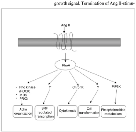
Sm all G pro te ins (Rho fam ily) (Figure 4)
In addition to signaling through hetero-trimeric G proteins, recent evidence sug-gests that AT1 receptors activate monomeric
small (21 kDa) guanine nucleotide-binding proteins (small G proteins) in VSMC. The small G protein superfamily comprises five subfamilies (Ras, Rho, ADP ribosylation fac-tors, Rab and Ran) that act as molecular switches to regulate cellular responses (50). Of these, the Rho subfamily (RhoA, Rac1 and Cdc42) has been associated with Ang II signaling. In its GDP-bound form, RhoA is inactive and is mainly cytoplasmic. RhoA is activated upon the exchange of bound GDP for GTP and for its activation, requires post-translational modification (geranylgeranyla-tion) (50). One of the major downstream targets of RhoA is Rho kinase, which pro-motes VSMC contraction via the phospho-rylation of the myosin-binding subunit of myosin light chain phosphatase, thereby in-hibiting phosphatase activity (51). This con-tributes to increased Ca2+ sensitization.
Vari-ous vasoactive agonists signaling through G protein-coupled receptors enhance the sen-sitivity of the contractile machinery to changes in [Ca2+]
i via RhoA/Rho kinase.
Whether this system contributes to contrac-tion following AT1 receptor activation awaits
clarification. However, RhoA seems to play a role in AT1-stimulated growth signaling
pathways and in processes associated with atherosclerosis. Yamakawa et al. (52) dem-onstrated in rat VSMC that Ang II increases c-fos expression and protein synthesis via RhoA/Rho kinase-dependent mechanisms that do not involve ERK1/2 or p70S6-kinase activation. Funakoshi et al. (53) reported that Rho kinase mediates Ang II-induced monocyte chemoattractant protein-1
expres-RhoA Ang II
Actin organization
SRF regulated transcription
Cytokinesis Cell
transformation Phosphoinositidemetabolism PIP5K ?
CitronK ?
Rho kinase (ROCK) M BS PRK2
Figure 4. Rho-regulated signaling pathw ays by angiotensin II (Ang II). Rho is activated through AT1 receptors. Rho regulates the activity of many signaling pathw ays that influence
sion in rat VSMC. RhoA has also been shown to be important in AT1-mediated PLD
acti-vation.
Ang II also activates Rac1, another small G protein. Rac1 participates in cytoskeletal organization, cell growth, inflammation and regulation of NAD(P)H oxidase (50). In VSMC, Ang II activates Rac1, which is an upstream regulator of p21-activated kinase and JNK. Rac1 also plays a role in Ang II-induction of gene transcription and in the regulation of NAD(P)H oxidase, which mediates generation of superoxide anions (.O2-) in vascular cells (50).
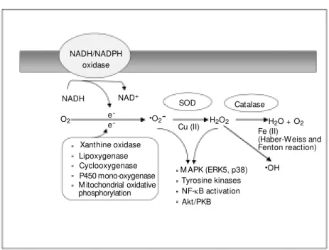
Ge ne ratio n o f re active o xyge n spe cie s (Figure 5)
Reactive oxygen species such as .O2- and
hydrogen peroxide (H2O2) act as
intercellu-lar and intracelluintercellu-lar second messengers that may play a physiological role in vascular tone and cell growth, and a pathophysiologi-cal role in inflammation, ischemia-reperfu-sion, hypertension and atherosclerosis. Xan-thine oxidase, mitochondrial oxidases and arachidonic acid are the major sources of oxidative molecules in non-vascular tissue, whereas a nonmitochondrial, membrane-as-sociated NAD(P)H oxidase appears to be the most important source of .O2- in vascular
cells (54). This enzyme transfers electrons from NADH or NADPH to molecular oxy-gen, producing .O2-. The complete
molecu-lar structure of the vascumolecu-lar oxidase is un-known, but it shares some features with the neutrophil oxidase, which comprises five subunits: a 22-kDa a-subunit (p22phox), a
glycosylated 91-kDa ß-subunit (gp91phox),
which together make up cytochrome b558,
the electron transfer element; cytosolic com-ponents p47phox and p67phox, and a
low-mo-lecular weight G protein, Rac1 or Rac2. Recent studies by Rueckschloss et al. (55) demonstrated complete homology of the cDNA sequence of gp91phox in human
um-bilical vein endothelial cells and neutrophils.
Alternatively, differences in the levels of gene expression of gp91phox and p67phox were
observed, with human umbilical vein endo-thelial cells exhibiting 1.1 and 2.5%, respec-tively, of the levels of expression of these genes, compared with those of neutrophils. By contrast, gene expression for p22phox and
p67phox was comparable between these two
cell types. These observations suggest that the levels of expression of gp91phox and
p67phox genes may be rate-limiting factors
for human vascular cell .O2- production.
Upon activation, the p47phox and p67phox
pro-teins are translocated to the membrane and associate with the cytochrome b558, creating
the active oxidase. In VSMC, Ang II in-creases .O2- production by activating
NAD(P)H oxidase. This effect is sustained and probably contributes to long-term sig-naling events such as cell growth.
NADH/NADPH oxidase
NADH NAD+
SOD Catalase
H2O + O2
H2O2 ·O2
-·OH
O2 e
-e
-Cu (II) Fe (II)
(Haber-Weiss and Fenton reaction) Xanthine oxidase
Lipoxygenase Cyclooxygenase P450 mono-oxygenase M itochondrial oxidative phosphorylation
M APK (ERK5, p38) Tyrosine kinases NF-kB activation Akt/PKB
Figure 5. Generation of reactive oxygen species in the vasculature. M any enzyme systems stimulate production of superoxide anion (.O2-) from O2. These include NADH/NADPH
oxidase, xanthine oxidase, lipoxygenase, cyclooxygenase, P450 mono-oxygenase and mito-chondrial oxidative phosphorylation. NADH/NADPH oxidase is a multisubunit enzyme that is the major regulated source of reactive oxygen species in endothelial and vascular smooth muscle cells. Dismutation of .O2- spontaneously or enzymatically by superoxide dismutase
(SOD) produces hydrogen peroxide (H2O2) that can undergo further reactions to generate
the highly reactive hydroxyl radical (.OH). H2O2 may be metabolized by catalase or
peroxi-dases to H2O and O2. Dow nstream targets of .O2- and H2O2 include ERK5, p38, tyrosine
Generation of reactive oxygen species is regulated by various cytokines and growth factors, including Ang II, which increases .O
2- and H2O2 production in cardiac,
vascu-lar smooth muscle, endothelial, adventitial and mesangial cells (21,54,56). Increased production of reactive oxygen species has been implicated in the pathogenesis of Ang II-induced but not catecholamine-induced hypertension (57). Mechanisms underlying oxidative stress-induced hypertension may be associated with the vascular mitogenic effects of .O
2- and H2O2, decreased
bioavail-ability of endothelium-derived nitric oxide and contractile actions of .O2- and H2O2.
Growth of VSMC by Ang II has an essential redox-sensitive component, which appears to be mediated in part via activation of p38 MAPK and JNK (48). Another redox-sensi-tive process cascade whereby Ang II influ-ences cell function is through phosphoryla-tion of the cell survival protein kinase Akt/ PKB and activation of NF-kB, important in inflammatory responses (54).
Ang II-induce d e xpre ssio n o f
pro to -o nco ge ne s and gro wth facto rs
Long-term control of Ang II-regulated cellular growth, adhesion, migration, fibro-sis and collagen deposition within the vascu-lature involves protein synthesis. Ang II in-duces the expression of several proto-onco-genes in human and rat VSMC, including c -fos, c-jun, c-myc, erg-1, VL-30, and proto-oncogene/activator protein 1 complex (58). Stimulation of early response genes by Ang II is associated with increased gene expres-sion and production of growth factors, such as PDGF, EGF, transforming growth factor-ß, IGF-1, basic fibroblast growth factor and platelet activating factor, vasoconstrictor agents, such as ET-1, adhesion molecules such as ICAM-1, VCAM-1 and E-selectin, and integrins anß3 and ß5, and chemotactic factors such as tumor necrosis factor-a and monocyte chemoattractant protein-1 (58).
These agents may contribute indirectly to the trophic and inflammatory actions of Ang II in cardiovascular tissue.
Ang II influences the architecture and integrity of the vascular wall by modulating cell growth as well as regulating extracellu-lar matrix composition. Ang II increases ex-pression and production of fibronectin, col-lagen type 1, tenascin, glycosaminoglycans, chondroitin/dermatan sulfates and proteo-glycans, major constituents of the extracel-lular matrix in the vessel wall. In VSMC, mesangial cells and endothelial cells, Ang II increases levels and activity of plasminogen activator inhibitor-1, influencing fibrinoly-sis, extracellular matrix turnover and degra-dation and regulation of cell migration. Some of these actions have been linked to the AT4
receptor subtype. However, this remains to be clarified. Ang II also stimulates the activ-ity of matrix metalloproteinases (58) respon-sible for extracellular matrix degradation. Accordingly, Ang II influences vascular structure by stimulating synthesis of struc-tural components of the extracellular matrix and by increasing production of factors that degrade the extracellular matrix proteins.
Co nclusio ns
down-stream targets of AT1 receptors. Alterations
of these highly regulated signaling pathways in cardiovascular cells may be pivotal in structural and functional abnormalities that underlie vascular pathological processes such as hypertension and atherosclerosis. Al-though there has been significant progress in the last few years in the elucidation of aber-rations in Ang II-induced signal transduc-tion in hypertension, we still know very little about the processes that underlie these
phe-nomena, and at what point some pathways become more important than others. With the availability today of molecular and phar-macological tools that allow manipulation of specific signaling molecules, identification of distinct abnormalities in intracellular sig-naling should be possible. This will further our understanding of the role of Ang II in physiological and pathophysiological pro-cesses and will help define novel therapeutic targets in cardiovascular disease.
Re fe re nce s
1. M atsusaka T & Ichikaw a I (1997). Biologi-cal functions of angiotensin and its recep-tors. Annual Review of Physiology, 59: 395-412.
2. Kim S & Iw ao H (2000). M olecular and cellular mechanisms of angiotensin II-me-diated cardiovascular and renal diseases.
Pharmacological Review s, 52: 11-34. 3. Ardaillou R (1999). Angiotensin II
recep-tors. Journal of the American Society of Nephrology, 10: S30-S39.
4. de Gasparo M , Husain A, Alexander W, Catt KJ, Chiu AT, Drew M , Goodfriend T, Harding JW, Inagami T & Timmermans BM WM (1995). Proposed update of an-giotensin receptor nomenclature. Hyper-tension, 25: 924-927.
5. Sw anson GN, Hanesw orth JM , Sardinia M F, Coleman JK, Wright JW, Hall KI, M iller-Wing AV, Cook VI, Harding ECE & Harding JW (1992). Discovery of a distinct binding site for angiotensin II (3-8), a puta-tive angiotensin IV receptor. Regulatory Peptides, 40: 409-419.
6. deGasparo M & Levens N (1998). Does blockade of angiotensin II receptors offer clinical benefits over inhibition of angio-tensin-converting enzyme? Pharmacology and Toxicology, 82: 257-271.
7. Allen AM , Zhuo J & M endelsohn FAO (2000). Localization and function of angio-tensin AT1 receptors. American Journal of
Hypertension, 13: 31S-38S.
8. Berk BC & Corson M A (1997). Angio-tensin II signal transduction in vascular smooth muscle. Role of tyrosine kinases.
Circulation Research, 80: 607-616. 9. Nahmias C & Strosberg D (1995). The
angiotensin AT receptor: searching for sig-nal-transduction pathw ays and physiologi-cal function. Trends in Pharmacological
Sciences, 16: 223-225.
10. Horiuchi M , Hayashida W , Kam be T, Yamada T & Dzau VJ (1997). Angiotensin type 2 receptor dephosphorylates Bcl-2 by activating mitogen-activated protein ki-nase phosphatase-1 and induces apopto-sis. Journal of Biological Chemistry, 272: 19022-19026.
11. Touyz RM , Endemann D, He G, Li J-S & Schiffrin EL (1999). Role of AT2 receptors
in angiotensin II-stimulated contraction of small arteries in young SHR. Hyperten-sion, 33 (Part 2): 366-373.
12. Phoon S & How es LG (2001). Role of angiotensin type 2 receptors in human forearm vascular responses of normal vol-unteers. Clinical and Experimental Phar-macology and Physiology, 28: 734-736. 13. Ichihara S, Senbonmatsu T, Price E, Ichiki
T, Gaffney A & Inagami T (2001). Ang II type 2 receptor is essential for left ven-tricular hypertrophy fibrosis in chronic Ang II-induced hypertension. Circulation, 104: 346-351.
14. Horiuchi M , Akishiita M & Dzau VJ (1999). Recent progress in angiotensin II type 2 receptor research in the cardiovascular system. Hypertension, 33: 613-621. 15. Alexander RW, Brock TA, Gimbrone Jr
M A & Rittenhouse SE (1985). Angiotensin increases inositol trisphosphate and cal-cium in vascular smooth muscle. Hyper-tension, 7: 447-451.
16. Ushio-Fukai M , Griendling KK, Akers M , Lyons PR & Alexander RW (1998). Tem-poral dispersion of activation of phospho-lipase C-beta 1 and -gamma isoforms by angiotensin II in vascular smooth muscle cells. Role of alphaq/11, alpha12, and beta gamma G protein subunits. Journal of Bio-logical Chemistry, 273: 19772-19777.
17. Touyz RM & Schiffrin EL (1997). Role of calcium influx and intracellular calcium stores in angiotensin II-mediated calcium hyperresponsiveness in smooth muscle from spontaneously hypertensive rats.
Journal of Hypertension, 15: 1431-1439. 18. Billah M M (1993). Phospholipase D and
cell signaling. Current Opinion in Immu-nology, 5: 114-123.
19. Ushio-Fukai M , Alexander RW, Akers M , Lyons PR, Lassègue B & Griendling KK (1999). Angiotensin II receptor coupling to phospholipase D is mediated by the ßg
subunits of heterotrimeric G proteins in vascular smooth muscle cells. M olecular Pharmacology, 55: 142-149.
20. Dhalla NS, Xu Y-J, Sheu S-S, Tappia PS & Panagia V (1997). Phosphatidic acid: a po-tential signal transducer for cardiac hyper-trophy. Journal of M olecular and Cellular Cardiology, 29: 2865-2871.
21. Touyz RM & Schiffrin EL (1999). Ang II-stimulated generation of reactive oxygen species in human vascular smooth muscle cells is mediated via PLD-dependent path-w ays. Hypertension, 34 (Part 2): 976-982. 22. Bonventre JV (1992). Phospholipase A2
and signal transduction. Journal of the AmericanSociety of Nephrology, 3: 128-150.
23. Nasjletti A (1997). The role of eicosanoids in angiotensin-dependent hypertension.
Hypertension, 31 (Part 2): 194-200. 24. Oyekan A, Balazy M & M cGiff JC (1997).
Renal oxygenases: differential contribu-tion to vasoconstriccontribu-tion induced by endo-thelin-1 and angiotensin II. American Jour-nal of Physiology, 273 (Part 2): R293-R300.
Annual Review of Cell Division, 13: 513-609.
26. Erpel T & Courtneidge SA (1995). Src fam-ily protein tyrosine kinases and cellular signal transduction pathw ays. Current Opinion in Cell Biology, 7: 176-182. 27. Touyz RM , Wu XH, He G, Park JB, Chen
X, Vacher J, Rajapurohitam V & Schiffrin EL (2001). Role of c-Src in the regulation of vascular contraction and Ca2+ signaling
by angiot ensin II in hum an vascular smooth muscle cells. Journal of Hyper-tension,19: 441-449.
28. Sayeski PP, Ali M S, Harp JB, M arrero M B & Bernstein KE (1998). Phosphorylation of p130Cas by angiotensin II is depen-dent on c-Src, intracellular Ca2+, and
pro-tein kinase C. Circulation Research, 82: 1279-1288.
29. Ihle JN (1995). Cytokine receptor signal-ling. Nature, 377: 591-594.
30. M arrero M B, Schieffer B, Li B, Sun J, Harp JB & Ling BN (1997). Role of Janus kinase/signal transducer and activator of transcription and mitogen-activated pro-tein kinase cascades in angiotensin II and platelet-derived grow th factor-induced vascular smooth muscle cell proliferation.
Journal of Biological Chem istry, 272: 24684-24690.
31. Leduc I & M eloche S (1995). Angiotensin II stimulates tyrosine phosphorylation of the focal adhesion protein paxillin in aortic smooth muscle cells. Journal of Biologi-cal Chemistry, 270: 4401-4404.
32. Guan JL (1997). Role of focal adhesion kinase in integrin signaling. International Journal ofBiochemistry and Cell Biology, 29: 1085-1096.
33. Tang H, Zhao ZJ, Landon EJ & Inagami T (2000). Regulation of calcium-sensitive ty-rosine kinase Pyk2 by angiotensin II in endothelial cells. Roles of yes tyrosine kinase and tyrosine phosphatase shp-2.
Journal of Biological Chem istry, 275: 8389-8396.
34. Cary LA, Han DC, Polte TR, Hanks SK & Guan JL (1998). Identification of p130Cas as a mediator of focal adhesion kinase-promoted cell migration. Journal of Cell Biology, 140: 211-221.
35. Takahashi T, Kaw ahara Y, Taniguchi T & Yokoyama M (1998). Tyrosine phosphory-lation and association of p130Cas and
c-Crk II by Ang II in vascular smooth muscle cells. American Journal of Physiology, 274 (Part 2): H1059-H1065.
36. Leevers SJ, Vanhaesebroek B & Water-field M D (1999). Signaling through phos-phoinositide 3-kinases: the lipids take cen-tre stage. Current Opinion in Cell Biology,
11: 219-225.
37. Saw ard L & Zahradka P (1997). Angio-tensin II activates phosphatidylinositol 3-kinase in vascular smooth muscle cells.
Circulation Research, 81: 249-257. 38. Takahashi T, Taniguchi T, Konishi H,
Kikkaw a U, Ishikaw a Y & Yokoyama M (1999). Activation of Akt/PKB after stimu-lation w ith Ang II in vascular smooth muscle cells. American Journal of Physi-ology, 276 (Part 2): H1927-H1934. 39. Saito Y & Berk BC (2001). Transactivation:
a novel signaling pathw ay from Ang II to tyrosine kinase receptors. Journal of M o-lecular and Cellular Cardiology, 33: 3-7. 40. Prenzel N, Zw ick E, Daub H, Leserer M ,
Abraham R, Wallasch C & Ullrich A (1999). EGF receptor transactivation by G protein-coupled receptors requires metallopro-teinase cleavage of proHB-EGF. Nature, 402: 884-888.
41. Eguchi S, Numaguchi K, Iw asaki H, M at-sumoto T, Yamakaw a T, Utsunomiya H, M otley ED, Kaw akatsu H, Ow ada KM , Hirata Y, M arumo F & Inagami T (1998). Calcium-dependent epidermal grow th fac-tor recepfac-tor transactivation mediates the angiotensin II-induced mitogen-activated prot ein kinase act ivat ion in vascular smooth muscle cells. Journal of Biologi-cal Chemistry, 273: 8890-8896.
42. Robinson M J & Cobb M H (1997). M ito-gen-activated protein kinase pathw ays.
Current Opinion in Cell Biology, 9: 180-186.
43. Touyz RM , He G, Deng LY & Schiffrin EL (1999). Role of extracellular signal-regu-lated kinases in angiotensin II-stimusignal-regu-lated contraction of smooth muscle cells from human resistance vessels. Circulation, 99: 392-399.
44. Touyz RM , He G, El M abrouk M , Diep Q, M ardigyan V & Schiffrin EL (2001). Differ-ential activation of ERK1/2 and p38M AP kinase by angiotensin II type I receptor in vascular smooth muscle cells from WKY and SHR. Journal of Hypertension, 19: 553-559.
45. Ip YT & Davis RJ (1998). Signal transduction by the cJun Nterminal kinase (JNK) -from inflammation to development. Cur-rent Opinion in Cell Biology, 10: 205-219. 46. Schmitz U, Ishida T, Ishida M , Surapisit-chat J, Hasham M I, Pelech S & Berk BC (1998). Angiotensin II stimulates p21-acti-vated kinase in vascular smooth muscle cells. Role in activation of JNK. Circulation Research, 82: 1272-1278.
47. Liao DF, M onia B, Dean N & Berk BC (1997). Protein kinase C-zeta mediates angiotensin II activation of ERK1/2 in
vas-cular smooth muscle cells. Journal of Bio-logical Chemistry, 272: 6146-6150. 48. Ushio-Fukai M , Alexander RW, Akers M &
Griendling KK (1998). p38 M itogen-acti-vated protein kinase is a critical compo-nent of the redox-sensitive signaling path-w ays activated by angiotensin II. Role in vascular smooth muscle cell hypertrophy.
Journal of Biological Chem istry, 273: 15022-15029.
49. Duff JL, M onia BP & Berk BC (1995). M itogen-activated protein kinase is regu-lated by the M AP kinase phosphatase (M KP-1) in vascular smooth muscle cells.
Journal of Biological Chem istry, 270: 7161-7166.
50. Laufs U & Liao JK (2000). Targeting Rho in cardiovascular disease. Circulation Re-search, 87: 526-528.
51. Somlyo AP & Somlyo AV (2000). Signal transduction by G protein, Rho kinase and protein phosphatase to smooth muscle and non-muscle myosin II. Journal of Physiology, 522: 177-185.
52. Yamakaw a T, Tanaka S-I, Numaguchi K, Yamakaw a Y, M otley ED, Ichihara S & Inagami T (2000). Involvement of Rho ki-nase in Ang II-induced hypertrophy of rat vascular smooth muscle cells. Hyperten-sion, 35: 313-318.
53. Funakoshi Y, Ichiki T, Shim okaw a H, Egashira K, Takeda K, Kaibuchi K, Takeya M , Yoshimura T & Takeshita A (2001). Rho kinase m ediat es Ang II-induced monocyte chemoattractant protein-1 ex-pression in rat vascular smooth muscle cells. Hypertension, 38: 100-104. 54. Griendling KK & Ushio-Fukai M (1997).
NADH/NADPH oxidase and vascular func-tion. Trends in Cardiovascular M edicine, 7: 310-317.
55. Rueckschloss U, Galle J, Holtz J, Zerkow -ski H & M oraw ietz H (2001). Induction of NAD(P)H oxidase by oxidised low -density lipoprotein in human endothelial cells. Antioxidative potential of hydroxymethyl-glutaryl coenzyme A reductase inhibitor therapy. Circulation, 104: 1767-1772. 56. Berry C, Hamilton CA, Brosnan M J, M agill
FG, Berg G, M cM urray JJV & Dominiczak AF (2000). An invest igat ion int o t he sources of superoxide production in hu-man blood vessels: Angiotensin II in-creases superoxide production in human internal mammary arteries. Circulation, 101: 2206-2212.
58. Hsueh WA, Do YS, Anderson PW & Law RE (1995). Angiotensin II in cell grow th and matrix production. Advances in Ex-perimental M edicine and Biology, 377: 217-223.
59. Berry C, Touyz RM , Dominiczak AF, Webb
RC & Johns DG (2001). Angiotensin re-ceptors: Signaling, vascular pathophysiol-ogy and int eract ions w it h ceram ide.
American Journal of Physiology, 281: H2337-H2365.
60. Touyz RM & Schiffrin EL (2000). Signal