Angio te nsin II-m e diate d vascular
sm o o th m uscle ce ll gro wth signaling
Department of Biochemistry, Vanderbilt University, Nashville, TN, USA T. Inagami
and S. Eguchi
Abstract
The mechanism by which Ang II stimulates the growth of vascular smooth muscle cells was investigated by measuring the phosphoryla-tion of mitogen-activated protein kinases ERK 1 and ERK 2. Ca2+ ionophore was found to have effects practically analogous to Ang II. We found that the signaling pathway involves the activation of epidermal growth factor receptor (EGFR) kinase, activation of the adaptor proteins Shc and Grb2, and the small G-protein Ras. Although the mechanism of AT1- (or Ca2+)-induced activation of EGFR is not yet clear, we have found that calcium-dependent protein kinase CAKß/ PYK2 and c-Src are involved in this process. These studies indicate a transactivation mechanism that utilizes EGFR as a bridge between a Gq-coupled receptor and activation of phosphotyrosine generation.
Co rre spo nde nce
T. Inagami
Department of Biochemistry Vanderbilt University Nashville, TN 37232-0146 USA
Fax: + 1-615-322-3201 E-mail:
inagamit@ ctrvax.vanderbilt.edu
Presented at the III International Symposium on Vasoactive Peptides, Belo Horizonte, MG, Brasil, O ctober 8-10, 1999.
Research supported by the National Institutes of Health, USA (grants HL58205 and HL35323).
Received November 26, 1999 Accepted February 11, 2000
Ke y wo rds
·Angiotensin II
·Type 1 receptor
·Epidermal growth factor receptor
·Vascular smooth muscle cells
Intro ductio n
Heptahedral receptors have been consid-ered to be coupled to various heterotrimeric G-proteins and transmit their signals through the classical 3,5-cyclic adenosine mono-nucleotide (cAMP) by Gs or Gi, or by eleva-tion of cytoplasmic Ca2+ or stimulation of
protein kinase C (PKC) through the activa-tion of Gq/11. On the other hand, signals
regu-lating cellular growth have been considered to go through growth factor or cytokine re-ceptors, which have a single transmembrane domain, like receptors for epidermal growth factor (EGFR) and platelet-derived growth factor (PDGFR). However, some of the heptahedral (heterotrimeric G-protein-coupled) receptors, such as the type 1 angio-tensin II (Ang II) receptor, AT1, have been
recognized to be one of the most potent stimulators of hypertrophic remodeling of
the vascular and cardiac ventricular wall (1-5). The mechanism for such a growth stimu-lation by Ang II, other than the well-known vascular contraction, regulation of renal tu-bular electrolyte handling, aldosterone re-lease and facilitation of adrenergic rere-lease, has not been clear.
However, in recent years observations of Ang II-stimulated activation of some of the mitogen-activated protein kinase (MAPK) such as extracellular signal-regulated kinase (ERK 1/2) have been reported, even though the pathway leading to ERK activation is, in many cases, mediated by protein tyrosine kinases and is thought to require the growth factor, or cytokine receptor signaling mechan-isms since it seems to require tyrosine phos-phorylation (6,7).
identified to reside in the type 1 receptor AT1. Following the cloning and structure
determination of AT1, by us (9) and Murphy
et al. (10), studies by Morrero et al. (11-13) and Dostal et al. (14) suggested possible direct binding of phospholipase C-g(PLC-g) and Janus kinase (JAK) to a phosphotyrosyl residue in the carboxyterminal segment of AT1, possibly Tyr
319
of Tyr319
-Ile-Pro-Pro-322
, and subsequent activation of these enzymes.
However, we entertained a different hy-pothesis since we found that the C-terminus truncation of rat AT1 at residue Lys
318
or eliminating the Tyr-Ile-Pro-Pro did not elimi-nate the activation of ERK. Another ration-ale for the hypothesis of the direct activation of the tyrosine kinase system by Tyr319
was due to the abundant presence of PLC-g and failure to observe PLC-ß in certain vascular smooth muscle cell (VSMC) lines which was consistently not the case in explanted VSMC we had used for a long time.
Ele vatio n o f Ca2+ mimics Ang II in MAPK/ERK activatio n
We hypothesized that the PLC-ß-medi-ated increase in Ca2+ or PKC may be the
mechanism of Ang II-mediated MAPK/ERK activation and demonstrated that calcium ionophore A23187 increased MAPK. Fur-thermore, the intracellular calcium chelator
BAPTA-AM or calcium release inhibitor TMB8 abolished the MAPK activation by Ang II, as shown in Figure 1. Interestingly, similar patterns of regulation were also seen in the activation of the small G-protein Ras which is located upstream of ERK. Thus, we were able to show that the activation of ERK by intracellular calcium proceeds through the Ras-dependent pathway (15). The pre-cise mechanism of activation of Ras or MAPK by Ca2+
is not known. The possibility of PKC-mediated signaling was eliminated for VSMC pretreated with a PKC inhibitor GF109203X or by PKC depletion by over-night exposure to phorbol ester. This is a unique and important feature of VSMC in contrast to cardiomyocytes in which a PKC-mediated mechanism seems to be involved (16).
Invo lve m e nt o f EGFR in the activatio n o f Ras and MAPK
However, the role of Ca2+
(but not PKC) in the activation of Ras and MAPK was not clear. To clarify the mechanism we exam-ined steps upstream of Ras, which were most likely growth factor receptors.
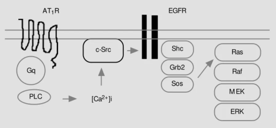
As shown in Figure 2, activated EGFR transmits its signal by tyrosine phosphoryla-tion of several tyrosine residues by EGFR kinase, subsequently forming a complex with Shc and then Grb2, or directly with Grb2. Grb2, in turn, activates the GDP-GTP ex-change protein mSOS for Ras. This pathway was revealed by experiments using a fusion protein formed between glutathione-S-trans-ferase (GST) and Grb2 for affinity precipita-tion of complex of Grb2 with Shc, EGFR and/or Sos.
By this technique we have demonstrated that activation of EGFR, as reflected by a complex formation with Grb2-GST, is in-duced by elevated cytoplasmic Ca2+
, and the activated EGFR leads to the activation of MAPK/ERK (17). Interestingly, the transac-tivation of EGFR occurs by an intracellular
E
R
K
a
c
ti
v
it
y
(
p
m
o
l
m
in
-1 m
g
-1) 2500
2000
1500
1000
500
0
Control
BAPTA-AM
Basal Ang II
mechanism rather than by EGF, which may have been produced and released from the Ang II-activated VSMC because the condi-tioned medium of Ang II-activated VSMC was not capable of activating EGFR in VSMC (17). This is a new concept of EGFR activa-tion by an intracellular mechanism through a Ca2+
-dependent system.
If EGFR mediates Ang II-dependent acti-vation of ERK, it is tempting to examine if a similar but distinct PDGFR could function as a similar mediator. EGFR kinase and PDGFR kinase specific inhibitors tyrphostin AG1478 and AG1295, respectively, became available (18). The expected inhibition of the activation of ERK by Ang II was ob-served only with the EGFR kinase inhibitor AG1478 (75% reduction), whereas the PDGFR kinase inhibitor AG1295 showed no significant effect (17). At present the mechanism for the selective activation of EGFR by Ang II but not PDGFR is not clear. However, it is interesting to note that the quantity of PDGFR in VSMC is much greater than that of EGFR. For understanding the mechanism of the specific activation of EGFR, a major void in our knowledge exists for the mechanism between increased cyto-plasmic Ca2+
and EGFR activation. How-ever, it is important to recognize that G-protein-coupled receptors are capable of transactivating a receptor tyrosine kinase sys-tem in many cells (19-21). This new concept is now supported by a similar mechanism mediated by a variety of heptahedral recep-tors.
Calcium-activate d tyro sine kinase
Recently, several investigators reported a new Ca2+
-activated tyrosine kinase in the cytoplasmic fraction, which was named CAKß (22)/PYK2 (23)/RAFTK (24)/ CADTK (25). Although the structure of this kinase indicates that it belongs to the family of focal adhesion kinase (FAK), this Ca2+
-activated proline-rich tyrosine kinase was
different from FAK.
The CAKß/PYK2 type soluble tyrosine kinase is a good candidate to mediate the signal of an elevated cytoplasmic Ca2+
to EGFR and/or ERK 1/2. We and others were able to show that such a mechanism is acti-vated by Ang II or Ca2+
in VSMC (26,27), liver epithelial cells (25), cardiac fibroblasts (28), and pheochromocytoma cells (23). The cytoplasmic tyrosine kinase was activated by Ang II through AT1 or the Ca
2+
ionophore A23187, but not by PKC (26).
Interestingly, a member of an oncogene and a soluble protein tyrosine kinase, c-Src, is also activated in this process of Ang II (AT1)-stimulated transactivation of EGFR,
as detected by the monoclonal antibody P28 specific for the active form of c-Src (17). The activation of PYK2 and c-Src precedes the transactivation of EGFR, since the inhi-bition of the EGFR kinase by tyrphostin AG1478 does not affect the activation of PYK2 or c-Src (26). Active c-Src and PYK2 also form a coprecipitable complex in agree-ment with the observation of Dikic et al. (29) in a neuronal cell system. This system is very complex and we are not yet able to describe it as a linear vertical flow of signaling events. This mechanism involving at least three ki-nases seems to be much more complex than we have imagined. It is possible that this area may also involve mechanisms and systems
Figure 2 - In VSM C, Ca2+-dependent transactivation of epidermal grow th factor receptor (EGFR) mediates Ang II-induced Ras/ERK M APK activation. AT1R, Type 1 angiotensin II receptor; PLC, phospholipase C; ERK, extracellular signal-regulated kinase; M EK, M APK kinase.
AT1R
Gq
ERK M EK Raf Ras Shc
Grb2
Sos c-Src
PLC
EGFR
that may be stimulated by reactive oxygen species.
Ro le s o f activate d ERKs in VSMC
ERKs are a focal point for diverse cell growth and proliferating responses. Activa-tion of ERK induces the transcripActiva-tion of the
fos gene. Indeed, we were able to directly demonstrate c-Fos expression in a rapid re-sponse to Ang II-induced EGFR transactiva-tion and subsequent ERK activatransactiva-tion. Ang
II-induced protein synthesis is also mediated by the EGFR and ERK (30).
D ual signals fo r S6 kinase activatio n
In the hypertrophic responses of VSMC, the net effects of multiple effectors such as Ang II, insulin, etc., are syntheses of two discrete S6 kinases (p90 and p70 S6 ki-nases), which regulate the function of ribo-somes. p90S6k
is subject to direct activation by ERK, but the upstream of p70S6k
still remains unclear.
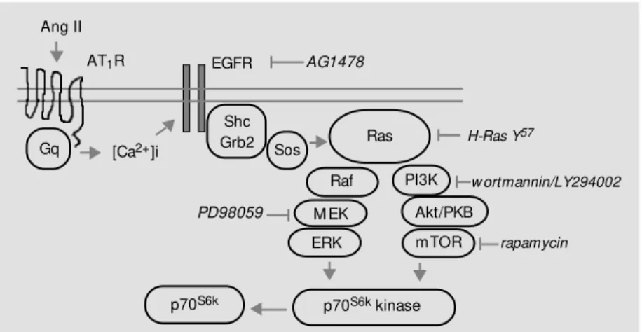
Quite recently, we found that regulation of p70S6k
is complex, since it is activated by Ang II through two separate but related signaling pathways (31), as discussed be-low. We have shown that Ras-dependent ERK activation results in the activation of p70S6k
. A dominant negative mutant of Ras was found to inhibit not only ERK 1/2, but also p70S6k
(Figure 3). Inhibition of MEK (MAPK kinase) by PD98059 (the ERK path-way on the left side below Ras in Figure 4) was sufficient to block p70S6k
, whereas phos-phatidyl inositol-3-kinase (PI3K) inhibitor blocked the p70S6k
activation without affect-ing the ERK. This observation suggested the presence of a second pathway. Indeed, we observed that Ang II activates Akt/protein kinase B (PKB). Akt/PKB was known to be activated by insulin through the activation of PI3K (32). However, we have found that Ang II activates wortmannin/LY294002-sen-sitive PI3K and its downstream components Akt/PKB and p70S6k
by phosphorylating the Ser411
residue of the latter, shown on the right side of Figure 4.
Activation of this pathway by insulin does not activate ERK. Of particular inter-est is that the PI3K-activating insulin ef-fect shares a common signaling pathway with Ang II. The role of Ras is also intri-guing in that it activates two bifurcating pathways. Although, Akt/PKB is a multi-functional activator of various signaling pathways, its activation of p70S6k
results
Ang II (min)
IB: p70S6k (S411P)
IB: p70S6k
IB: H-Ras 66
66
-0 2 5 10 20 40 0 2 5 10 20 40
H-Ras Y57 LacZ
Figure 3 - Effect of dominant-negative H-Ras transfection of angiotensin II (Ang II)-induced Ser411 phosphorylation (S411P) of p70S6k. VSM C w ere transfected w ith AdRas Y57 or control AdLacZ as indicated before stimulation. Cells w ere stimulated w ith Ang II (100 nM ) for indicated durations. Cell lysates w ere analyzed by immunoblotting (IB) w ith anti-[phospho-Ser411]-p70S6k, anti-p70S6k, and anti-H-Ras antibodies as indicated by repeated reprobing.
Gq
M EK Raf
Ras Shc
Grb2 [Ca2+]i
w ortmannin/LY294002
rapamycin H-Ras Y57
Sos
PI3K
Akt/PKB
mTOR
p70S6k kinase p70S6k
PD98059 Ang II
AT1R EGFR AG1478
Figure 4 - Hypothetical pathw ays involved in the phosphorylation of p70S6k by angiotensin II (Ang II). According to this model, activation of the putative S6 kinase (S6k) by Ang II requires tw o separate pathw ays w hich bifurcate at the point of Ras activation through transactivated epidermal grow th factor receptor (EGFR). AT1R, Type 1 Ang II receptor; ERK, extracellular signal-regulated kinase; PKB, protein kinase B; PI3K, phosphatidyl inositol-3-kinase; M EK, M APK kinase.
in resynthesis of the pathways leading to the hypertrophy of vascular smooth muscle cells (Figure 4).
Co nclusio n
We tried to summarize recent findings from our laboratory and others focusing on the hypertrophic effects of Ang II mediated by p70S6k
in quiescent cultured vascular smooth muscle cells. Although there are some large gaps in our knowledge, we have pro-vided unequivocal evidence of transactiva-tion of EGFR by a Gq/11-coupled receptor in
a unique fashion since PDGFR is not acti-vated.
Some of the components of pivotal im-portance in this system are Src family ty-rosine kinase, EGFR, Ras-ERK, or Ras-PI3K and Akt, ending at p70S6k
. CAKß/PYK2 may
be a potential candidate for bridging Ca2+
to EGFR.
These studies are of particular relevance to vascular remodeling since this system seems to provide possible common sites of actions of two major components of vascular degenerative change, insulin and Ang II. Furthermore, it is important to note that the Ang-II-to-ERK pathway is mediated by re-active oxygen species (Frank GD, Eguchi S, Yamakawa T, Tanaka S, Inagami T and Motley ED, unpublished results).
Ackno wle dgm e nts
We are indebted to Drs. Kotaro Numagu-chi, Hiroaki Iwasaki, Yukio Hirata, Gerald D. Frank, and Evangeline D. Motley for valuable scientific input, and to Tina Stack for preparation of the manuscript.
Re fe re nce s
1. Geisterfer AA, Peach M J & Ow ens GK (1988). Angiotensin II induces hypertro-phy, not hyperplasia, of cultured rat aortic sm ooth m uscle cells. Circulation Re-search, 62: 749-756.
2. Gibbons GH, Pratt RE & Dzau VJ (1992). Vascular smooth muscle cell hypertrophy vs hyperplasia of cult ured rat aort ic smooth muscle cells. Journal of Clinical Investigation, 90: 456-461.
3. Weber H, Taylor DS & M olloy CJ (1994). Angiotensin II induces delayed mitogen-esis and cellular proliferation in rat aortic smooth muscle cells: correlation w ith the expression of specif ic endogenous grow th factors and reversal by suramin. Journal of Clinical Investigation, 93: 788-798.
4. Sadoshima J & Izumo S (1993). M olecular characterization of angiotensin II-induced hypertrophy of cardiac myocytes and hy-perplasia of cardiac fibroblasts; critical role of the AT1 receptor subtype. Circulation Research, 73: 413-423.
5. Schorb W, Booz GW, Dostal DE, Conrad KM , Chang KC & Baker KM (1993). Angio-tensin II is mitogenic in neonatal rat car-diac fibroblast. Circulation Research, 72: 1245-1254.
6. Griendling KK, Ushio FM , Lassegue B & Alexander RW (1997). Angiotensin II
sig-naling in vascular smooth muscle: new concepts. Hypertension, 29: 366-373. 7. Berk BC & Corson M A (1997).
Angio-tensin II signal transduction in vascular smooth muscle: role of tyrosine kinases. Circulation Research, 80: 607-626. 8. Timmermans PBM WM , Wong PC, Chiu
AT, Herblin WF, Benefield P, Carini DJ, Lee RJ, Wexler RR, Saye JAM & Smith RD (1993). Angiotensin II receptors and angiotensin II receptor antagonist. Phar-macological Review s, 45: 205-251. 9. Sasaki K, Yamano Y, Bardhan S, Iw ai N,
M urray JJ, Hassegaw a M , M atsuda Y & Inagami T (1991). Cloning and expression of a complementary DNA encoding a bo-vine adrenal angiotensin II type-1 recep-tor. Nature, 351: 230-233.
10. M urphy TJ, Alexander RW, Griendling KK, Runge M S & Bernstein KE (1991). Isola-tion of a cDNA encoding the vascular type-1 angiotensin II receptor. Nature, 351: 233-236.
11. M orrero M B, Schieffer B, Paxton WG, Heerdt L, Berk BC, Delafontaine P & Bernstein KE (1995). Direct stimulation of Jak/STAT pathw ay by the angiotensin II AT1 receptor. Nature, 375: 247-255. 12. Ali SM , Sayeski PP, Dirksen LB, Hayzer
DJ, M orrero M B & Bernstein KE (1997). Dependence on the motif YIPP for the
physical association of Jak 2 kinase w ith the intracellular carboxyl tail of the angio-tensin II AT1 receptor. Journal of Biologi-cal Chemistry, 272: 23382-23388. 13. Venema RC, Ju H, Venema VJ, Schieffer
B, Harp JB, Ling BN, Eaton DC & M orrero M (1998). Angiotensin II-induced associa-tion of phospholipase Cg1 w ith the G-protein-coupled AT1 receptor. Journal of Biological Chemistry, 273: 7703-7708. 14. Dostal DE, Hunt RA, Kule CE, Bhat GJ,
Karoor V, M cWhinney CD & Baker KM (1997). M olecular mechanisms of angio-tensin II in modulating cardiac function: intracardiac effects and signal transduc-tion pathw ays. Journal of M olecular and Cellular Cardiology, 29: 2893-2902. 15. Eguchi S, M atsum oto T, M otley ED,
Utsunomiya H & Inagami T (1996). Identi-fication of an essential signaling cascade for mitogen-activated protein kinase acti-vation by angiotensin II in cultured rat vas-cular smooth muscle cells: possible re-quirement of Gq-mediated p21ras activa-tion coupled to a Ca2+/calmodulin sensi-tive tyrosine kinase. Journal of Biological Chemistry, 271: 14169-14175.
trans-duction pathw ays. Circulation Research, 82: 337-345.
17. Eguchi S, Num aguchi K, Iw asaki H, M atsumoto T, Yamakaw a T, Utsunomiya H, M otley ED, Kaw akatsu H, Ow ada KN, Hirata Y, M urumo F & Inagami T (1998). Calcium-dependent epidermal grow th fac-tor recepfac-tor transactivation mediates the angiotensin II-induced mitogen-activated prot ein kinase act ivat ion in vascular smooth muscle cells. Journal of Biologi-cal Chemistry, 273: 8890-8896.
18. Levitzki A & Gazit A (1995). Tyrosine ki-nase inhibition: an approach to drug de-velopment. Science, 267: 1782-1788. 19. Zw ick E, Hackel PO, Norbert P & Ullrich A
(1999). The EGF receptor as central trans-ducer of heterologous signaling systems. Trends in Pharmacological Sciences, 20: 408-412.
20. Carpenter G (1999). Employment of the epiderm al grow t h f act or recept or in grow th factor-independent signaling path-w ays. Journal of Cell Biology, 146: 697-702.
21. Luttrell LM , Kaaka Y & Lefkow itz RJ (1999). Regulation of tyrosine kinase cas-cades by G-protein-coupled receptors. Current Opinion in Cell Biology, 11: 177-183.
22. Sasaki H, Nagura K, Ishino M , Tobioka H, Kotani K & Sasaki T (1995). Cloning and characterization of cell adhesion kinase ß, a novel protein-tyrosine kinase of the
fo-cal adhesion kinase subfamily. Journal of Biological Chemistry, 270: 21206-21219. 23. Lev S, M oreno H, M artinez R, Canoll P,
Peles E, M ussacchio JM , Plow man GD, Rudy B & Schlessinger J (1995). Protein tyrosine kinase PYK2 involved in Ca2+ -induced regulation of ion channel and M AP kinase functions. Nature, 376: 737-745.
24. Avaham S, London R, Fu Y, Ot a S, Hiregow dara D, Li J, Jiang S, Pasztor LM , White RA, Groopman JE & Avaham H (1995). Identification and characterization of a novel related adhesion focal tyrosine kinase (RAFTK) from megakaryocytes and brain. Journal of Biological Chemistry, 270: 27742-27751.
25. Yu H, Li X, M archetto GS, By R, Hunter D, Calvo B, Daw son TL, Wilm M , Anderegg RJ, Graves LM & Earp HS (1996). Activa-tion of a novel calcium-dependent pro-tein-tyrosine kinase: correlation w ith c-Jun N-terminal kinase but not mitogen acti-vated protein kinase activation. Journal of Biological Chemistry, 271: 29993-29998. 26. Eguchi S, Iw asaki H, Inagami T,
Numagu-chi K, Yamakaw a T, M otley ED, Ow ada KM , M arumo F & Hirata Y (1999). Involve-ment of PYK2 in angiotensin II signaling of vascular smooth muscle cells. Hyper-tension, 33 (Part II): 201-206.
27. Brinson AE, Harding T, Diliberto PA, He Y, Li X, Hunter D, Herman B, Earp HS & Graves LM (1998). Regulation of a
cal-cium-dependent tyrosine kinase in vascu-lar smooth muscle cells by angiotensin II and platelet-derived grow th factor: de-pendence on calcium and the actin cyto-skeleton. Journal of Biological Chemistry, 273: 1711-1718.
28. M urasaw a S, M ori Y, Nozaw a Y, M asaki H, M aruyama K, Tustsumi Y, M origuchi Y, Shibasaki Y, Tanaka Y, Iw asaka T, Inada M & M atsubara H (1998). Role of calcium-sensitive tyrosine kinase PYK2/CAKß/ RAFTK in angiotensin II-induced Ras/ERK signaling. Hypertension, 32: 668-675. 29. Dikic I, Tokiw a G, Lev S, Courteneige SA
& Schlessinger J (1996). A role for Pyk 2 and Src in linking G-protein-coupled re-ceptors w ith M AP kinase activation. Na-ture, 383: 547-550.
30. Eguchi S, Iw asaki H, Hirata Y, Frank GD, M otley ED, Yamakaw a T, Numaguchi K & Inagami T (1999). Epidermal grow th fac-tor recepfac-tor is indispensable for c-Fos ex-pression and protein synthesis by angio-tensin II. European Journal of Pharmacol-ogy, 376: 203-206.
31. Eguchi S, Iw asaki H, Ueno H, Frank GD, M otley ED, Eguchi K, M arumo F, Hirata Y & Inagami T (2000). Possible requirement of EGF receptor, RAS, ERK and AKT. Jour-nal of Biological Chemistry (in press). 32. Alessi DR & Cohen P (1998). M echanism