Cyclophosphamide in the treatment
of focal segmental glomerulosclerosis
Serviço de Nefrologia, Departamento de Medicina, Faculdade de Medicina, Universidade Federal da Bahia, Salvador, BA, Brasil
R. Martinelli, L.J. Pereira, O.M.M. Silva, A.S. Okumura and H. Rocha
Abstract
Prednisone is the initial treatment of primary focal segmental glomer-ulosclerosis. However, when immunosuppressive agents in combina-tion with steroids are used in the treatment of prednisone-dependent and prednisone-resistant patients the remission rate is variable. We report a long-term trial using cyclophosphamide (2.0 to 3.0 mg/kg body weight for 12 weeks) in combination with prednisone (1.0 to 2.0 mg/kg body weight), as compared with prednisone alone for the treatment of prednisone-resistant and frequently relapsing nephrotic syndrome and focal segmental glomerulosclerosis. Fifty-four patients (34 males and 20 females) with a diagnosis of idiopathic nephrotic syndrome and focal segmental glomerulosclerosis, followed-up for an average of 86.1 ± 82.4 months, were evaluated. Complete remission occurred in 20.4% and partial remission in 14.8% of the patients treated with steroids and in 26.7 and 20.0% of the patients treated with cyclophosphamide + prednisone, respectively. Of the 24 prednisone-resistant patients treated with steroids in combination with cyclophos-phamide, 33.3% obtained a complete/partial response. At the time of final evaluation, 25% of the patients treated with prednisone and 10.0% of those treated with prednisone in combination with cyclo-phosphamide had reached end-stage renal disease. Persistent neph-rotic syndrome and progressive renal insufficiency were more fre-quently observed among the patients treated with prednisone alone (50.0 vs 33.3% and 33.3 vs 16.7%, respectively). The treatments were well tolerated and no patient experienced adverse reactions requiring discontinuation of medications. Although open-label and non-ran-domized, the present trial showed that cyclophosphamide is a reason-able choice for the treatment of primary focal segmental glomerulo-sclerosis and prednisone-resistant nephrotic syndrome.
Correspondence R. Martinelli
Hospital Universitário, UFBA Rua João das Botas, s/n 40110-160 Salvador, BA Brasil
Fax: +55-71-235-8854 E-mail:rm@ufba.br
Received July 22, 2003 Accepted March 11, 2004
Key words
•Cyclophosphamide •Prednisone
•Nephrotic syndrome •Primary glomerular disease •Focal segmental
glomerulosclerosis
Introduction
Focal segmental glomerulosclerosis is an important primary glomerular disease caus-ing nephrotic syndrome and chronic renal failure in both adults and children (1-3). Although its natural course has been well described, its treatment is still a matter of debate (4-12). Prednisone is the initial
are usually much lower ones than those achieved in initially prednisone-responsive patients (9,16-19).
Cyclophosphamide is a well-known cy-totoxic agent used in the treatment of fre-quently relapsing, steroid-dependent and ste-roid-resistant nephrotic syndrome and of immunologic diseases with secondary glo-merular involvement. Although it has been used in the treatment of focal segmental glomerulosclerosis for many years, the ben-efit of the combination of cyclophospha-mide along with prednisone has not been fully characterized (6,9,16-18,20,21). The trials have been limited, the studies have been non-randomized and open-label and the series have included small numbers of patients with a short-term follow-up.
We report an open-label, non-random-ized long-term trial using cyclophosphamide in combination with prednisone, as com-pared to prednisone alone, for the treatment of focal segmental glomerulosclerosis and prednisone-resistant and frequently relaps-ing nephrotic syndrome.
Subjects and Methods
A total of 54 patients, 34 males and 20 females with a mean age of 18.6 ± 12.9 years (30 of them younger than 18 years) fol-lowed-up on average for 86.1 ± 82.4 months, were included in the present study.
All patients had a diagnosis of idiopathic nephrotic syndrome and focal segmental glo-merulosclerosis and have been followed at the Renal Service of the University Hospital, University of Bahia, Salvador, BA, Brazil, for at least 6 months. All patients underwent a percutaneous renal biopsy and sections containing at least 8 glomeruli were exam-ined by a nephropathologist, classified ac-cording to Churg et al. (22) and D’Agati (23) and characterized by a segmental area of solidification of some glomeruli. In the af-fected areas the capillary loops were seg-mentally collapsed and the capillary lumina
were obliterated by matrix material, with adhesion of the tuft to the adjacent Bowman’s capsule. The remainder of the capillary tuft and the unaffected glomeruli were normal in appearance. The extent of interstitial fibrosis was graded on a scale of 0, 1 and 2 respec-tively indicating absent, mild and moderate/ severe disease.
At the time of initial evaluation a full his-tory was taken and a physical examination performed, in addition to the following labora-tory tests: urinalysis, 24-h urinary protein ex-cretion, creatinine clearance, serum levels of creatinine, cholesterol, albumin and fasting blood glucose, and other tests to exclude sys-temic diseases, as appropriate. Patients diag-nosed as being obese or having reflux ne-phropathy, HIV infection or systemic diseases that could cause renal disease were excluded. For the purpose of the present study, patients were considered to be adults if the renal disease was diagnosed at the age >18 years and as children if it was diagnosed at age ≤18 years. Nephrotic syndrome was di-agnosed by the presence of edema, urinary protein excretion equal to or greater than 3.5 g/24 h for adults or greater than 30 mg/kg body weight/24 h for children, serum albu-min below 3.0 g/dl and serum cholesterol greater than 250 mg/dl. Hypertension was defined as blood pressure above 140/90 mmHg in adult patients and readings ex-ceeding the 95th percentiles for systolic or diastolic blood pressure for age and gender, in patients younger than 12 years. Renal disease was considered to be progressive when an age-related increase in serum creati-nine to levels above normal occurred during the follow-up. Renal insufficiency was de-fined by serum creatinine levels above 1.4 mg/dl or the age-related normal levels and end-stage renal disease was defined as se-rum creatinine levels higher than 7.5 mg/dl or five times the age-related normal upper limits or by the need for dialysis therapy.
di-vided into two to three doses for 4 to 6 weeks, followed by a single dose of pred-nisone on alternate days for 4 additional weeks, which was then gradually discontin-ued over 2 to 3 months. Remission was defined as the absence of proteinuria (com-plete) or reduction of proteinuria to non-nephrotic levels (partial), and resistance as the persistence of nephrotic range proteinu-ria during the 6 weeks of daily treatment. Relapse was defined as recurrence of the nephrotic range proteinuria after urine had been protein-free for at least 4 weeks. The occurrence of frequent relapse was defined as 3 or more relapses within 12 months in an initially steroid-responsive patient. The clini-cal response to prednisone was classified as: steroid responsive - complete or partial re-mission of proteinuria during the steroid therapy persisting for at least 8 weeks after therapy; steroid dependent - remission of proteinuria during therapy but recurrence when the dosage was reduced below a criti-cal level or relapse of proteinuria within the first month after the end of prednisone thera-py; resistant - no remission of proteinuria during 8 consecutive weeks of daily therapy. All steroid-resistant patients and patients with frequently relapsing nephrotic syndrome (N = 30) received a daily oral dose of 2.0 to 3.0 mg cyclophosphamide/kg body weight for 12 weeks, concurrently with the same steroid regimen. Because the addition of cyclophosphamide to the steroid regimen is a standard treatment for steroid dependent and frequently relapsing nephrotic syndrome
in current practice, it was not necessary to obtain the approval of the Ethics Committee nor the written informed consent from the patients.
The clinical course and final outcome of each patient were evaluated by recording current medication, blood pressure, serum creatinine and albumin levels, urinalysis, and 24-h urinary protein excretion.
Data are reported as means ± SD or as proportions. Comparisons were performed using the Student t-test for continuous data
and the chi-square test or the Fisher exact test for categorical data, as appropriate. The renal survival curves were plotted by the Kaplan-Meier method (24). The log-rank test was used for comparison of survival curves.
Spearman correlation coefficients were calculated to examine the relationship be-tween the variables and the development of end-stage renal disease. The analyses were two-tailed and the level of significance was set at P < 0.05 in all cases (25).
All statistical analyses were performed with the Statistical Package for the Social Sciences (SPSS), version 10.0.
Results
Demographic data and clinical findings at the time of initial evaluation for the two treatment groups are presented in Table 1. The patients treated with steroids alone were older, more frequently males, and 50% had renal insufficiency at the time of the
diagno-Table 1. Demographic data and clinical findings at the time of initial evaluation for patients with nephrotic syndrome in the two treatment groups.
Groups Age Gender Renal Hypertension Follow-up
(M/F) failure (months)
Prednisone 24.0 ± 12.0* 19/5 12 (50.0)* 7 (29.2) 51.5 ± 59.1*
Prednisone + cyclophosphamide 14.2 ± 12.1 15/15 8 (26.7) 7 (23.3) 113.8 ± 88.6
Total 18.6 ± 12.9 34/20 20 (37.0) 14 (25.9) 86.1 ± 82.4
sented in Table 2: complete remission was recorded in 20.4% and partial remission in 14.8% of the patients treated with prednisone and in 26.7 and 20.0%, respectively, of the patients treated with cyclophosphamide + prednisone. Remissions were less frequent among children than among adults for pred-nisone (23.3 vs 50.0%; P = 0.04), but there
was no statistical difference for prednisone plus cyclophosphamide. Four patients (2 treated with prednisone and 2 treated with cyclophosphamide in conjunction with pred-nisone) who were resistant to the immuno-suppressive treatment subsequently devel-oped a clinical course characterized by per-sistent non-nephrotic proteinuria. In the 50 patients who had the renal interstitium ex-amined, there was no association between the degree of interstitial fibrosis and the response to therapy (Table 3). A decrease in the frequency of relapses was observed in all 6 patients with frequently relapsing neph-rotic syndrome. Despite mild signs of hyper-cortisolism, no patient presented an adverse reaction requiring discontinuation of treat-ment.
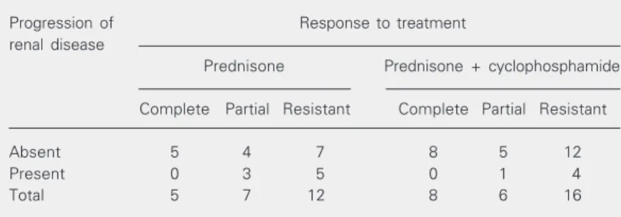
The influence of the response to therapy on the progression of renal disease is shown in Table 4 and itwas more frequent among patients resistant to therapy, although 1 pa-tient who initially showed complete remis-sion of nephrotic syndrome during treatment with prednisone progressed to persistent non-nephrotic proteinuria and mild renal insuffi-ciency.
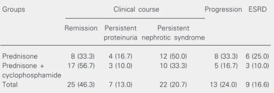
Persistent nephrotic syndrome and pro-gressive renal insufficiency were more fre-quent among patients treated with prednisone alone than among patients treated with pred-nisone plus cyclophosphamide (Table 5). The renal survival rate (Figure 1) of the group was 88% at 5 years, 75% at 10 and 75% at 15 years. At the time of final clinical evaluation, 18 of 54 patients had hyperten-sion. Seven patients with hypertension were treated with angiotensin converting enzyme inhibitors (ACEi). Nine patients (16.6%) had
Table 2. Response to treatment of nephrotic syn-drome with immunosuppressive agents.
Response to Prednisone Prednisone +
treatment cyclophosphamide
Complete 11 (20.4) 8 (26.7) Partial 8 (14.8) 6 (20.0) Resistant 30 (55.5) 16 (53.3) Dependent 5 (9.3) 0 (00.0)
Total 54 (100) 30 (100)
Data are reported as number of patients and per-cent values are given in parentheses.
Table 4. Effect of the response to treatment on the progression of renal disease in patients with focal segmental glomerulosclerosis.
Progression of Response to treatment
renal disease
Prednisone Prednisone + cyclophosphamide
Complete Partial Resistant Complete Partial Resistant
Absent 5 4 7 8 5 12
Present 0 3 5 0 1 4
Total 5 7 12 8 6 16
Table 3. Response to treatment and degree of interstitial fibrosis in patients with prednisone-resistant nephrotic syndrome.
Interstitial fibrosis Response to treatment
Prednisone Prednisone + cyclophosphamide
Complete Partial Resistant Complete Partial Resistant
0 3 5 6 7 2 8
1 1 2 4 3 2 6
2 1
Total 5 7 10 10 4 14
There was no statistical significance between the results for prednisone compared to prednisone + cyclophosphamide (χ2 test; P > 0.05).
sis of nephrotic syndrome. The follow-up of this group of patients was also significantly shorter. Fifteen patients (68%) treated with prednisone and 14 (50%) treated with cyclo-phosphamide in combination with prednisone had a mild to moderate degree of interstitial fibrosis.
pre-reached end-stage renal disease, correspond-ing to 25% of the patients treated with pred-nisone alone and 10% of the patients treated with prednisone + cyclophosphamide.
Discussion
In recent years prednisone has became the treatment of choicefor focal segmental glomerulosclerosis and its therapeutic re-sponse has been recognized as a predictor of long-term prognosis (7,12,16,26-29). In con-trast, the persistence of nephrotic syndrome portends a reserved prognosis (15,30). Since spontaneous remission of focal segmental glomerulosclerosis is rare (7,12-14) and an improved prognosis is associated with the remission of nephrotic syndrome, it is in the group of patients with steroid-resistant focal segmental glomerulosclerosis that cytotoxic drugs have been used in combination with prednisone.
In the present series, remission of the proteinuria associated with prednisone was documented in 35.2% of the patients, and was more frequent in adults than in children. This finding was not unexpected since only nephrotic children resistant to steroids or
Table 5. Clinical course of patients with nephrotic syndrome in the two treatment groups.
Groups Clinical course Progression ESRD
Remission Persistent Persistent proteinuria nephrotic syndrome
Prednisone 8 (33.3) 4 (16.7) 12 (50.0) 8 (33.3) 6 (25.0) Prednisone + 17 (56.7) 3 (10.0) 10 (33.3) 5 (16.7) 3 (10.0) cyclophosphamide
Total 25 (46.3) 7 (13.0) 22 (20.7) 13 (24.0) 9 (16.6)
Data are reported as number of patients and percent values are given in parentheses. ESRD = end-stage renal disease.
with atypical manifestations are submitted to renal biopsies in our service. In the pres-ent study steroid resistance was defined as persistence of proteinuria in the nephrotic range over a period of 8 weeks of daily steroid therapy. It is possible that other pa-tients might experience remission of pro-teinuria with a more prolonged course of prednisone therapy (9). Recently, Ponticelli et al. (19) showed that patients treated for more than 16 weeks attained complete re-mission more frequently than patients treated for 16 weeks or less. Remission of proteinu-ria, either complete (26.7%) or partial
Figure 1. Renal survival of patients with focal segmental glomerulosclerosis and nephrotic syndrome treated with immunosup-pressive agents. Number of patients reported at bottom of figure.
Renal survival (%)
100
80
60
40
20
0
Total Steroid
Steroid + cyclophosphamide Follow-up (months)
0 24 48 72 96 120 144 168 192 216 240 264
30 28 25 24 23 22 21 18 17 17 16 15 14 9 8 8 8 7 6 6 6 6
54 50 40 34 38 34 28 26 22 20 19 17 16 15 10 9 9 9 8 7 7 7
(20.0%), was demonstrated in 46.7% of the patients, treated with cyclophosphamide in combination with prednisone, resulting in more sustained remission and preservation of renal function. Although cyclophospha-mide did not completely prevent relapses, it was associated with a clinical course charac-terized by remission of the nephrotic syn-drome: no patient with complete remission and 16.7% of those with partial remission after treatment with prednisone in combina-tion with cyclophosphamide subsequently evolved persistently nephrotic syndrome. The addition of cyclophosphamide, however, was less effective in patients with prednisone-resistant nephrotic syndrome, since remis-sion was observed in 33.3% of them, a rate similar to that observed by others (31). Nev-ertheless, in the present series, remission of proteinuria was associated with preservation of renal function, with all patients with com-plete remission of nephrotic syndrome and 3 of the 8 patients with partial remission con-tinuing to have normal renal function at the time of final evaluation. Banfi et al. (16) and Ponticelli et al. (19) also concluded that achieving complete or partial remission was a strong clinical predictor of outcome in patients with focal segmental glomeruloscle-rosis and nephrotic syndrome.
Unexpectedly, the preservation of renal function was more frequently observed among the patients treated with cyclophos-phamide in combination with prednisone than with prednisone alone. This finding could not berelated to the degree of intersti-tial fibrosis, even though the small number of patients in the present series does not permit us to reach a definitive conclusion. However, it could be a consequence of a selection bias since renal insufficiency at initial evaluation, a predictor of poor out-come, was more frequent in the group of patients treated with prednisone alone than in the group treated with both drugs together. Also, the use of ACEi and dietary protein restriction, which are well-known reducers
of urinary protein excretion in glomerular diseases, and the current levels of systolic blood pressure (32) were not controlled. An ACEi was used in 40% of hypertensive pa-tients, evenly distributed through both treat-ment groups.
Tarshish et al. (21), in a prospective study, found no additional benefit for renal func-tion after the treatment of children with cyclo-phosphamide plus prednisone. In contrast to this observation but consistent with our find-ings, Geary et al. (20), Tufro-McReddie et al. (33), and Tune et al. (34) found progres-sion to renal failure to be less frequent in children treated with cyclophosphamide, and Banfi et al. (16) and Ponticelli et al. (19) found the same to be true in adults. Better preservation of renal function was also re-ported by Cattran et al. (18) in patients treated with cyclosporine.
chil-dren have been reported infrequently. Cyclosporine (18) and mycophenolate mofetil (35) have been used in the treatment of focal segmental glomerulosclerosis. So far, no comparative study has been pub-lished although Cattran et al. (18) reported remission of proteinuria in 70% of the pa-tients with steroid-resistant nephrotic syn-drome treated with cyclosporine, a greater proportion than that achieved in the present study using cyclophosphamide. The benefi-cial effects afforded by the use of ACEi in the treatment of proteinuric nephropathies, including focal segmental glomerulosclero-sis, also suggest that they can be used as supportive treatment for this glomerular dis-ease (36-38) although definite evidence that
ACEi alter the natural course of focal seg-mental glomerulosclerosis is still lacking (39). The role of other agents used in the treatment of focal segmental glomerulosclerosis is still poorly defined (40).
Although open-label and non-random-ized, the therapeutic regimens were stan-dardized. The present trial showed that cyclo-phosphamide in combination with steroids could be a reasonable choice in the treatment of steroid-resistant focal segmental glomer-ulosclerosis. Randomized placebo-controlled trials are needed to define the benefit of cyclophosphamide or other cytotoxic agents or of ACEi in the long-term treatment of focal segmental glomerulosclerosis.
References
1. D’Agati V (1994). The many masks of focal segmental glomerulo-sclerosis. Kidney International, 46: 1223-1241.
2. Haas M, Meehan SM, Karrison TG & Spargo BH (1997). Changing etiologies of unexplained adult nephrotic syndrome: a comparison of renal biopsy findings from 1976-1979 and 1995-1997. American Journal of Kidney Diseases, 30: 621-631.
3. Srivastava T, Simon SD & Alon US (1999). High incidence of focal segmental glomerulosclerosis in nephrotic syndrome of childhood. Pediatric Nephrology, 13: 13-18.
4. Aviles DH, Irwin KC, Dublin LS & Vehaskari VM (1999). Aggressive treatment of severe idiopathic focal segmental glomerulosclerosis. Pediatric Nephrology, 13: 298-300.
5. Beaufils H, Alphonse JC, Guedon J & Legrain M (1978). Focal glomerulosclerosis: Natural history and treatment. Nephron, 21: 75-85.
6. Cameron JS (1996). The enigma of focal segmental glomeruloscle-rosis. KidneyInternational, 50 (Suppl 57): S119-S131.
7. Cattran DC & Rao P (1998). Long-term outcome in children and adults with classic focal segmental glomerulosclerosis. American JournalofKidneyDiseases, 32: 72-79.
8. Korbet SM, Schwartz MM & Lewis EJ (1994). Primary focal seg-mental glomerulosclerosis: clinical course and response to therapy. AmericanJournalofKidneyDiseases, 23: 773-783.
9. Matalon A, Valeri A & Appel GB (2000). Treatment of focal segmen-tal glomerulosclerosis. Seminars in Nephrology, 20: 309-317. 10. Mongeau JG, Robitaille PO, Clermont MJ, Merouani A & Russo P
(1993). Focal segmental glomerulosclerosis (FSG) 20 years later. From toddler to grown up. ClinicalNephrology, 40: 1-6.
11. Rennert WP, Kala UK, Jacobs D, Goetsch S & Verhaat S (1999). Pulse cyclophosphamide for steroid-resistant focal segmental glo-merulosclerosis. PediatricNephrology, 13: 113-116.
12. Rydel JJ, Korbet SM, Borok RZ & Schwartz MM (1995). Focal segmental glomerular sclerosis in adults: Presentation, course and
response to treatment. American JournalofKidney Diseases, 25: 534-542.
13. Newman WJ, Tisher CC, McCoy RC, Gunnells JC, Krueger RP, Clapp JR & Robinson RR (1976). Focal glomerular sclerosis: con-trasting clinical patterns in children and adults. Medicine, 55: 67-87. 14. Pei Y, Cattran D, Delmore T, Katz A, Lang A & Rance P (1987). Evidence suggesting under-treatment in adults with idiopathic focal segmental glomerulosclerosis. American Journalof Medicine, 82: 938-944.
15. Yoshikawa N, Ito H, Akamatsu R, Matsuyama S, Hasegawa O, Nakahara C & Matsuo T (1986). Focal segmental glomerulosclerosis with and without nephrotic syndrome in children. Journalof Pediat-rics, 109: 65-70.
16. Banfi G, Moriggi M, Sabadini E, Fellin G, D’Amico G & Ponticelli C (1991). The impact of prolonged immunosuppression on the out-come of idiopathic focal segmental glomerulosclerosis. A collabora-tive retrospeccollabora-tive study. ClinicalNephrology, 36: 53-59.
17. Burgess E (1999). Management of focal segmental glomeruloscle-rosis: evidence-based recommendations. Kidney International, 55 (Suppl 70): S26-S32.
18. Cattran DC, Appel GB, Hebert LA, Hunsicker LG, Pohl MA, Hoy WE, Maxwell DR & Kunis CL (1999). A randomized trial of cyclosporine in patients with steroid-resistant focal segmental glomerulosclerosis. KidneyInternational, 56: 2220-2226.
19. Ponticelli C, Villa M, Banfi G, Cesana B, Pozzi C, Pani A, Passerini P, Farina M, Grassi C & Baroli A (1999). Can prolonged treatment improve the prognosis in adults with focal segmental glomerulo-sclerosis? AmericanJournalofKidneyDiseases, 34: 618-625. 20. Geary DF, Farine M, Thorner P & Baumal R (1984). Response to
cyclophosphamide in steroid-resistant focal segmental glomerulo-sclerosis: a reappraisal. ClinicalNephrology, 22: 109-113. 21. Tarshish P, Tobin JN, Bernstein J & Edelman Jr CM (1996).
glo-merulosclerosis. A report of the International Study of Kidney Dis-ease in Children. PediatricNephrology, 10: 590-593.
22. Churg J, Bernstein J & Glassock RJ (1995). Renal Disease. Classifi-cation and Atlas of Glomerular Diseases. 2nd edn. Igaku-Shoin, New York.
23. D’Agati V (2003). Pathologic classification of focal segmental glo-merulosclerosis. Seminars in Nephrology, 23: 117-134.
24. Kaplan EL & Meier P (1958). Nonparametric estimation for incom-plete observations. American Statistical Association Journal, 53: 457-481.
25. Armitage P & Berry G (1994). Statistical MethodsinMedical Re-search. 3rd edn. Blackwell Science, Oxford, UK.
26. Arbus GS, Poucell S, Bacheyie GS & Baumal R (1982). Focal seg-mental glomerulosclerosis with idiopathic nephrotic syndrome: Three types of clinical response. JournalofPediatrics, 101: 40-45. 27. Chan PCK, Chan KW, Cheng IKP & Chan MK (1991). Focal
scleros-ing glomerulopathy: Risk factors of progression and optimal mode of treatment. InternationalUrologyandNephrology, 239: 619-629. 28. Chitalia VC, Wells JE, Robson RA, Searle M & Lynn KL (1999).
Predicting renal survival in primary focal segmental glomeruloscle-rosis from the time of presentation. KidneyInternational, 56: 2236-2242.
29. Korbet SM (1999). Clinical picture and outcome of primary focal segmental glomerulosclerosis. Nephrology, Dialysis, Transplanta-tion, 14 (Suppl 3): 68-73.
30. Velosa JA, Holley KE, Torres VE & Offord KP (1983). Significance of proteinuria on the outcome of renal function in patients with focal segmental glomerulosclerosis. MayoClinic Proceedings, 58: 568-577.
31. Ponticelli C & Passerini P (2003). Other immunosuppressive agents for focal segmental glomerulosclerosis. Seminars in Nephrology, 23: 242-248.
32. Jafar TH, Stark PC, Schmid CH, Landa M, Maschio G, de Jong P, de Zeeuw D, Shahinfar S, Toto R & Levey A (2003). Progression of
chronic kidney disease: The role of blood pressure control, proteinu-ria, and angiotensin-converting enzyme inhibition. A patient-level meta-analysis. Annals of Internal Medicine, 139: 244-252. 33. Tufro-McReddie A, Alvarez E, Arrizurieta E & Repetto H (1992).
Focal glomerulosclerosis in children: an Argentinean experience. PediatricNephrology, 6: 158-161.
34. Tune BM, Kirpekar R, Sibley RK, Reznik VM, Griswold WR & Mendoza AS (1995). Intravenous methylprednisolone and oral alky-lating agent therapy of prednisone-resistant pediatric focal segmen-tal glomerulosclerosis: a long-term follow-up. Clinical Nephrology, 43: 84-88.
35. Choi MJ, Eustace JÁ, Gimenez LF, Atta MG, Scheel PJ, Sothinathan R & Briggs WA (2002). Mycophenolate mofetil treatment of primary glomerular diseases. Kidney International, 61: 1098-1114. 36. Ruggenenti P, Perna A, Gherardi G, Benini R & Remuzzi G (2000).
Chronic proteinuric nephropathies: outcomes and response to treat-ment in a prospective cohort of 352 patients with different patterns of renal injury. American Journal of Kidney Diseases, 35: 1155-1165.
37. Crenshaw G, Bigles S, Salem M & Crook ED (2000). Focal segmen-tal glomerulosclerosis in African-Americans: effects of steroids and angiotensin converting enzyme inhibitors. AmericanJournalof the MedicalSciences, 319: 320-325.
38. Huissoon A, Meehan S & Keogh J (1991). Reduction of proteinuria with captopril therapy in patients with focal segmental glomerulo-sclerosis and IgA nephropathy. Irish Journal of Medical Science, 160: 319-321.
39. Korbet SM (2003). Angiotensin antagonists and steroids in the treatment of focal segmental glomerulosclerosis. Seminars in Ne-phrology, 23: 219-228.