A sim ple RT-PCR-base d strate gy fo r
scre e ning co nne xin ide ntity
Departments of 1Neuroscience, 2Anesthesiology and 3Medicine,
Albert Einstein College of Medicine, New York, NY, USA
4Departamento de Medicina Interna and Instituto de Patologia Tropical e Saúde
Pública, Universidade Federal de Goiás, Goiânia, GO , Brasil M. Urban1,
R. Rozental1,2,4 and
D.C. Spray1,3,4
Abstract
Vertebrate gap junctions are aggregates of transmembrane channels which are composed of connexin (Cx) proteins encoded by at least fourteen distinct genes in mammals. Since the same Cx type can be expressed in different tissues and more than one Cx type can be expressed by the same cell, the thorough identification of which connexin is in which cell type and how connexin expression changes after experimental manipulation has become quite laborious. Here we describe an efficient, rapid and simple method by which connexin type(s) can be identified in mammalian tissue and cultured cells using endonuclease cleavage of RT-PCR products generated from multi primers (sense primer, degenerate oligonucleotide corresponding to a region of the first extracellular domain; antisense primer, degenerate oligonucleotide complementary to the second extracellular domain) that amplify the cytoplasmic loop regions of all known connexins except Cx36. In addition, we provide sequence information on RT-PCR primers used in our laboratory to screen individual connexins and predictions of extension of the multi primer method to several human connexins.
Co rre spo nde nce
R. Rozental
Department of Neuroscience Albert Einstein College of Medicine Kennedy Center Room # 724 1300 Morris Park Avenue New York 10461, NY USA
Fax: + 1-718-430-8682 E-mail: rozental@ aecom.yu.edu
Research supported by the National Institutes of Health.
Received September 18, 1998 Accepted April 5, 1999
Ke y wo rds
·Gap junctions
·Intercellular communication ·cDNA sequences
·Universal primers ·Multi primers
Intro ductio n
Gap junction channels are found in most tissues where they provide conduits for dif-fusion of ions and small molecules between neighboring cells. Cloning studies have thus far revealed fourteen distinct connexin (Cx) cDNA sequences encoded by different genes in rodent tissues with highly homologous isoforms in other mammals. In mammals, group I (ß) connexins include Cx26, Cx30, Cx30.3, Cx31, Cx31.1 and Cx32, and group II (a) connexins include Cx33, Cx37, Cx40,
Cx43, Cx45, Cx46 and Cx50 (1). In addition, a new connexin type (named Cx36) has been identified in the mammalian brain and as-signed to a new group of connexins termed group III (g) (2,3).
commer-cially available). To circumvent these draw-backs, we have developed a rapid method for screening connexin identity using RT-PCR techniques. Because nucleotide se-quences of connexin cDNAs are highly con-served in both intramembrane and extracel-lular domains, a pair of degenerate oligo-nucleotide primers were previously designed to amplify the cytoplasmic loop regions of all known connexins (4). Here, we have utilized this pair of multi primers to am-plify connexin DNA sequences starting from total RNA and identified individual con-nexin species through the use of specific endonucleases; amplification of the cyto-plasmic loop regions results in major bands that distinguish group I from group II con-nexins. We show that group I connexins display sizes on gels of 350-390 bp, while
group II connexin sequences amplified by these primers are 420-520 bp. We also show that certain connexins are not readily identi-fied by this technique and propose connexin-specific RT-PCR primers to overcome this limitation.
In the present study we amplified cloned cDNAs representing connexins from groups I and II (Figures 1 and 2) and identified connexins generated by reverse-transcribed RNA samples from a neural cell line (Figure 3); the identity of the connexin was con-firmed by sequencing. Finally, we demon-strate the applicability of our method by detecting the connexins expressed in the liver (i.e., Cx26, Cx32 and Cx43; Figure 4 and Table 1). We provide a detailed bench-top protocol and a succinct description of our methodology, discuss its potential
applica-587
518
431
381
267
234
213
184
M rCx26 rCx32 rCx43 mCx45
Group II Group I
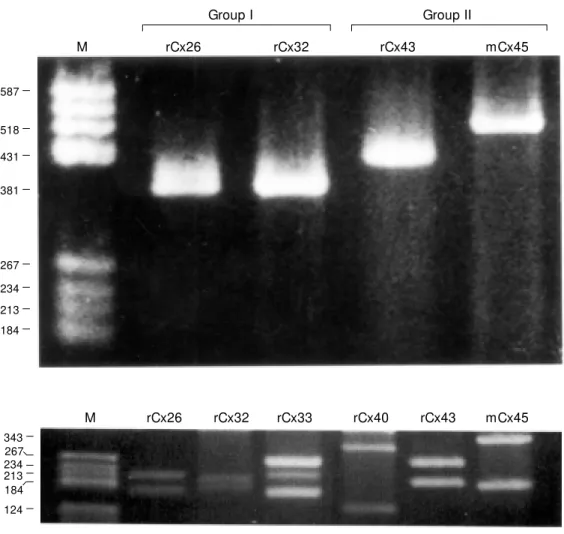
Figure 1 - Detection of connex-ins belonging to groups I (rCx26, rCx32) and II (rCx43, mCx45) us-ing “ multi primers” w ith con-nexin-cloned cDNA. PCR prod-ucts corresponding to 382 bp for Cx26, 381 bp for Cx32, 431 bp for Cx43, 518 bp for Cx45. M o-lecular markers are indicated on the left (M ). r, Rat; m, mouse.
343 267 234 213 184
124
M rCx26 rCx32 rCx33 rCx40 rCx43 mCx45
C
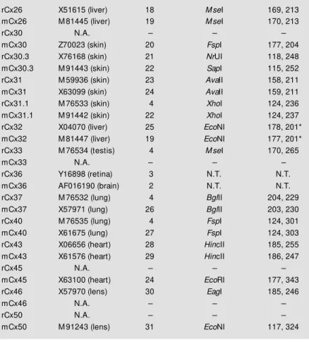
Figure 3 - Connexin detection in a neural cell line (M K31 cell line). Bench-top protocol of our methodology and potential applications. A, Screening connexin identity in a cell line using RT-PCR assays. First generation of PCR products indicates that the amplified DNA sequences belong to group II (A2,4). B2, Second generation of PCR products indicates the expression of Cx43 in these cells. Lane 4 is the PCR product of Cx43 from rat heart and lane 5 is its corresponding negative control. B, Lanes 2,4 show the presence of restriction products of Cx43 after HincII w as used. Lane 3 is a negative control for the cells. The negative controls w ere run in PCR w ithout reverse transcriptase to verify that genomic DNA w as not present (lanes 3,5). C, Sequencing the RT-PCR products revealed the presence of Cx43 (99.7% identical) in these cells. Terminology: M ouse heart Cx43 sequence (M USCX43) and sequence found in the neural cell line (M K31CX43). M olecular markers (A1, B1).
A
587 450
267 184
1 2 3 4
255 185
1 2 3 4
B
M USCX43
M K31CX43
M USCX43
M K31CX43
M USCX43
M K31CX43
M USCX43
M K31CX43
M USCX43
M K31CX43
M USCX43
M K31CX43
M USCX43
M K31CX43
M USCX43
M K31CX43
M USCX43
M K31CX43
300
57
350
107
400
157
450
207
500
257
550
307
600
357
650
407
700
440
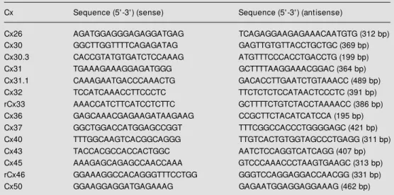
Figure 4 - Determination of the connexin types expressed in rat liver. A, First generation of PCR products indicates that the amplified DNA sequences belong to groups I and II (lane 2), as expected, and its corresponding negative control (lane 3). M olecular markers (lane 1). B, Second generation of PCR products. For this figure, first round PCR bands w ere isolated from the gel and PCR products belonging to Cx26 (lane 2), Cx32 (lane 3) and Cx43 (lane 4) w ere identified, as expected. M olecular markers (lane 1). The endonucleases used w ere M seI (Cx26; lane 2), EcoNI (Cx32; lane 3) and HincII (Cx43; lane 4), respectively.
587 540
458 434
267
234 213 184
1 2 3 1 2 3 4
5
5
tions and limitations and provide advice on avoiding potential pitfalls. In addition, we also provide GenBank access numbers for connexin types expressed in rodents and humans (Tables 1 and 2), a list of specific restriction enzymes to be used and sequences for connexin-specific primers (Table 3) rou-tinely used in our laboratory. Thus, this com-munication provides a starting point for those new to the gap junction field and should also serve as a useful reference for more experi-enced researchers.
Mate rial and Me tho ds
Pre paratio n and tre atme nt o f RNA
Total RNA was isolated from cultured cells or tissues by a procedure based on that of Chomczynski and Sacchi (5), quantitated by absorbance measurements at 260 nm and 280 nm (Hitachi U-1100) with 1 OD unit considered equal to 40 µg/ml. Ten U of DNase I (Boehringer Mannheim, Indianapo-lis, IN, USA) was added to each 5 µg of RNA and incubated at 25o
C for 15 min to digest residual genomic DNA. DNase was heat-inactivated at 65o
C for 15 min before the RT-PCR analyses.
RT-PCR analyse s
First strand cDNA was synthesized from total RNA templates using random primers and the superscript preamplification system (Gibco BRL-Life Technologies, Inc., Grand Island, NY, USA) (6). One to two micro-grams of total RNA was brought to 11 µl in diethyl pyrocarbonate (DEPC)-treated water and combined with 1 µl random hexamers (50 ng/µl). The mixture was heated at 70o
C for 10 min and then incubated on ice. The remaining components for reverse transcrip-tion were then added and incubated for 10 min as follows: 2 µl of 10x synthesis buffer, 2 µl 0.1 M DTT, 2 µl 25 mM MgCl2, 1 µl 10 mM dNTP mix and 1 µl (200 units)
super-script reverse transuper-scriptase. The reaction mix was left at room temperature (RT) for 10 min, incubated at 42o
C for 50 min and the reaction terminated by incubating at 70o
C for 15 min.
Degenerate oligonucleotides correspond-ing to conserved regions of the first and second extracellular connexin domains were synthesized on an ABI model 391 Sequencer (Applied Biosystems, Foster City, CA, USA). The sequences of the 24-mer sense and 21-mer antisense multi pri21-mers (4) are given below (using IUB group codes):
A) Sense 5' GGC TGT RAV AAY GTC TGC TAY GAC 3'
B) Antisense 5' TGG GVC KGG AVA BGA AGC AGT 3'
PCR reactions contained (in a final volume of 50 µl): 5 µl of RT reaction (0.5-1 µg of first strand cDNA), 1 µM of sense and anti-sense primers, 5 µl of 10x PCR buffer (100 mM Tris-HCl; 500 mM KCl, pH 8.3), 0.2 mM dNTP, 3 mM MgCl2 and 2.5 U Taq polymerase (Gibco BRL-Life Technologies, Inc.). Thirty cycles were performed on the samples using a PTC-100 Thermocycler (M.J. Research Inc., Watertown, MA, USA) as follows: 1) denaturation at 94oC for 30 s; 2) annealing at 55o
C for 30 s; 3) extension at 72o
C for 30 s. This was followed by a final extension cycle at 72oC for 8 min and a soak cycle at 4o
C. Reaction products were ana-lyzed by electrophoresis on 2% agarose gels in order to detect contaminants. The corresponding bands were isolated from gels and purified (Qiagen Inc., Valencia, CA, USA). The DNA was reamplified, and re-striction digestion analysis with specific en-zymes (New England Biolabs Inc., Beverly, MA, USA) was performed to identify the connexins expressed in the rat liver and in a mouse neural cell line (MK31 cell line; 7,8).
D NA se que ncing and analysis
RT-PCR products detected in hippocam-pal cells were sequenced and compared to murine cytoplasmic loop connexin sequences available at GenBank (Figure 3C).
Re sults and D iscussio n
This report describes an improved tech-nique for the screening of tissue- or cell-specific RNA samples for the presence of mRNAs encoding members of the connexin multigene family. Previous studies have uti-lized RT-PCR techniques to detect connexin expression in different preparations (9-17). However, our methodology is novel in the ability to detect all known connexins be-longing to groups I and II through the use of multi primers with only a single PCR reac-tion followed by specific restricreac-tion diges-tions. The previous methods required the use of specific conditions and specific primers for each connexin type and the reported methods lacked detailed experimental pro-tocols. In addition, our study presents theo-retical cleavage patterns of connexin PCR products specific for each connexin type as well as the GenBank access numbers for each connexin.
In contrast to previous publications, we have used the terminology multi primers instead of universal primers, due to the fact that these degenerate oligonucleotides do not amplify the cytoplasmic loop regions of Cx36 (group III) and do not detect Cx37, Cx40 and Cx45 in a background where Cx43 is present.
The protocol we developed evolved from the use of degenerate oligonucleotides for PCR amplification of connexins reported by Haefliger et al. (4). Optimization of our prepa-ration was achieved by reducing the duprepa-ration of denaturation at 94o
C from 1 min to 30 s, annealing at 55oC and extension at 72oC for 30 s and the final extension to 8 min. By reducing the duration of the cycles used by
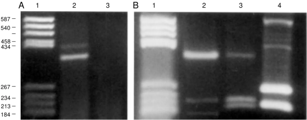
Haefliger et al. (4) by 50% (from 1 min to 30 s) and by increasing the concentration of MgCl2 from 1.5 to 3 mM we have detected 8 connexin types in various cell and tissue preparations (Figures 1, 2, 3A,B, 4; Table 1). Multi primers amplified sequences con-served among connexins, resulting in major bands that distinguished group I (350-390 bp) from group II (420-520 bp) connexins; these results are illustrated in Figure 1 for the group I connexins with Cx26 and Cx32 and for the group II connexins Cx43 and Cx45. Unique restriction sites within the amplicons generated by multi primers for each rodent and human connexin for which sequence information exists were deduced from the program PCGene (IntelliGenetics, Inc., Campbell, CA, USA). These predicted frag-ment sizes for rodent and human connexins are presented in Tables 1 and 2. Our predic-tions for the sizes of PCR product fragments after endonuclease treatment were confirmed using amplicons obtained from PCR with multi primers using cloned cDNAs encod-ing Cx26, Cx32, Cx33, Cx40, Cx43 and Cx45 (Figure 2); so far the only anomaly has been the presence of a canonical site for EcoNI in rat Cx32 DNA leading to an unex-pected cleavage product of 213 bp (see Fig-ure 2, rCx32).
and Cx43 (Figure 4B4) (Table 1).
Pre cautio ns
Problems with RT-PCR sensitivity gener-ally are associated with duration and tempera-ture of incubation, numbers of cycles and reagents (in particular, Mg2+
). Incubation times should be kept as short as possible in order to optimize the protocol and minimize the occur-rence of non-specific amplification. Since one of the key variables in this protocol is the concentration of Mg2+
and Mg2+
concentra-tion requirements can vary dramatically de-pending on the sample to be studied, we rec-ommend that a concentration curve for Mg2+ be established before completely implement-ing this protocol in order to avoid nonspecific priming and suboptimal enzymatic activity. For preliminary characterization of the con-nexins expressed in a specific tissue, we sug-gest the use of the Q-solution (Qiagen Inc.) which acts similarly to DMSO by modifying the melting behavior of DNA. More impor-tantly, it has been shown that the concentra-tion of Mg2+ does not need to be previously adjusted, providing an efficient amplification at a uniform concentration.
Although this method is now optimized to the point where it is reliable in discrimi-nating many different connexin types, we have thus far been unable to detect Cx37, Cx40 and Cx45 in tissues where Cx43 is co-expressed. Nevertheless, one cannot rely only on the electrophoretic separation for identi-fication of amplicons in agarose DNA gels. To substantiate the identity of the connexins, we strongly recommend one of the following strategies: i) all amplicons must be sequenced (as illustrated in Figure 3C), or ii) use of specific primers (Table 3), as still required for Cx37, Cx40, Cx45 and Cx36 (see 13,17). In our laboratory, immunocytochemical and functional electrophysiological assays have been routinely carried out in parallel to fur-ther substantiate the results obtained using the RT-PCR strategy.
Table 1 - PCR products obtained from the 14 connexins expressed in rodents. Detec-tion of connexin type identity from PCR products. PCR products of cDNA of Cx26, Cx30.3, Cx31, Cx31.1, Cx32, Cx33, Cx37, Cx40, Cx43, Cx45, Cx46 and Cx50.
r, Rat; m, mouse. Tissues w ith high expression, references for molecular cloning and GenBank database accession numbers are listed below . * A cleavage product of 213 bp, corresponding to a canonical site for EcoNI, is routinely found in rat liver. N.A., Not available; N.T., not tested.
Connexin (Cx) GenBank access # References Restriction enzymes Products (bp)
rCx26 X51615 (liver) 18 M seI 169, 213
mCx26 M 81445 (liver) 19 M seI 170, 213
rCx30 N.A. - -
-mCx30 Z70023 (skin) 20 FspI 177, 204
rCx30.3 X76168 (skin) 21 NrUI 118, 248
mCx30.3 M 91443 (skin) 22 SapI 115, 252
rCx31 M 59936 (skin) 23 AvaII 158, 211
mCx31 X63099 (skin) 24 AvaII 159, 211
rCx31.1 M 76533 (skin) 4 XhoI 124, 236
mCx31.1 M 91442 (skin) 22 XhoI 124, 237
rCx32 X04070 (liver) 25 EcoNI 178, 201*
mCx32 M 81447 (liver) 19 EcoNI 177, 201*
rCx33 M 76534 (testis) 4 M seI 170, 265
mCx33 N.A. - -
-rCx36 Y16898 (retina) 3 N.T. N.T.
mCx36 AF016190 (brain) 2 N.T. N.T.
rCx37 M 76532 (lung) 4 BglII 204, 229
mCx37 X57971 (lung) 26 BglII 203, 230
rCx40 M 76535 (lung) 4 FspI 124, 301
mCx40 X61675 (lung) 27 FspI 124, 303
rCx43 X06656 (heart) 28 HincII 185, 255
mCx43 X61576 (heart) 29 HincII 186, 247
rCx45 N.A. - -
-mCx45 X63100 (heart) 24 EcoRI 177, 343
rCx46 X57970 (lens) 30 EagI 185, 246
mCx46 N.A. - -
-rCx50 N.A. - -
-mCx50 M 91243 (lens) 31 EcoNI 117, 324
Table 2 - Predicted unique cleavage sites for human gap junction proteins deduced from cDNA sequences.
References for GenBank database accession numbers, molecular cloning, restriction enzymes and predicted cleavage patterns.
Connexin (Cx) GenBank access # References Restriction enzymes Products (bp)
Cx26 M 86849 32 M seI 170, 213
Cx32 X04325 33 DdeI 100, 279
Cx37 M 96789 34 BglII 203, 229
Cx40 U03486 35 DraIII 119, 307
Cx43 M 65188 36 ScrFI 99, 333
D iffe re nce s amo ng co nne xin iso fo rms in
m o use , rat and hum ans
Connexin sequences are found to be more conserved when comparing one isoform in different species (e.g., human and mouse Cx43 differ by only 12% at the nucleotide level and 2.5% at the amino acid level) than when com-paring different isoforms in the same species (e.g., mouse Cx40 and Cx43 differ by 40% at the nucleotide level). Thus, the unique sites for endonucleases and the restriction fragment lengths might be conserved from mice to hu-mans. However, evolutionary rates vary for different connexin genes among species (1). In rodents, the most striking difference that we have observed is related to rat/mouse Cx30.3 (Table 1). In humans, several connexins have also been cloned; their GenBank access num-bers and predicted cleavage patterns are pro-vided in Table 2. Note that Cx26 and Cx37 express endonuclease sites (and lengths of products) similar to those present in rat/mouse Cx26 and mouse Cx37, respectively.
Co nclusio ns
In summary, the RT-PCR assay followed by connexin-specific endonucleases is sim-ple, sensitive and reliable. Although initially developed for detection of small amounts of cDNA, we have found the method to be highly effective when preceding routine Northern blot analysis, enabling rapid dis-crimination of connexins belonging to group I from those belonging to group II in both large and small amounts of samples. In addi-tion, the present methodology could be a-dapted to other gene families with high se-quence conservation.
Ackno wle dgm e nts
We thank Ms. M. Kremer for her techni-cal expertise, Mrs Camilla Heinzmann (Qiagen Inc.) for technical support and Drs. R. Dermietzel and X. Zheng for their helpful comments.
Table 3 - Connexin-specific primers routinely used to verify the expression of connexins expressed in mice (* except Cx33 and Cx46).
* M ouse Cx33 and Cx46 sequences are not yet available (see Table 1); Cx33- and Cx46-specific primers are listed for rat sequences (e.g., rCx33 and rCx46).
Cx Sequence (5' -3' ) (sense) Sequence (5' -3' ) (antisense)
Cx26 AGATGGAGGGAGAGGATGAG TCAGAGGAAGAGAAACAATGTG (312 bp)
Cx30 GGCTTGGTTTTCAGAGATAG GAGTTGTGTTACCTGCTGC (369 bp)
Cx30.3 CACCGTATGTGATCTCCAAAG ATGTTTCCCACCTGACCTG (199 bp)
Cx31 TGAAAGAAAGGAGATGGG GCTTTTAAGGAAACGGAC (364 bp)
Cx31.1 CAAAGAATGACCCAAACTG GACACCTTGAATCTGTAAACC (489 bp)
Cx32 TCCATCAAACCTTCCCTC TTCTCTCTCCATAACTCCCTC (391 bp)
rCx33 AAACCATCTTCATCCTCTTC GCTTTTCTGTCTACCTAAAACC (386 bp)
Cx36 GAGCAAACGAGAAGATAAGAAG CCGCTTCTACATCATCCA (195 bp)
Cx37 GGCTGGACCATGGAGCCGGT TTTCGGCCACCCTGGGGAGC (421 bp)
Cx40 TTTGGCAAGTCACGGCAGGG TTGTCACTGTGGTAGCCCTGAGG (311 bp)
Cx43 TACCACGCCACCACTGGC AATCTCCAGGTCATCAGG (407 bp)
Cx45 AAAGAGCAGAGCCAACCAAA GTCCCAAACCCTAAGTGAAGC (313 bp)
rCx46 GGAAAGGCCACAGGGTTTCCTGG GGGTCCAGGAGGACCAACGG (331 bp)
Re fe re nce s
1. Bennett M VL, Zheng X & Sogin M L (1995). The connexin fam ily tree. In: Kanno Y, Kataoka K, Shiba Y, Shibata Y & Shimazu T (Editors), Intercellular Commu-nication Through Gap Junctions. Progress in Cell Research. Elsevier Science, B.V., The Netherlands, 3-8.
2. Condorelli DF, Parent i R, Spinella F, Salinaro AT, Belluardo N, Cardile V & Cicirata F (1998). Cloning of a new gap junction gene (Cx36) highly expressed in m am m alian brain neurons. European Journal of Neuroscience, 10: 1202-1208. 3. Sohl G, Degen J, Teubner B & Willecke K
(1998). The murine gap junction gene con-nexin36 is highly expressed in mouse retina and regulated during brain develop-ment. FEBS Letters, 428: 27-31. 4. Haefliger JA, Bruzzone R, Jenkins N,
Gil-bert DJ, Copeland NG & Paul DL (1992). Four novel members of the connexin fam-ily of gap junction proteins. M olecular cloning, expression and chrom osom e mapping. Journal of Biological Chemistry, 267: 2057-2064.
5. Chomczynski P & Sacchi N (1987). Single-step method of RNA isolation by acid guanidinium thiocyanate-phenol-chloro-form extraction. Analytical Biochemistry, 162: 156-159.
6. Saiki RK, Gelfand DH, Stoffel S, Scharf SJ, Higuchi R, Horn GT, M ullis KB & Erlich HA (1988). Primer-directed enzymatic amplifi-cation of DNA w ith a thermostable poly-merase. Science, 239: 487-491. 7. M ehler M F, Rozental R, Dougherty M ,
Spray DC & Kessler JA (1993). Cytokine regulation of neuronal differentiation of hippocam pal progenitor cells. Nature, 362: 62-65.
8. Rozental R, M ehler M F, M orales M , Andrade-Rozental AF, Kessler JA & Spray DC (1995). Differentiation of hippocampal progenitor cells in vitro: Temporal expres-sion of intercellular communication cou-pling and voltage- and ligand-gated re-sponses. Developmental Biology, 167: 350-362.
9. Carter TD, Chen XY, Carlile G, Kalapotha-kis E, Ogden D & Evans WH (1996). Por-cine aortic endothelial gap junctions: iden-tification and permeation by caged InsP3.
Journal of Cell Science, 109: 1765-1773. 10. Chandross KJ, Spray DC & Kessler JA
(1996). Regulation of connexin expression in Schw ann cells. In: Spray DC & Dermietzel R (Editors), Gap Junctions in the Nervous System. Landes Bioscience Publishers, Austin, TX, 229-241.
11. Davies TC, Barr KJ, Jones DH, Zhu D & Kidder GM (1996). M ultiple members of the connexin gene family participate in preim plant at ion developm ent of t he mouse. Developmental Genetics, 18: 234-243.
12. Dermietzel R, Farooq M , Kessler JA, Althaus H, Hertzberg EL & Spray DC (1997). Oligodendrocytes express gap junction proteins connexin32 and Cx45.
Glia, 20: 101-114.
13. Hellmann P, Winterhager E & Spray DC (1996). Properties of connexin 40 gap junct ion channels endogenously ex-pressed and exogenously overexex-pressed in hum an choriocarcinom a cell lines.
Pflügers Archiv, 432: 501-509.
14. Oyamada Y, Komatsu K, Kimura H, M ori M & Oyamada M (1996). Differential regu-lation of gap junction protein (connexin) genes during cardiomyocytic differentia-tion of mouse embryonic stem cells in vitro. Experimental Cell Research, 229: 318-326.
15. Pozzi A, Risek B, Kiang DT, Gilula NB & Kumar NM (1995). Analysis of multiple gap junction gene products in the rodent and human mammary gland. Experimen-tal Cell Research, 220: 212-219. 16. Valdimarsson G, De Sousa PA & Kidder
GM (1993). Coexpression of gap junction proteins in the cumulus-oocyte complex.
M olecular Reproduction and Develop-ment, 36: 7-15.
17. Rozental R, M orales M , M ehler M F, Ur-ban M , Kremer M , Dermietzel R, Kessler JA & Spray DC (1998). Changes in the properties of gap junctions during neu-ronal differentiation of hippocampal pro-genitor cells. Journal of Neuroscience, 18: 1753-1762.
18. Zhang JT & Nicholson BJ (1989). Se-quence and tissue distribution of a sec-ond protein of hepatic gap junctions, Cx26, as deduced from its cDNA. Journal of Cell Biology, 109: 3391-3401. 19. Willecke K, Kozjek G, Dahl E, Nicholson
BJ & Hennemann H (1992). M olecular cloning of mouse connexin26 and -32: Similar genomic organization but distinct promoter sequence of tw o gap junction genes. European Journal of Cell Biology, 58: 81-89.
20. Dahl E, M anthey D, Chen Y, Schw arz HJ, Chang YS, Lalley PA, Nicholson BJ & Willecke K (1996). M olecular cloning and functional expression of mouse connexin-30, a gap junction gene highly expressed in adult brain and skin. Journal of
Biologi-cal Chemistry, 271: 17903-17910. 21. Tucker M A & Barajas L (1994). Rat
con-nexins 30.3 and 31 are expressed in the kidney. Experimental Cell Research, 213: 224-230.
22. Hennemann H, Dahl E, White JB, Schw arz HJ, Lalley PA, Chang S, Nicholson BJ & W illecke K (1992). Tw o gap junction genes, connexin31.1 and 30.3, are closely linked on mouse chromosome 4 and pref-erentially expressed in skin. Journal of Biological Chemistry, 267: 17225-17233. 23. Hoh JH, John SA & Revel JP (1991). M
o-lecular cloning and characterization of a new member of the gap junction gene family, connexin31. Journal of Biological Chemistry, 266: 6524-6531.
24. Hennemann H, Schw arz HJ & Willecke K (1992). Characterization of gap junction genes expressed in F9 embryonic carci-noma cells: molecular cloning of mouse connexin31 and -45 cDNAs. European Journal of Cell Biology, 57: 51-58. 25. Paul DL (1986). M olecular cloning of
cDNA for rat liver gap junction protein.
Journal of Cell Biology, 103: 123-134. 26. W illecke K, Heynkens R, Dahl E,
Stutenkemper R, Hennemann H, Jung-bluth S, Suchyna T & Nicholson BJ (1991). M ouse connexin37: cloning and functional expression of a gap junction gene highly expressed in lung. Journal of Cell Biology, 14: 1049-1057.
27. Hennemann H, Suchyna T, Lichtenberg-Frate H, Jungbluth S, Dahl E, Schw arz J, Nicholson BJ & Willecke K (1992). M olec-ular cloning and functional expression of mouse connexin40, a second gap junc-tion gene preferentially expressed in lung.
Journal of Cell Biology, 117: 1299-1310. 28. Beyer EC, Paul DL & Goodenough DA
(1987). Connexin43: A protein from rat heart homologous to a gap junction pro-tein from liver. Journal of Cell Biology, 105: 2621-2629.
29. Ruangvoravat CP & Lo CW (1992). Con-nexin 43 expression in the mouse em-bryo: localization of transcripts w ithin de-velopmentally significant domains. Devel-opmental Dynamics, 194: 261-281. 30. Paul DL, Ebihara L, Takem ot o LJ,
Sw enson KI & Goodenough DA (1992). Connexin46, a novel lens gap junction pro-tein, induces voltage gated current in non-junctional plasma membrane of Xenopus oocytes. Journal of Cell Biology, 115: 1077-1089.
functional member of the connexin family of gap junction proteins, is the lens fiber protein M P70. M olecular Biology of the Cell, 3: 711-720.
32. Lee SW, Tomasetto C, Paul D, Keyimarsi K & Sager R (1992). Transcriptional dow n regulation of gap-junction proteins blocks junctional communication in human mam-mary tumor cell lines. Journal of Cell Biol-ogy, 118: 1213-1221.
33. Kumar NM & Gilula NB (1986). Cloning
and characterization of human and rat liver cDNAs coding for a gap junction protein.
Journal of Cell Biology, 103: 767-776. 34. Reed KE, Westphale EM , Larson DM ,
Wang H-Z, Veenstra RD & Beyer EC (1993). M olecular cloning and functional expression of human connexin37, an en-dothelial cell gap junction protein. Journal of Clinical Investigation, 91: 997-1004. 35. Kanter HL, Saffitz JE & Beyer EC (1994).
M olecular cloning of tw o human cardiac
gap junction proteins, connexin40 and connexin45. Journal of M olecular Cell Car-diology, 26: 861-868.