Case report
Urachal carcinoma: imaging findings
Vanessa Monteiro
1and Teresa Margarida Cunha
2 1Unidade Local de Sau´de do Baixo Alentejo, EPE;2Instituto Portugueˆs de Oncologia de Lisboa Francisco Gentil, Portugal Correspondence to: Vanessa Monteiro. Email: [email protected] 4
Abstract
Urachal carcinoma is a rare neoplasm, which accounts for only 0.5 – 2% of bladder malignancies, and arises from a remnant of the fetal genitourinary tract. A 46-year-old woman presented with a history of pelvic pain and frequent daytime urination. Ultrasound (US), computed tomography (CT), and magnetic resonance (MR) demonstrated a supravesical heterogeneous mass with calcifications. The patient underwent a partial cystectomy with en-bloc resection of the mass and histopathological examination revealed the diagnosis of urachal adenocarcinoma. Urachal carcinomas are usually associated with poor prognosis and early diagnosis is fundamental. CT and MR are useful to correctly diagnose and preoperatively staging.
Keywords:Urinary, CT, MR imaging, ultrasound, bladder, neoplasms – primary
Submitted November 14, 2011; accepted for publication December 6, 2011
The urachus is an embryological remnant of urogenital sinus and allantois that usually involutes before birth and remains as the median umbilical ligament. Pathogenesis of urachal carcinoma is not fully understood. The prognosis is generally poor, because these tumors often are diagnosed late in the course of the disease and consequently the majority present with local invasion or metastatic disease. Although there are no specific imaging findings of urachal carcinomas, the most characteristic imaging feature is a midline supravesical mass with low-attenuation component and calcifications reflecting the mucin content. CT and MR also provide important information about the local exten-sion of the tumor and distant metastases. Surgery is the mainstay of therapy. We report a case of primary adenocar-cinoma of the urachus and describe and illustrate US, CT, and MR imaging findings.
Case report
A 46-year-old woman presented at our hospital with recur-rent pelvic pain and frequent daytime urination of three months duration. Her medical history was unremarkable.
Despite suprapubic tenderness on palpation, physical examination was normal.
Complete blood count revealed marked increase in CEA to 1603 ng/mL (reference value ,4 ng/mL) and slight increase in CA 125 to 46.5 U/mL (reference value ,35 U/mL).
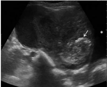
Pelvic ultrasound revealed a supravesical complex cystic mass with foci of increased echogenicity, suggestive of cal-cifications (Fig. 1).
On contrast-enhanced CT it was seen a heterogeneous mass, of approximately 12.5 cm, extending through the anterosuperior wall of the bladder to the right flank. The lesion revealed low-attenuation and multiple dystrophic cal-cifications, suggestive of mucinous adenocarcioma. There was no evidence of local or distant metastases (Fig. 2).
Fig. 1 Pelvic ultrasonography revealed a large complex cystic mass with cal-cifications (arrow) located superiorly to the bladder ()
Acta Radiologica Short Reports 2012;1:4. DOI: 10.1258/arsr.2011.110018
by guest on August 27, 2014
arr.sagepub.com
MR was performed to better characterize the lesion and revealed a large inhomogeneous mass in contact with the dome and superior wall of the bladder and
occupying the right flank. The lesion showed high signal intensity on T2-weighted images and intermediate signal on T1-weighted images. After intravenous contrast
Fig. 3 MR revealed an inhomogeneous mass with intermediate signal intensity on T1-weighted images (a) and high signal intensity on T2-weighted images (b). After the administration of gadolinium (c), the mass enhanced slightly peripherally and on solid vegetations ()
Fig. 2 On axial contrast-enhanced CT scan (a) was seen a large heterogeneous solid mass, with areas of low attenuation and dystrophic calcifications, sugges-tive of mucin content. Coronal (b) and sagittal (c) reformatted CT images, revealed the mass located superior to the bladder and extending through the right flank. The lesion contact the bladder but no focal thickening of the bladder wall was evident
2 V Monteiro and TM Cunha
. . . .
by guest on August 27, 2014
arr.sagepub.com
administration the mass enhanced slightly peripherally (Fig. 3).
Open laparotomy revealed that the lesion was strongly adhered to the wall of the bladder and partial cystectomy with en-bloc resection of the mass was performed.
Histologic analysis revealed an urachal mucinous
adenocarcinoma.
Discussion
The urachus is an embryologic remnant of the fetal geni-tourinary tract seen as a tubular structure extending in the midline between the dome of the bladder and the umbilicus that usually involutes before birth and remains in adults as the median umbilical ligament.
Primary adenocarcinomas of the urachus are extremely rare, representing only 0.5 –2% of all bladder malignancies, although 34% of bladder adenocarcinomas are urachal in origin (1). It is believed to arise from malignant transform-ation of columnar or glandular metaplastic epithelium and mucin production is found in up to 75% of cases on histo-logical analysis.
Urachal carcinomas mainly affect patients between 40 and 70 years old and have a male predilection (2). These tumors can be silent because of their extraperitoneal location, and consequently usually presents at an advanced stage, and often shows local invasion or metastases to the pelvic lymph nodes, lung, brain, liver, or bone. Hematuria, dysuria, abdominal pain, and umbilical discharge are the most frequent symptoms.
The diagnostic evaluation for urachal carcinoma should include a careful history and physical examination. A urine analysis with cytology may be helpful.
Although carcinoma of the urachus can occur in any site along the urachal tract, the most common locations are the dome of the bladder and the umbilicus.
US may demonstrate a supravesical complex cystic mass. CT or MR confirm the US findings and provide more infor-mation about local extent of the disease, pelvic lymph-node involvement, and systemic metastases. On CT urachal
carcinoma is usually seen as a midline mass anterosuperior to the dome of the bladder that can be solid, cystic, or mixed. In 60% of cases low-attenuation components are seen, representing mucin content. Like other mucinous ade-nocarcinomas, urachal carcinomas may produce calcifica-tions, which may occur in 50 –70% of cases and are considered nearly pathognomonic for urachal adenocarci-noma (1, 3). On MR imaging a mass with increased signal intensity is usually seen on T2-weighted images because of the presence of mucin within the tumor (4).
The radiologic differential diagnosis includes benign urachal tumors that may mimic malignancy, like adenomas, fibromas, fibromyomas, and hamartomas, infected urachal remnants, and other malignant tumors like adenocarcino-mas of non-urachal origin, transitional cell carcinoadenocarcino-mas, and metastases originating from primary lesions of the colon, prostate, or female genital tract (1, 5).
Primary treatment of localized disease includes wide local excision of the urachus, umbilicus, and surrounding soft tissue combined with partial or radical cystectomy and bilateral pelvic lymphadenectomy (6).
A cystoscopy is necessary to evaluate whether the carci-noma has penetrated the urothelium of the bladder and a transurethral biopsy should be performed if possible.
Conflict of interest:None.
REFERENCES
1 Yu JS, Kim KW, Lee HJ,et al.Urachal remnant diseases: spectrum of CT and US findings.Radiographics2001;21:451 – 61
2 Ashley RA, Inman BA, Sebo TJ,et al.Urachal carcinoma: clinicopathologic features and long-term outcomes of an aggressive malignancy.Cancer
2006;107:712 –20
3 Koster IM, Cleyndert P, Giard RW. Best cases from the AFIP: urachal carcinoma.Radiographics2009;29:939 –42
4 Wong-You-Cheong JJ, Woodward PJ,et al.Neoplasms of the urinary bladder: radiologic-pathologic correlation.Radiographics2006;26:553 – 80 5 Mengiardi B, Wiesner W, Stoffel F,et al.Case 44: adenocarcinoma of the
urachus.Radiology2002;222:744 –7
6 Henly DR, Farrow GM, Zincke H. Urachal cancer: role of conservative surgery.Urology1993;42:635 –9
#2012 The Foundation Acta Radiologica
This is an open-access article distributed under the terms of the Creative Commons Attribution License (http://creativecommons. org/licenses/by-nc/2.0/), which permits non-commercial use, distribution and reproduction in any medium, provided the original
work is properly cited.
Urachal carcinoma 3
. . . .
by guest on August 27, 2014
arr.sagepub.com