242 Radiol Bras. 2013 Jul/Ago;46(4):242–246
Mucinous carcinoma of the breast: iconographic essay
with histopathological correlation
*
Carcinoma mucinoso da mama: ensaio iconográfico com correlação histopatológica
Gustavo Nunes Medina Coeli1, Henrique Ferreira dos Reis1, Dayse Ribeiro Bertinetti1,
Francesca Maia Faria2, Daniel Guimarães Tiezzi3, Tatiane Mendes Gonçalves de Oliveira4
The present essay is aimed at describing the most characteristic imaging findings of mucinous carcinoma of the breast, with emphasis on the patterns related to better prognosis. The authors selected cases of mucinous carcinoma of the breast whose images were available, highlighting the imaging findings suggestive of this subtype of breast cancer, either at mammography, ultrasonography or magnetic resonance imaging.
Keywords: Breast neoplasm; Mucinous carcinoma; Mammography; Ultrasonography; Magnetic resonance imaging. O objetivo deste artigo é descrever os aspectos de imagem mais característicos do carcinoma mucinoso de mama, destacando-se os padrões relacionados a melhor prognóstico. Foram selecionados casos de carcinoma mucinoso de mama enfatizando as características de imagem que sugiram esse subtipo de neoplasia mamária, seja na mamografia, ultrassonografia ou ressonância magnética.
Unitermos: Neoplasia mamária; Carcinoma mucinoso; Mamografia; Ultrassonografia; Ressonância magnética.
Abstract
Resumo
* Study developed at Centro de Ciências das Imagens e Física Médica – Faculdade de Medicina de Ribeirão Preto da Universidade de São Paulo (FMRPUSP), Ribeirão Preto, SP, Bra-zil.
1. Radiologists, MDs, Residents at Centro de Ciências das Imagens e Física Médica – Hospital das Clínicas da Faculdade de Medicina de Ribeirão Preto da Universidade de São Paulo (HC-FMRPUSP), Ribeirão Preto, SP, Brazil.
2. Pathologist, Physician Assistant, Department of Pathol-ogy, Faculdade de Medicina de Ribeirão Preto da Universidade de São Paulo (FMRPUSP), Ribeirão Preto, SP, Brazil.
3. PhD, Professor, Department of Gynecology and Obstet-rics, Mastology Division, Faculdade de Medicina de Ribeirão Preto da Universidade de São Paulo (FMRPUSP), Ribeirão Preto, SP, Brazil.
4. MD, Radiologist, Centro de Ciências das Imagens e Física Médica – Hospital das Clínicas daFaculdade de Medicina de beirão Preto da Universidade de São Paulo (HC-FMRPUSP), Ri-beirão Preto, SP, Brazil.
Mailing Address: Dr. Gustavo Nunes Medina Coeli. Avenida Anna Cardoso de Miranda, 185, Jardim Inconfidência. Uberlân-dia, MG, Brazil, 38411-231. E-mail: gustavonmc@yahoo. com.br.
Received September 26, 2012. Accepted after revision March 7, 2013.
Coeli GNM, Reis HF, Bertinetti DR, Faria FM, Tiezzi DG, Oliveira TMG. Mucinous carcinoma of the breast: iconographic essay with histo-pathological correlation. Radiol Bras. 2013 Jul/Ago;46(4):242–246.
highlighting findings associated with prog-nostic prediction.
Histological diagnosis
The diagnosis of mucinous neoplasm may be suspected at fine-needle aspiration biopsy (FNAB), although with low accu-racy in the differentiation between benign (mucoceles) and malignant mucinous le-sions (MMC).
Core biopsy exhibits high sensitivity and high positive predictive value (PPV) for the diagnosis of mucinous neoplasm, particularly in cases where such biopsy is imaging-guided, reaching values close to 100%(8,9). However, the differentiation be-tween pure and mixed types can only be established after excision and evaluation of the lesion in its whole extent(6,9).
Prognosis
The better prognosis of MMCs, com-pared with NS-IDC, explains the diagnos-tic importance of these tumors(7). Pure type shows indolent growth, while the mixed type presents variable biological behavior, sometimes similar to NS-IDCs(10).
Thus, MMCs, specially the pure type, demonstrated a lower histological degree (well differentiated tumors), higher hor-tribution, but with higher incidence in
eld-erly(5).
Histologically, such variant is character-ized by a tumor-like arrangement of neo-plastic cells involved by extracellular mu-cin, in most cases associated with periph-eral ductal carcinoma in situ (DCIS)(6). Cellularity as well as the amount of mucin has large variation among mucinous tu-mors. The greater the amount of mucin, the better the prognosis is(6).
There are two histological presentations with different imaging features and prog-noses: pure and mixed types. Pure type comprises tumors with a mucinous arrange-ment in their whole extent, with less than 10% of non-mucinous component, or even a poorly differentiated non-mucinous com-ponent(6,7). The mixed type presents a greater amount of neoplastic cells not in-volved by mucin, generally in association with a smaller amount of extracellular mucin, which implies intermediate charac-teristics between the pure type and nonspe-cific invasive ductal carcinoma (NS-IDC), for this reason sometimes referred to as “IDC with mucinous differentiation”(6,7).
The present pictorial essay focus on the most typical imaging findings of such spe-cific type of carcinoma and its subtypes,
INTRODUCTION
dis-mone receptor (HR) expression, lower in-cidence of adverse oncogenes, lower rate of axillary lymph node involvement at di-agnosis, and longer disease-free survival (with no significant difference in overall survival)(7,11,12).
Imaging findings
MMC is part of a group of 10% to 20% of the malignant tumors which present as
circumscribed lesions, sometimes initially interpreted as benign lesions, which delays the diagnosis and appropriate treatment(5, 8,12) decisions.
In such a context, mammography, ultra-sonography and magnetic resonance imag-ing represent fundamental tools in the at-tempt to recognize findings not only sug-gesting the diagnosis of MMC, but also helping to differentiate between the two
his-tological types of the neoplasm directly re-lated to different prognostic factors(11,13–15). Typically, a MMC presents as an ovoid or round nodule with circumscribed mar-gins. At mammography, the pure type cor-relates with circumscribed or micro-lobu-lated margins, which present a direct rela-tionship with the amount of extracellular mucin(16), as demonstrated on Figure 1. The mixed type presents more indistinct or spiculated contours secondary to a higher degree of fibrosis and peripheral desmopla-sia, similarly to a NS-IDC. Microcalcifi-cations are not common, and when they occur, rarely represent psammomatous cal-cifications, most times associated with the presence of peripheral component of DCIS(6,16,17).
Ultrasonography has higher sensitivity than mammography in the detection of MMCs(16,18). Characteristically, MMCs are visualized as an ovoid nodule with circum-scribed or microlobulated margins, and may be either hypo- or isoechoic, with pos-terior acoustic shadowing in 37% to 71% of the cases, as demonstrated on Figure 2. Similarly to morphological findings at mammography and ultrasonography, MMCs, at magnetic resonance imaging, are visualized as ovoid or lobulated masses with predominantly regular contours. The signal intensity is variable on T1-weighted images and with strong signal (similar to that of water or vessels) on T2-weighted images (Figure 3). Such high signal of the MMCs on T2-weighted images shows di-rect correlation with the degree of
extracel-Figure 2. A: Female, 69-year-old patient with pure MMC, with typical sonographic findings: ovoid and microlobulated nodule, isoechoic to fat tissue and with posterior acoustic enhancement. B: Female, 58-year-old patient with an ovoid nodule with indistinct and angulated margins, with no noticeable microlobulation, with posterior acoustic enhancement, diagnosed as mixed MMC. C: Histology from this patient demonstrating mucinous neoplasia with peripheral DCIS foci at left.
*
*
➤ ➤
Figure 1. A: Female, 69-year-old patient. Craniocaudal view demonstrating nodule with microlobulated margins in the medial quadrant of the right breast, diagnosed as pure MMC. B: Histological section of the lesion, identifying mucinous tumor with neoplastic cells involved by moderate amount of extracellular mucin.
*
➤
Figure 3. Female, 32-year-old patient with pure MMC. A: Mediolateral, oblique mammographic view demonstrating microlobulated ovoid nodule at the junction of the medial quadrants of the right breast. B: Axial MRI STIR image of the described nodule, demonstrating intense hypersignal onT2-weighted sequence with hypointense septa, common findings in mucinous carcinomas. C: Histological section at microscopy identifying mucinous tumor with a remarkable amount of mucin and little cellularity. D,E: Dynamic contrast-enhanced, T1-weighted images with fat suppression and subtraction, at the second and fifth minutes, dem-onstrating predominantly ring-shaped and progressive enhancement, and also marked enhancement of the septa within the lesion. F: Sagittal 3D reconstruc-tion.
*
➤➤
*
*
lular mucin and has high diagnostic sensi-tivity, although not pathognomonic, since it is also found in other lesions (secondary to necrosis, hemorrhage, edema, myxoid matrix or cystic component)(19). In addittion, the presence of intermediate signal on T2-weighted images suggests the mixed type of MMC.
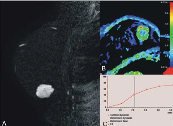
On dynamic contrast-enhanced se-quences, any enhancement morphology may occur, however, peripheral, ring-shaped or heterogeneous enhancement are more characteristic, and progressive along time (Figure 3). The pure type of MMC generally presents mild to moderate en-hancement at the early phases, with centrip-etal tendency determining type 1
(progres-sive) curves, a feature demonstrated on Figure 4, or type 2 curve (plateau)(19). The progressive enhancement pattern is related to the tumor cellularity, nuclear grading, and amount of extracellular mucin. Thus, an intense enhancement in the first two minutes after gadolinium injection, or a type 3 curve (washout) must raise suspicion of mixed type MMC or, an even rarer pure tumor with high cellularity(10).
As compared with other subtypes of breast cancer, MMCs, in general, present low signal intensity on diffusion-weighted images. The quantitative analysis with measurement of the apparent diffusion co-efficient (ADC) demonstrates high ADC values in relation to NS-IDCs. High ADC
values may be associated with the presence of extracellular mucin and low tumor cel-lularity. However, such characteristic seems to be related to a larger extent to pure MMCs, which show high ADC value, simi-lar or greater than benign lesions. Mixed mucinous tumors may exhibit lower ADC values, similarly to other breast cancer sub-types. Diffusion-weighted sequences and ADC calculation represent additional tools to increase the specificity of magnetic reso-nance imaging(19–21) (Figure 5).
CONCLUSION
imag-das Associações de Ginecologia e Obstetrícia para rastreamento do câncer de mama por métodos de imagem. Radiol Bras. 2012;45:334–9.
2. Azevedo AC, Canella EO, Djahjah MCR, et al. Conduta das funcionárias de um hospital na ade-são ao programa de prevenção do câncer de mama. Radiol Bras. 2012;45:215–8.
3. Moreira BL, Lima ENP, Bitencourt AGV, et al. Metástase na mama originada de carcinoma ova-riano: relato de caso e revisão da literatura. Radiol Bras. 2012;45:123–5.
4. Vieira SC, Silva JS, Madeira EB, et al. Heman-gioma de mama simulando metástase no PET-CT. Radiol Bras. 2011;44:401–2.
5. Yoo JL, Woo OH, Kim YK, et al. Can MR imag-ing contribute in characterizimag-ing well-circum-scribed breast carcinomas? Radiographics. 2010; 30:1689–702.
6. Rosen PP. Rosen’s breast pathology. 3rd ed. Phila-delphia: Lippincott Williams & Wilkins; 2009. 7. Bae SY, Choi MY, Cho DH, et al. Mucinous
car-cinoma of the breast in comparison with invasive ductal carcinoma: clinicopathologic characteris-tics and prognosis. J Breast Cancer. 2011;14:308– 13.
8. Bode MK, Rissanen T. Imaging findings and ac-curacy of core needle biopsy in mucinous car-cinoma of the breast. Acta Radiol. 2011;52:128– 33.
9. Gobbi H. Carcinoma mucinoso invasor da mama e seus diagnósticos diferenciais na biópsia per-cutânea com agulha grossa [Editorial]. J Bras Patol Med Lab. 2010;46(2).
10. Monzawa S, Yokokawa M, Sakuma T, et al. Mu-cinous carcinoma of the breast: MRI features of pure and mixed forms with histopathologic
cor-Figure 4. Female, 54-year-old patient with anatomopathological diagnosis of pure MMC. A: Sag-ittal T2-weighted SPAIR image demonstrating lobulated lesion with high signal on T2-weighted sequence. Color mapping of wash-in (B) and kinetic curve (C) showing slight enhancement at the early phase with progressive enhancement pattern.
Figure 5. Female, 54-year-old patient with pure mucinous tumor represented on diffusion-weighted im-ages, with b value = 0 on A and b = 700 on B, revealing minimum restriction, corroborated by the high signal intensity on C (ADC map).
ing findings, as the good correlation with histopathology has important implication in patient’s prognosis.
The imaging findings may suggest both subtypes of MMCs, pure and mixed ones. Mammography and ultrasonography allow a satisfactory evaluation of the lesion’s morphology, but magnetic resonance imag-ing, besides providing data on morphology, also provides clues about tissue
composi-tion and along with the enhancement pat-tern after administration of gadolinium-based contrast medium, contribute to a better characterization of the lesion and to improve diagnostic accuracy.
REFERENCES
relation. AJR Am J Roentgenol. 2009;192:W125– 31.
11. Wilson TE, Helvie MA, Oberman HA, et al. Pure and mixed mucinous carcinoma of the breast: pathologic basis for differences in mammographic appearance. AJR Am J Roentgenol. 1995;165: 285–9.
12. Cao AY, He M, Liu ZB, et al. Outcome of pure mucinous breast carcinoma compared to infil-trating ductal carcinoma: a population-based study from China. Ann Surg Oncol. 2012;19: 3019–27.
13. Vianna AD, Gasparetto TD, Torres GC, et al. Can-cerização de lóbulos: correlação de achados ma-mográficos e histológicos. Radiol Bras. 2011;44: 275–8.
14. Oliveira FGFT, Fonseca LMB, Koch HA.
Respon-sabilidade civil do radiologista no diagnóstico do câncer de mama através do exame de mamogra-fia. Radiol Bras. 2011;44:183–7.
15. Miranda CMNR, Santos CJJ, Maranhão CPM, et al. A tomografia computadorizada multislice é fer-ramenta importante para o estadiamento e segui-mento do câncer de mama? Radiol Bras. 2012; 45:105–12.
16. Conant EF, Dillon RL, Palazzo J, et al. Imaging findings in mucin-containing carcinomas of the breast: correlation with pathologic features. AJR Am J Roentgenol. 1994;163:821–4.
17. Lam WW, Chu WC, Tse GM, et al. Sonographic appearance of mucinous carcinoma of the breast. AJR Am J Roentgenol. 2004;182:1069–74. 18. Calas MJG, Alvarenga AV, Gutfilen B, et al.
Ava-liação de parâmetros morfométricos calculados a
partir do contorno de lesões de mama em ultrasso-nografias na distinção das categorias do sistema BI-RADS. Radiol Bras. 2011;44:289–96.
19. Woodhams R, Kakita S, Hata H, et al. Diffusion-weighted imaging of mucinous carcinoma of the breast: evaluation of apparent diffusion coeffi-cient and signal intensity in correlation with his-tologic findings. AJR Am J Roentgenol. 2009; 193:260–6.
20. Marques EF, Medeiros MLL, Souza JA, et al. In-dicações de ressonância magnética das mamas em um centro de referência em oncologia. Radiol Bras. 2011;44:363–6.