Diagnosis of patients with Prader-Willi and Angelman Syndromes: the
importance of an overall investigation
Monica Castro Varela, Cintia Fridman and Célia Priszkulnik Koiffmann
Centro de Estudos do Genoma Humano, Departamento de Biologia, Instituto de Biociências,
Universidade de São Paulo, São Paulo, SP, Brazil.
Abstract
Seventy-two patients with clinical diagnoses of Prader-Willi (PWS; n = 28patients) or Angelman syndromes (AS; n = 44 patients) were submitted to chromosome analysis, SNRPN-SNURF exon 1 methylation assay, and microsatellite genotyping. Analysis of the methylation pattern confirmed the PWS diagnosis in 18out of 28patients and the AS diagnosis in 20 out of 44 patients. FISH and microsatellite analysis detected a deletion in 30 patients (14 PWS and 16 AS). Eight patients had normal FISH results (4 PWS and 4 AS); microsatellite markers showed that these patients had a uniparental disomy (UPD). Based on this study, we propose a strategy for the routine diagnosis of these syndromes that consists of the following steps: 1) methylation analysis, which does not require parental samples; 2) microsatellite genotyping of patient and parents to differentiate deletions, UPD and imprinting mutations; and 3) FISH for otherwise uninformative cases, and whenever parental samples are not available. Of the 34 patients whose PWS or AS diagnoses were not confirmed by laboratory tests, five presented a small extra marker chromosome, identified in three of them as an inv dup(15). One AS patient carried a balanced t(15;15) translocation associated with paternal UPD. Therefore G-banded chromosome analysis should be performed on all such patients, to detect possible structural rearrangements.
Key words: Angelman syndrome, Prader-Willi syndrome, diagnosis, 15q deletion, uniparental disomy, genomic imprinting.
Received: March 5, 2002; accepted: March 25, 2002.
Introduction
The Prader-Willi (PWS) and Angelman (AS) syn-dromes are clinically distinct developmental and neuro-be-havioral disorders resulting from the loss of imprinted gene expression within chromosome 15q11-q13 (Nichollset al., 1998; Nicholls and Knepper, 2001). PWS patients show neonatal hypotonia, with poor sucking and failure to thrive, hyperphagia with onset at 1-6 years of age, severe obesity, mild mental retardation, hypogonadism and characteristic facies and behavior (Praderet al., 1956; Holmet al., 1993; Cassidy, 1997, Fridmanet al., 2000a). AS patients present delayed psychomotor development, severe mental retarda-tion, absence of speech, typical happy disposition with outbursts of laughter, ataxia, seizures, microcephaly, ma-crostomia, and prognathism (Angelman, 1965; Williamset al., 1995, Fridmanet al., 2000b, Lossieet al., 2001). The prevalence of these syndromes has been reported to be 1/15-20,000 for PWS (Cassidy, 1997) and 1/20,000 for AS (Clayton-Smith, 1993).
PWS and AS are clear examples of genomic imprint-ing in humans since, the clinical manifestations of these syndromes depend on the parental origin of the mutations within the 15q11-q13 segment. Approximately 70-75% of individuals with PWS and AS have 15q11-q13 deletions, which are of paternal origin in PWS and of maternal origin in AS (Knollet al., 1989; Mageniset al., 1990). Maternal uniparental disomy (UPD) of chromosome 15is found in about 25% of PWS patients (Mascariet al., 1992), whereas paternal UPD occurs in only 2-3% of patients with AS (Mageniset al., 1990). About 1-5% of patients with PWS and AS have biparental inheritance of chromosome 15, but show abnormal methylation pattern and gene expression due to mutations in the imprinting center (Buitinget al., 1995; Saitohet al., 1996; Ohta et al., 1999). Some AS patients (~8%) may also have mutations in the UBE3A gene (Kishinoet al., 1997; Matsuuraet al., 1997; Malzacet al., 1998).
The recurrence risk in deletion and UPD cases is less than 1%, whereas for familial imprinting mutation and UBE3A mutation the risk can be as high as 50% (Bürgeret al., 1997; Saitohet al., 1997). Therefore, the identification of the genetic mechanism involved in each patient is a
requisite for genetic counseling and depends on the effi-ciency and reliability of genetic tests.
We report here on 72 patients with a clinical diagno-sis of PWS or AS, who underwent chromosome analydiagno-sis, SNRPN-SNURF exon 1 methylation assay and microsa-tellite genotyping. A strategy for the diagnosis of these syn-dromes is proposed.
Material and Methods
Patients
Genetic studies were carried out on 72 patients with a clinical diagnosis of PWS (28 patients, 12 males and 16 fe-males) and AS (44 patients, 19 males and 25fefe-males). These patients were referred for genetic tests by the Depart-ments of Neurology, Endocrinology and The Children’s In-stitute, Hospital das Clínicas of the University of São Paulo, from December 1996 to December 1998 (Varela, 1999; 2000).
Most of these patients were examined by at least one of the authors, but in some cases only blood samples were provided, with a brief clinical description. The clinical diagnosis of PWS was based on the presence of mild to moderate mental retardation, obesity and hyperphagia in adolescents, and hypotonia, poor sucking and hypogona-dism in infants and children. For AS patients, the diagnostic features were delayed psychomotor development, severe mental retardation, absence of speech, typical happy dispo-sition with outbursts of laughter, and ataxia. Ages ranged from 9 months to 18 years in PWS patients, and from 17 months to 11 years in AS patients.
Cytogenetic studies
Chromosome studies of patients and their parents were performed on peripheral blood lymphocytes, after GTG-banding. FISH was performed with the SNRPN and GABRB3 probes (ONCOR, Gaithersburg, MD). For the identification of extra marker chromosomes, a chromo-some 15α-satellite probe (D15Z; ONCOR, Gaithersburg, MD) was used.In situhybridization and immunochemical detection were carried out according to the manufacturer’s instructions. At least 20 metaphases were analyzed per case.
Methylation analysis
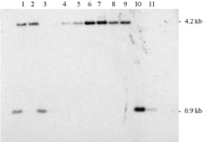
DNA was extracted from peripheral blood leukocytes by standard procedures. The methylation status of the PWS/AS region was assessed by Southern blotting. Geno-mic DNA double digested withXbaI andNotI was separated by electrophoresis on a 1.0% agarose gel, and transferred to a nylon membrane. A32P labeled 0.6 kbEcoRI-NotI fragment, which contains exon 1 ofSNRPN-SNURF,was used as a
probe (Glennet al., 1996). In this assay, PWS patients pres-ent a single 4.2 kb methylated maternal band, whereas AS patients have a 0.9 kb non-methylated paternal band. Normal individuals have both bands.
Dinucleotide repeat (CA)npolymorphisms
Microsatellite analysis was performed using three markers within the critical region 15q11-q13 [4-3RCA (D15S11), LS6-1CA (D15S113) and GABRB3RCA (GABRB3)], after multiplex PCR and polyacrylamide gel eletrophoresis (Mutiranguraet al.1993). The genotyping of two loci outside the PWS/AS region (D15S117 and D15S984) allowed to distinguish a deletion from a UPD: uniparental inheritance within the PWS/AS region to-gether with biparental inheritance outside this region identifies a deletion; the presence of uniparental inheri-tance both within and outside the critical region reveals a UPD.
Results
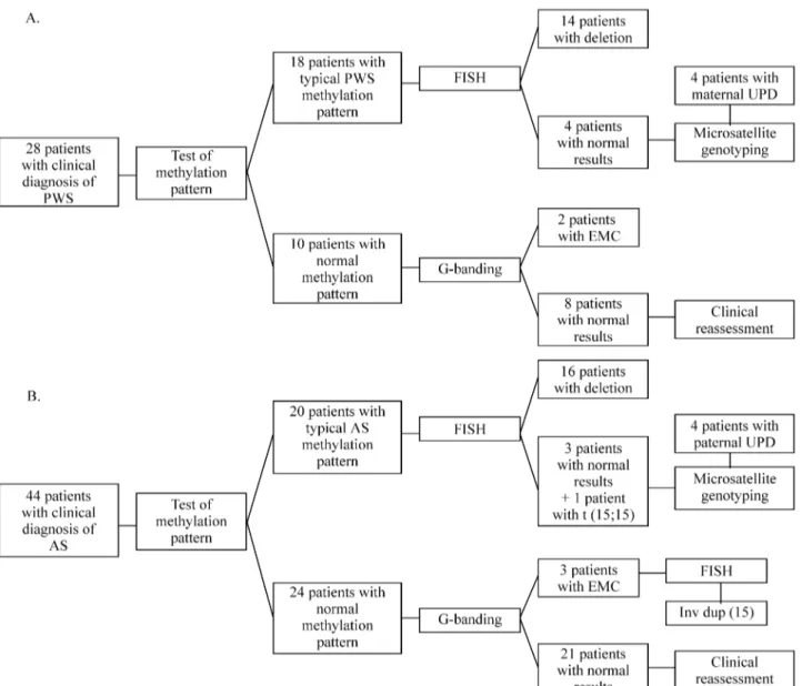
The methylation pattern analysis confirmed the diag-nosis of PWS syndrome in 18 out of 28 patients, and of AS syndrome in 20 out of 44 patients (Figure 1). The results are summarized in Figure 2.
The patients with a typical PWS or AS methylation pattern (n = 38) had their chromosomes analyzed after high resolution banding (GTG). The 15q11-q13 microdeletion was detected in seven (20%) patients (4 PWS and 3 AS); in five (14.28%) patients (3 PWS and 2 AS) the presence of a microdeletion was incertain; 22 patients (62.8%) had nor-mal karyotypes (10 PWS and 12 AS), and in one (2.8%), a Robertsonian translocation [t(15;15)] was identified. All parents had normal karyotypes.
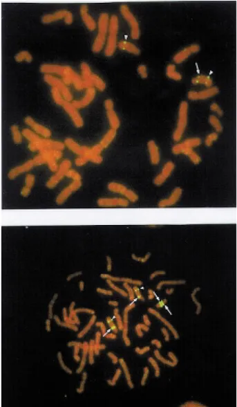
Afterin situhybridization, deletions were detected in 14 PWS patients (77.8%; four of them previously detected by GTG banding) and in 16 AS patients (80%; three of
them detected previously by GTG banding) (Figure 3a). In the As patient with the t(15;15) translocation, no deletion was apparent. No abnormalities were observed in the seven remaining cases, who also presented normal karyotypes on GTG-banding analysis.
Microsatellite genotyping was performed in 33 of the 38 patients with a typical methylation pattern, since paren-tal samples were not available for the other five cases. A de-letion was detected in 20 (60.6%) patients (7 with PWS and 13 with AS). Eight (24.3%) patients had UPD (Figure 4), including seven patients with normal FISH results and the AS patient with the t(15;15). In five cases (15.1%) the re-sults were uninformative.
Among the 34 patients with normal methylation pat-terns, chromosome analysis after GTG banding revealed five cases (2 “PWS patients” and 3 “AS patients”) with an extra marker chromosome (EMC) with the size of a
G-group chromosome. In the two “PWS patients”, the EMC did not show FISH signals of theα-satellite D15Z probe nor of the single-copy sequences GABRB3 and SNRPN. Two FISH signals were observed on the EMCs in the three “AS patients”, allowing these markers to be iden-tified as inv dup (15) (Figure 3b). The clinical description of these three patients can be found in Varelaet al.(1999). Microsatellite genotyping with markers within the PWS/AS critical region was performed on these inv dup(15) carriers. The presence of three alleles (two mater-nal and one patermater-nal) of the marker GABRB3 confirmed that the FISH signals observed on the EMCs were produced by probe GABRB3 and not by the control probe. The ma-ternal origin of the markers was established. All of the 29 remaining cases with normal methylation patterns presen-ted normal karyotypes.
Discussion
This work is part of an ongoing research on children with PWS and AS. The diagnostic hypothesis of PWS or AS was initially investigated by SNRPN-SNURF methy-lation analysis. This test has been used as a diagnostic tool for PWS and AS, since the methylation pattern is par-ent-specific in this region (Glennet al., 1996; Kubotaet al., 1996) and detects patients with deletion, UPD and imprint-ing mutations, which account for 99% of PWS and 85% of AS cases.
Classical cytogenetic techniques have low sensitivity for the detection of microdeletions, even at higher banding levels. Differences in condensation of band 15q12 in homo-logues make it difficult to detect a deletion in this region: in our study, only seven of the 30 patients (23.3%) with a dele-tion were detected by this method. FISH with chromosome 15q11-q13 probes was the most efficient cytogenetic
Figure 3- FISH with GABRB3 probe (arrows) shows (a) a deletion and (b) a duplication in an inv dup(15). The signal of a control probe located at 15q22 is seen (arrowheads).
diagnostic test for PWS and AS, identifying a deletion in about 79% (30/38) of the patients.
Since FISH does not detect UPD, imprinting or UBE3A mutations, the remaining patients require DNA analysis using microsatellite markers within and outside the PWS/AS region. Eight patients with UPD (four PWS and four AS) were detected in this sample. The disadvan-tage of this methodology is that it requires parental blood samples and, even when they are available, the results can be uninformative (as in 15.1% of our sample).
So, an efficient strategy for the routine diagnosis of PWS and AS patients (Figure 2) consists of: a) methylation analysis, that allows the diagnosis of 99% of PWS and 85% of AS patients, and does not require parental samples; b) microsatellite genotyping of the family (child, mother and father) to diagnose deletions, UPD or imprinting mutations; c) in uninformative cases or if parental samples are not available, the FISH technique is recommended, since it ap-pears to detect all the deletions (~75% of PWS and AS pa-tients). Routine cytogenetic studies after G-banding should be performed in all patients referred with a clinical diagno-sis of PWS or AS. Among the 20 patients whose diagnodiagno-sis of AS was confirmed, one carried a balanced t(15;15). In-deed, about 5% of PWS and AS of the literature present a chromosome rearrangement (Butler, 1990).
Patients with hypotonia, delayed psychomotor devel-opment, severe mental retardation, seizures and dys-morphic features, frequently referred as “suspected AS”, may be carriers of an extra marker derived from chromo-some 15[inv dup(15)]. In the present sample, three such carriers were found, and the incidence of this marker ap-pears to be as high as 1/5,000 births (Buckton et al., 1995). FISH using repetitive, single-copy, or whole-chromosome painting probes is a requisite for the proper identification of these extra chromosomes. Microsatellite genotyping is use-ful to disclose their parental origin, also allowing to better characterize their genetic content. The phenotypic variabil-ity observed among patients with inv dup(15) is influenced by the origin of the marker, by the extent of the euchromatic segment and by its isodysomic or heterodysomic nature (Mignonet al., 1996).
In patients with a normal methylation pattern and nor-mal chromosomes, a clinical reassessment is recommended to determine whether additional DNA investigations are in-dicated. Among “AS patients” with a normal methylation pattern, cases can be found who carry a UBE3A mutation that accounts for approximately 8% of patients with the clinical features of AS (Nichollset al., 1998).
Acknowledgments
This work was supported by FAPESP (M.C.V. 97/02383-0, C.F. 95/07161-0), CNPq and PRONEX. Dr Robert D. Nicholls kindly provided the SNRPN-SNURF exon 1 probe. We thank Ms. Roseli M. Zanelato for techni-cal assistance.
References
Angelman H (1965) “Puppet” children: a report on three cases. Develop Med Child Neurol 7:681-688.
Buiting K, Saitoh S, Gross S, Dittrich B, Schwartz S, Nicholls RD and Horsthemke B (1995) Inherited microdeletions in the Angelman and Prader-Willi syndromes define an imprinting center on human chromosome 15. Nature Genet 9:395-400. Buckton KE, Spowart G, Newton MS and Evans JH (1985)
Forty-four probands with an additional “marker” chromo-some. Hum Genet 69:353-370.
Bürger J, Buiting K, Dittrich B, Gro S, Lich C, Sperling K, Horsthemke B and Reis A (1997) Different mechanisms and recurrence risks of imprinting defects in Angelman syn-drome. Am J Hum Genet 61:88-93.
Butler MG (1990) Prader-Willi syndrome: current understanding of cause and diagnosis. Am J Hum Genet 35:319-332. Cassidy SB (1997) Prader-Willi syndrome. J Med Genet
34:917-923.
Clayton-Smith J (1993) Clinical research on Angelman syndrome in the United Kingdom: observations on 82 affected individ-uals. Am J Med Genet. 46:12-15.
de Vries BB, Fryns JP, Butler MG, Canziani F, Wesby-van SE, van Hemel JO, Oostra BA, Halley DJ and Niermeijer MF (1993) Clinical and molecular studies in fragile X patients with a Prader-Willi-like phenotype. J Med Genet 30:761-766.
Eugster EA, Berry SA and Hirsch B (1997) Mosaicism for dele-tion 1p36.33 in a patient with obesity and hyperphagia. Am J Med Genet 70:409-412.
Fridman C, Varela MC, Kok F, Setian N and Koiffmann CP (2000a) Prader-Willi syndrome: genetic tests and clinical findings. Genetic Testing 4:387-392.
Fridman C, Varela MC, Kok F, Diament A and Koiffmann CP (2000b) Paternal UPD15: further genetic and clinical studies in four Angelman syndrome patients. Am J Med Genet 92:322-327.
Glenn CC, Saitoh S, Jong MTC, Filbrandt MM, Surti U, Driscoll DJ and Nicholls RD (1996) Gene structure, DNA methy-lation, and imprinted expression of the human SNRPN gene. Am J Hum Genet 58:335-346.
Holm VA, Cassidy SB, Butler MG, Hanchett JM, Greenswag LR, Whitman BY and Greenberg F (1993) Prader-Willi syn-drome: consensus diagnostic criteria. Pediatrics 91:398-402. Kishino T, Lalande M and Wagstaff J (1997) UBE3A/E6AP mu-tations cause Angelman syndrome. Nature Genet 15:70-73. Knoll JHM, Nicholls RD, Magenis RE, Graham Jr JM, Lalande M and Latt SA (1989) Angelman and Prader-Willi syndromes share a common chromosome 15deletion but differ in pa-rental origin of the deletion. Am J Med Genet 32:285-290. Kubota T, Sutcliffe JS, Aradhya S, Gillessen-Kaesbach G,
Chris-tian SL, Horsthemke B, Beaudet AL and Ledbetter DH (1996) Validation studies of SNRPN methylation as a diag-nostic test for Prader-Willi syndrome. Am J Med Genet 66:77-80.
Lossie AC, Whitney MM, Amidon D, Dong HJ, Chen P, Theriaque D, Hutson A, Nicholls RD, Zori RT, Williams CA and Driscoll DJ (2001) Distinct phenotypes distinguish the molecular classes of Angelman syndromes. J Hum Genet 38:834-845.
Wag-staff J (1998) Mutation Analysis of UBE3A in Angelman syndrome patients. Am J Hum Genet 32:1353-1360. Magenis RE, Toth-Fejel S, Allen LJ, Black M, Brown MG,
Budden S, Cohen R, Friedman JM, Kalousek D, Zonana J, Lacy D, LaFranchi S, Lahr M, Macfarlane J and Williams CPS (1990) Comparison of the 15q deletions in Prader-Willi and Angelman syndromes: specific regions, extent of dele-tions, parental origin, and clinical consequences. Am J Med Genet 35:333-349.
Mascari MJ, Gottlieb W, Rogan PK, Butler MG, Waller DA, Ar-mour JAL, Jeffreys AJ, Ladda RL and Nicholls RD (1992) The frequency of uniparental disomy in Prader-Willi syn-drome. N Engl J Med 326:1599-607.
Matsuura T, Sutcliffe JS, Fang P, Galjaard R-J, Jiang Y-H, Benton CS, Rommens JM and Beaudet AL (1997) De novo truncat-ing mutations in E6-AP ubiquitin-protein ligase gene (UBE3A) in Angelman syndrome. Nature Genet 15:74-77. Mignon C, Malzac P, Moncla A, Depetris D, Roeckel N,
Cro-quette M-F and Mattei M-G (1996) Clinical heterogeneity in 16 patients with inv dup 15chromosome: cytogenetic and molecular studies, search for imprinting effect. Euro J Hum Genet 4:88-100.
Mutirangura A, Jayakumar A, Sutcliffe JS, Nakao M, McKinney MJ, Buiting K, Horsthemke B, Beaudet A, Chinault AC and Ledbetter DH (1993) A complete YAC contig of the Prader-Willi/Angelman chromosome region (15q11-q13) and refined localization of the SNRPN gene. Genomics 18:546-552.
Nicholls RD and Knepper JL (2001) Genome organization, func-tion, and imprinting in Prader-Willi and Angelman syn-dromes. Annu Ver Genomics Hum Genet 2:153-175. Nicholls RD, Saitoh S and Horsthemke B (1998) Imprinting in
Prader-Willi and Angelman syndromes. TIG 14:194-200. Ohta T, Buiting K, Kokkonen H, Mc Candless S, Heeger S, Leisti
H, Driscoll DJ, Cassidy SB, Horsthemke B and Nicholls RD (1999) Molecular mechanism of Angelman syndrome in two large families involves an imprinting mutation. Am J Hum Genet 64:385-396.
Prader A, Labhart A and Willi H (1956) Ein Syndrom von Adipo-sitas, Kleinwuchs, Kryptorchismus und Oligophrenie nach myotonieartigem Zustand im Neugeborenenalter. Schweiz Med Wochenschr 86:1260-1261.
Saitoh S, Buiting K, Cassidy SB, Conroy JM, Driscoll DJ, Gabriel JM, Gillessen-Kaesbach G, Glenn CC, Greenswag LR, Horsthemke B, Kondo I, Kuwajima K, Niikawa N, Rogan PK, Schwartz S, Seip J, Williams CA and Nicholls RD (1997) Clinical spectrum and molecular diagnosis of Angel-man and Prader-Willi syndrome imprinting mutation pa-tients. Am J Med Genet 68:195-206.
Saitoh S, Buiting K, Rogan PK, Buxton JL, Driscoll DJ, Arne-mann J, Konig R, Malcolm S, Horsthemke B and Nicholls RD (1996) Minimal definition of the imprinting center and fixation of a chromosome 15q11-q13 epigenotype by im-printing mutations. Proc Natl Acad Sci USA 93:7811-7815. Stein CK, Stred SE, Thomson LL, Smith FC and Hoo JJ (1996)
Intersticial 6q deletion and Prader-Willi-like phenotype. Clin Genet 49:306-310.
Varela MC (1999) Síndromes de Prader-Willi e Angelman: Aná-lise Cromossômica, Espectro da Variabilidade Fenotípica e o Papel dos Cromossomos Marcadores. Master’s Thesis. Instituto de Biociências, Universidade de São Paulo, São Paulo, Brasil.
Varela MC (2000) Prader-Willi and Angelman syndromes: chro-mosome analysis, the spectrum of the clinical variability and the study of marker chromosomes. Master’s Thesis Ab-stract. Genet Mol Biol 23 (1):249-250.
Varela MC, Fridman C, Matsumoto TE, Kim CA, Kok F, Diament A and Koiffmann CP (1999) A clinical, cytogenetic and mo-lecular study of three Brazilian patients with supernumerary inv dup(15) marker chromosomes. Am J Hum Genet 65 [suppl]:A347 (#1962).