Nasal septum changes in adolescent patients treated
with rapid maxillary expansion
Tehnia Aziz1, Francis Carter Wheatley2, Kal Ansari3, Manuel Lagravere4, Michael Major5, Carlos Flores-Mir6
Objective: To analyze cone-beam computed tomography (CBCT) scans to measure changes in nasal septal devia-tion (NSD) after rapid maxillary expansion (RME) treatment in adolescent patients. Methods: This retrospective study involved 33 patients presenting with moderate to severe nasal septum deviation as an incidental finding. Out of these 33 patients, 26 were treated for transverse maxillary constriction with RME and seven, who did not undergo RME treat-ment, were included in the study as control group. CBCT scans were taken before appliance insertion and after appliance removal. These images were analyzed to measure changes in nasal septum deviation (NSD). Analysis of variance for re-peated measures (ANOVA) was used. Results: No significant changes were identified in NSD regardless of the applica-tion or not of RME treatment and irrespective of the baseline deviaapplica-tion degree. Conclusion: This study did not provide strong evidence to suggest that RME treatment has any effect on NSD in adolescent patients; however, the results should be interpreted with caution, due to the small sample size and large variation amongst individual patient characteristics.
Keywords:Nasal septum. CBCT. Rapid maxillary expansion. Rapid palatal expansion.
1 Private practice, Edmonton, Alberta, Canada.
2 Graduate student in Computer Sciences, Boston University, Boston,
Massachusetts, USA.
3 Assistant professor, University of Alberta, Department of Surgery, Edmonton,
Alberta, Canada.
4 Assistant professor, University of Alberta, Department of Dentistry, Edmonton,
Alberta, Canada.
5 Clinical assistant professor, University of Alberta, Department of Dentistry,
Edmonton, Alberta, Canada.
6 Professor, University of Alberta, Department of Dentistry, Edmonton, Alberta,
Canada.
DOI: http://dx.doi.org/10.1590/2177-6709.21.1.047-053.oar
How to cite this article: Aziz T, Wheatley FC, Ansari K, Lagravere M, Ma-jor M, Flores-Mir C. Nasal septum changes in adolescent patients treated with rapid maxillary expansion. Dental Press J Orthod. 2016 Jan-Feb;21(1):47-53. DOI: http://dx.doi.org/10.1590/2177-6709.21.1.047-053.oar
Submitted: December 05, 2014 - Revised and accepted: April 24, 2015
» The authors report no commercial, proprietary or financial interest in the products or companies described in this article.
Contact address: Carlos Flores-Mir
Division of Orthodontics, University of Alberta, 5-528 Edmonton Clinic Health Academy 11405 87 Ave, T6G 1C9 Edmonton, Canada E-mail: [email protected]
Objetivo: analisar imagens de tomografia computadorizada de feixe cônico (TCFC) para mensurar as alterações no desvio de septo nasal (DSN) após o tratamento com expansão rápida da maxila (ERM) em pacientes adolescentes. Méto-dos: o presente estudo retrospectivo incluiu 33 pacientes com desvio de septo nasal de moderado a severo, diagnosticado como um achado incidental. Dos 33 pacientes analisados, 26 tiveram a constrição maxilar transversal tratada por meio de ERM; 7 pacientes não foram submetidos à ERM, sendo incluídos no estudo como grupo controle. As imagens de TCFC foram obtidas antes da instalação do aparelho e após sua remoção, sendo analisadas para mensurar as alterações no DSN. A análise de variância para medidas repetidas (ANOVA) foi empregada. Resultados: não foram identificadas alterações significativas no DSN, independentemente da realização ou não do tratamento com ERM e do grau inicial de desvio.
Conclusão: esse estudo não fornece evidências suficientes para sugerir que o tratamento com ERM produza qualquer efeito sobre o DSN em pacientes adolescentes. Porém, esses resultados devem ser interpretados com cautela, em virtude do tamanho reduzido da amostra e da grande variação das características individuais dos pacientes.
INTRODUCTION
The reciprocal efects of nasal breathing on cranio-facial development have been intensively investigated in the literature. According to Moss’ functional theory, nasal respiration enables normal growth and develop-ment of the craniofacial structures.1 Moss hypothesized
that undisturbed nasal airlow is a continuous stimulus for lowering the palate and for lateral maxillary growth, thereby indicating a close relationship between nasal breathing and dentofacial morphology.
Nasal septal deviation (NSD) is deined as devia-tion of either the bony or cartilaginous septum or both from the facial midline. In humans, it has been hy-pothesized that signiicant nasal obstruction caused by NSD can afect nasal airlow and increase nasal airway resistance.2 Resultant impaired nasal breathing can lead
to preferential mouth breathing which, if chronic, may cause craniofacial alterations. These potential changes consist of a long face syndrome characterized by nar-row maxilla, steep mandibular plane, retrognathic man-dible, increased lower facial height, lip incompetence, constricted alar bases and typically malocclusion con-sisting of a posterior crossbite.2
Rapid maxillary expansion (RME) is routinely used in Orthodontics to treat transverse maxillary constric-tion, posterior dental crossbite and crowding.3
Con-sidering that maxillary bones form the periphery of the nasal cavity, it has been proposed that opening the mid palatal suture through RME could also result in lateral displacement of the nasal walls, thereby increasing nasal cavity dimensions.4
The earliest report of RME having an efect on na-sal septal changes came in 1975 when it was discovered that RME treatment in a patient cohort also appeared to improve NSD.5 More recently, a 94% reduction in
septal deviation was reported in children aged 5-9 years old, presenting with transverse maxillary constriction treated with RME.6 NSD correction was noted in the
lower and middle half of the nasal cavity when com-pared to a nonexpansion control group.
Although several studies have investigated the efect of RME on nasal cavity size and airway,5,7-11 there is a
paucity of research on the changes caused by RME in the nasal septum. To our knowledge, only three stud-ies have conducted a two-dimensional cephalomet-ric analysis5,6,12 to assess nasal septal changes produced
during RME. All of them used coronal views from
posterior anterior radiographs. Two studies reported fa-vorable improvement of septal deviation ater RME5,6
treatment in growing patients, while one12 study
re-ported no change in nongrowing patients aged 15-19 years old. However, these studies had some major limi-tations. There was lack of standardization in study de-sign and the nasal septum was measured at one single radiographic image instead of its entirety by assessing diferent points along the septum. It was also unclear whether both pre- and postexpansion radiographs mea-sured the septal change at a set landmark. Although im-provement in nasal septum was reported ater expan-sion, it was not clear whether the change was the same at each anatomical location along the nasal septum.
Considering the importance of nasal breathing for the development of craniofacial structures, it would be beneicial to ascertain whether RME can reliably im-prove NSD and hence its detrimental efects on nasal breathing. Therefore, the objective of this study is to analyze three-dimensional changes of the nasal septum resulting from maxillary expansion in an adolescent patient sample. The use of three-dimensional imaging should overcome some of the limitations observed in previously conducted research.
MATERIAL AND METHODS
This retrospective study fulilled all ethical require-ments and was approved by the Health Research Ethics Board at the University of Alberta.
Patient samples were obtained from a previously con-ducted randomized clinical trial14 at the Department of
Dentistry at University of Alberta during an 18-month period. A total of 33 patients with varying degrees of NSD at T1 (prior to rapid maxillary expansion [RME] treatment) were selected from an available pool of CBCT scans of 120 patients through a brief visual inspection of the entire nasal septum of each patient. Patients with nasal septum deviation were identiied from transverse and coronal views of cone-beam computed tomographic (CBCT) records taken prior to treatment with RME (or without RME for control patients).
Based on a previous publication,13 septal deviation
The inal sample consisted of:
» 14 patients treated with RME with moderate to severe NSD at T1 (more than 9 degrees);
» 12 patients treated with RME with mild NSD at T1 (less than 9 degrees);
» 7 untreated patients with RME with moderate to severe NSD at T1 (control group).
The BAME (bone anchored maxillary expansion) sample had a mean age of 14.2 ± 1.3 years, the TAME (tooth anchored maxillary expansion) sample had a mean age of 14.1 ± 1.4 years and inally the control sample had a mean age of 12.9 ± 1.2 years. Individual matching was not possible due to unequal sample sizes.
RME was carried out until posterior dental cross-bite overcorrection by 20% was achieved (maxillary lingual cusps overlapping with lingual inclines of man-dibular buccal cusps). Ater active expansion treatment, the screw was ixated with a composite resin into the turn-key mechanism of the appliance. The appliance was retained for a total of six months from the time of insertion. CBCT scans taken at T1 (at baseline, before expansion) and T2 (ater appliance removal) were ana-lyzed for this study. (For more detailed information on the methods of the previously conducted randomized trial please refer to reference14).
All CBCT scans were taken with either a NewTom (18 patients) or an i-CAT (15 patients). Images were converted into DICOM format sotware with a voxel size of 0.25 mm. All images at T1 and T2 for each patient were then uploaded to OsiriX DICOM Viewer (v. 5.8, 32 bit, Pixmeo, Geneva, Switzerland).
Based on a previous publication,15 the following steps
were followed:
1. Landmarks were identiied in the 3-D viewer/2-D
orthogonal MPR mode in OsiriX for each pa-tient in sagittal view (Table 1, Fig 1).
2. Based on the landmarks identiied on sagittal
view, three axial (A1, A2, A3) and four coronal DICOM landmarks (C1, C2, C3, C4) for each patient at each time point were isolated (Fig 1).
3. These landmarks were: the most anterior point of
nasal bone (A1, C1), perpendicular plate and vomer junction (A2, C4), anterior nasal spine (C2), crista galli (C3), halfway point between anterior nasal spine and perpendicular plate/vomer junction (A3). The landmarks were chosen due to their ease of identiication based on anatomical locations and because
they could reasonably cover the boundaries of normal septal anatomy in anterior-posterior and inferior to superior directions. Landmark A3 was the only land-mark not identiied by an anatomical structure.15 A total
of 14 images for every patient was analyzed consider-ing T1 and T2. These images were then transferred to MATLAB (MathWorks R2013b, Natick, Massachu-setts) for NSD analysis. One researcher registered the time point (T1 or T2 DICOM image) in the MATLAB sotware, but was blinded to the degree of septal devia-tion or whether it was a treatment or a control patient. This sotware enabled the septum to be systematically traced and analyzed.
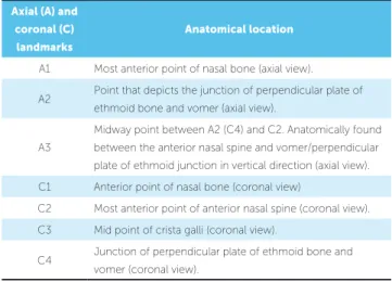
Table 1 - Descriptions of landmarks in sagittal view for image generation.
Figure 1 - Description and location of landmarks in sagittal view for axial and coronal image generation.
Axial (A) and coronal (C)
landmarks
Anatomical location
A1 Most anterior point of nasal bone (axial view).
A2 Point that depicts the junction of perpendicular plate of ethmoid bone and vomer (axial view).
A3
Midway point between A2 (C4) and C2. Anatomically found between the anterior nasal spine and vomer/perpendicular
plate of ethmoid junction in vertical direction (axial view).
C1 Anterior point of nasal bone (coronal view)
C2 Most anterior point of anterior nasal spine (coronal view).
C3 Mid point of crista galli (coronal view).
C4 Junction of perpendicular plate of ethmoid bone and vomer (coronal view).
During analysis in MATLAB, the axial images (im-ages A1, A2, A3) were traced from anterior to posterior direction. For example, for axial image A1, the nasal septum was systematically traced by placing points ap-proximately 1-2 mm apart along its anterior posterior course. Similarly, in coronal images (C1, C2, C3, C4), the nasal septum was traced in entirety from superior to inferior direction by placing points 1-2 mm apart.
The data from NSD measurements from MATLAB sotware were automatically transferred to a comma separated value (csv) spreadsheet. Once data analysis was complete, data were further copied to an Excel spread-sheet for ease of statistical analysis with SPSS program.
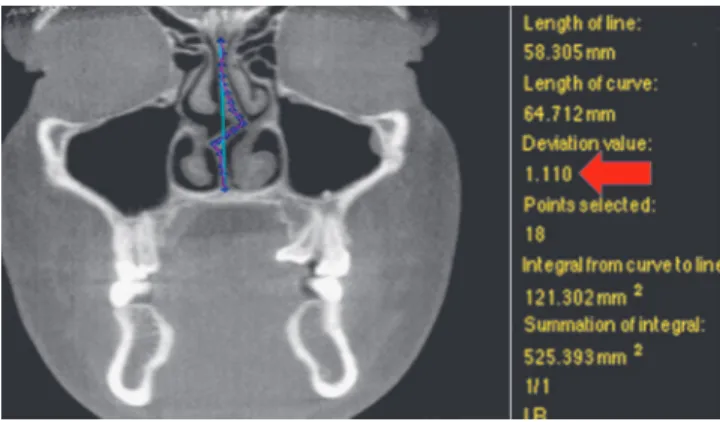
For the present study, NSD was quantiied based on the “degree of tortuosity” or the ratio of length of the curve to the length of an imaginary line in the mid sagittal plane (Fig 2 – red arrow points to ratio). In other words, the degree of tortuosity is an absolute measurement of the degree of septal deviation from the midline at each identiied landmark in both coro-nal and/or axial views.
STATISTICAL TESTS
Reliability and measurement error
Intrarater reliability and measurement error were conducted for identiication of landmarks in sagittal view in OsiriX and then as NSD tracing in MATLAB (from the selected DICOMs based on landmark iden-tiication/image isolation in OsiriX). All measurements were repeated three times with at least ive days apart.
Landmark identification was done in OsiriX with X, Y and Z coordinates noted for anterior point of the nasal bone (landmarks A1/C1), vomer and perpendicular plate junction (landmarks A2/ C4), anterior nasal spine (C2), crista galli (landmark C3), and halfway point between anterior nasal spine and vomer/perpendicular plate junction (A3). Na-sal septum tracing and NSD measurement ratio in MATLAB for each image (A1, A2, A3, C1, C2, C3 and C4) were also recorded.
Intraclass correlation with consistency under two-way mixed model was tabulated in SPSS for both land-mark identiication and nasal septum measurements.
Main study statistical tests
Statistical analysis was carried out with the aid of SPSS (version 21) using alpha = 0.05. Analysis of
variance for repeated measures (ANOVA) was per-formed with two within-subject factors and one between-subject factor. Baseline septal deviation of “mild” and “moderate to severe” was considered be-tween subjects factor. Time (T1 and T3, two levels) and landmark (A1, A2, A3, C1, C2, C3 and C4, 7 levels) were the two within-subject factors.
RESULTS
The intraclass correlation coefficients and cor-responding confidence interval (95%) for both OsiriX landmark identification and MATLAB NSD measurements are listed in Tables 2 and 3. Loca-tion of most landmarks indicated good agreement16
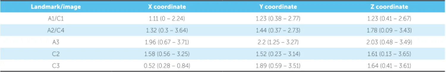
between parameters by high ICC values (> 0.8). Minimum, maximum and mean measurement error for both OsiriX landmark identification and MAT-LAB NSD ratios are listed in Tables 4 and 5. Mean measurement error in OsiriX was in the range of 0.5-2.2 mm with A3 having the largest error in all coordinates (1.96 to 2.2 mm). Difference between landmark coordinates in OsiriX measured at three different time points for reliability was not greater than 4 mm. MATLAB NSD ratios at all landmarks were less than 0.02.
There was no signiicant diference in NSD accord-ing to time [F (1.24) = 0.2, p = 0.659]. There was also lack of evidence for diferences in NSD at images A1, A2, A3, C1, C2, C3, C4 with time (time*landmark location) [F (2.93, 70.24) = 0.205, p = 0.889].
Landmark/image X coordinate Y coordinate Z-coordinate
A1/C1 0.980 (0.943 – 0.995) 0.974 (0.925 – 0.993) 0.994 (0.983 – 0.998)
A2/C4 0.963 (0.897 – 0.990) 0.974 (0.926 – 0.993) 0.986 (0.959 – 0.996)
A3 0.941 (0.839 – 0.984) 0.929 (0.810 – 0.980) 0.988 (0.965 – 0.997)
C2 0.980 (0.943 – 0.995) 0.978 (0.937 – 0.994) 0.990 (0.971 – 0.997)
C3 0.998 (0.993 – 0.999) 0.973 (0.924 – 0.993) 0.986 (0.959 – 0.996)
Table 2 - Intrarater reliability for OsiriX landmark/image identification.
Table 3 - Intrarater reliability for MATLAB NSD measurements.
Image ICC Confidence interval
A1 0.948 (0.871 – 0.983)
A2 0.993 (0.982 – 0.998)
A3 0.872 (0.702 – 0.957)
C1 0.947 (0.867 – 0.983)
C2 0.914 (0.791 – 0.972)
C3 0.904 (0.771 – 0.969)
C4 0.941 (0.854 – 0.981)
Table 4 - Measurement error in millimeters for OsiriX landmark/image identification.
Landmark/image X coordinate Y coordinate Z coordinate
A1/C1 1.11 (0 – 2.24) 1.23 (0.38 – 2.77) 1.23 (0.41 – 2.67)
A2/C4 1.32 (0.3 – 3.64) 1.44 (0.37 – 2.73) 1.78 (0.09 – 3.43)
A3 1.96 (0.67 – 3.71) 2.2 (1.25 – 3.27) 2.03 (0.48 – 3.49)
C2 1.58 (0.56 – 3.25) 1.52 (0.23 – 3.14) 1.61 (0.13 – 3.65)
C3 0.52 (0.28 – 0.84) 1.89 (0.59 – 3.51) 1.64 (0.41 – 3.61)
Table 5 - Measurement error for MATLAB NSD ratio..
Image Mean ratio
A1 0.0016
A2 0.0042
A3 0.0069
C1 0.0025
C2 0.0023
C3 0.0166
C4 0.0090
Partial eta square was 0.008 for time*spatial image accounting for only 0.8 % of variance explained by the efect of RME on NSD. Baseline septal deviation of mild or moderate to severe had no efect change at spatial landmarks with time (baseline deviation*spatial image*time) [F (2.93, 70.24) = 1.85, p = 0.147, ac-counting for only 7% of variance in NSD].
DISCUSSION
There is no “gold standard” test to diagnose sep-tal deviation17 and diferent protocols for measuring
septal deviation have been identiied in the literature. The degree of tortuosity measurement was used in this study to compare the length of the curve of the devi-ated septum to the length of an ideal straight septum. This measurement solely measured the nasal septum in isolation and did not classify or include other con-founding nasal pathology that could be the reason for septal deviation, such as turbinate hypertrophy or mu-cosal swelling. Therefore, this measurement method was well suited to the objective of our study.
Landmark identification in OsiriX and NSD ra-tios in MATLAB were indicative of good reliability. It was ascertained that identifying the location of landmark A3 with certainty was challenging. It was the only landmark that was not associated with a hard tissue anatomical structure, and rough approximation in space was made on all DICOMS without a ruler to accurately measure the half way point between anteri-or nasal spine and the vomer, and perpendicular plate junction. Although reliability at A3 for both OsiriX and MATLAB was suggestive of good reliability, a mean measurement error close to 4 mm was reported in x, y and z coordinates.
Owing to the retrospective nature of the study and the available CBCT records of patients that have undergone RME, sample size was less than ideal. Nevertheless, our findings were similar to a recently conducted study12 in two dimensions, whereby no
change in nasal septal deviation was identified pre- and postmaxillary expansion. On the other hand, this study was different than the previous one,12 since
the analysis of septal deviation was based on three-dimensional measurements on CBCT as opposed to two-dimensional on a posterior-anterior cephalo-gram. To date, this study appears to be the only one comparing the effects of RME on NSD using three-dimensional analysis of CBCT scans.
The main finding of this investigation was that rapid maxillary expansion had no effect on patients that had nasal septal deviation at baseline, as measured at images in axial and coronal views. Furthermore, mild or severe baseline deviation had no statistically significant effect on NSD change, as measured at set landmarks. The time difference between T1 and T2 in the treatment group was similar to that of the control
group; neither one of the groups had statistically significant changes in NSD over a 6-month period. It could be challenging to identify the true effect of RME treatment on NSD due to relatively small sam-ple size and individual patient variation. In fact, four patients out of a sample of 26 (15%) depicted subjec-tive visual improvement, as determined by one author in NSD from RME at mostly the coronal location of C3 (one at A3) (Figs 6 to 9). All four presented with baseline deviation of moderate to severe, but it is unclear as to why others with severe deviation and similar characteristics did not have a similar change. This is parallel to the conclusions of Harvolds pri-mate study18 whereby, even though the experiment
protocol and sample characteristics were standard-ized, the animals responded and adapted to nasal ob-struction quite differently. In fact, based on the sta-tistical model, only 7% of variance in NSD could be attributed to RME treatment.
It has been proposed19 that early intervention with
RME (i.e., before palatal suture starts closing) in pre-pubescence would result in greater skeletal than dental change. Given that all patients in this study were ado-lescents, it is possible that lack of statistically signii-cant change in NSD was the result of subjects having more advanced craniofacial development. In addition, increased bone density (calciication) of surrounding craniofacial structures in adolescence can ofer greater resistance to skeletal change from RME. In contrast, in patients with mixed and deciduous dentition, stud-ies20,21 have reported the efect of RME to be attributed
to between one half to two-thirds skeletal change..
In fact, the studies5,6 that reported favorable efects of
RME on NSD consisted of patients recruited prior to their adolescent growth spurt.
Although there is a lack of studies examining the effect of RME on NSD, there are several studies5,7-10
LIMITATIONS
This study did not provide strong evidence to suggest that RME treatment has any efect on NSD in adolescent patients, but the results should be interpreted with caution, due to the small sample size and large variation amongst individual patient characteristics. In this sense, this could be considered a pilot study testing this novel methodology.
Although potential diferences between bone- or tooth-anchoraged expansion appliances over NSD could not be considered because of the limited number of subjects per group, the reality is that the expansion anchorage site may be of little real impact, as stated in the previous RCT14 using the larger sample of 120
sub-jects in which dentoalveolar and skeletal changes were pretty similar regardless of expansion anchorage.
CONCLUSIONS
This study did not provide strong evidence to suggest that RME treatment has any efect on NSD in adoles-cent patients; however, the results should be interpreted with caution, due to the small sample size and large variation amongst individual patient characteristics.
Authors contribution
Conceived and designed the study: TA, KA, ML, MM, CFM. Drafted the study: TA, CFM. Data ac-quisition, analysis or interpretation: TA, FCW, KA, MM, CFM. Wrote the article: TA, ML, MM, CFM. Critical revision of the article: TA, FCW, KA, ML, MM, CFM.Final approval of the article: TA, FCW, KA, ML, MM, CFM.
1. Moss-Salentijn L. Melvin L. Moss and the functional matrix. J Dent Res. 1997 Dec;76(12):1814-7.
2. Vig KW. Nasal obstruction and facial growth: the strength of evidence for clinical assumptions. Am J Orthod Dentofacial Orthop. 1998 Jun;113(6):603-11. 3. Proit WR, Fields Jr HW, Sarver DM. Contemporary orthodontics. 5th ed.
St. Louis: Elsevier Mosby; 2013.
4. Warren DW, Hershey HG, Turvey TA, Hinton VA, Hairield WM. The nasal airway following maxillary expansion. Am J Orthod Dentofacial Orthop. 1987 Feb;91(2):111-6.
5. Gray LP. Results of 310 cases of rapid maxillary expansion selected for medical reasons. J Laryngol Otol. 1975 Jun;89(6):601-14.
6. Farronato G, Giannini L, Galbiati G, Maspero C. RME: inluences on the nasal septum. Minerva Stomatol. 2012 Apr;61(4):125-34.
7. Enoki C, Valera FC, Lessa FC, Elias AM, Matsumoto MA, Anselmo-Lima WT. Efect of rapid maxillary expansion on the dimension of the nasal cavity and on nasal air resistance. Int J Pediatr Otorhinolaryngol. 2006 Jul;70(7):1225-30.
8. Kiliç N, Oktay H. Efects of rapid maxillary expansion on nasal breathing and some naso-respiratory and breathing problems in growing children: a literature review. Int J Pediatr Otorhinolaryngol. 2008 Nov;72(11):1595-601.
9. Baratieri C, Alves M Jr, Souza MM, Araújo MTS, Maia LC. Does rapid maxillary expansion have long-term efects on airway dimensions and breathing? Am J Orthod Dentofacial Orthop. 2011 Aug;140(2):146-56.
10. Bicakci AA, Agar U, Sökücü O, Babacan H, Doruk C. Nasal airway changes due to rapid maxillary expansion timing. Angle Orthod. 2005 Jan;75(1):1-6.
11. Baccetti T, Franchi L, Cameron CG, McNamara JA Jr. Treatment timing for rapid maxillary expansion. Angle Orthod. 2001 Oct;71(5):343-50.
12. Altug-Atac AT, Atac MS, Kurt G, Karasud HA. Changes in nasal structures following orthopaedic and surgically assisted rapid maxillary expansion. Int J Oral Maxillofac Surg. 2010 Feb;39(2):129-35.
REFERENCES
13. Setlur J, Goyal P. Relationship between septal body size and septal deviation. Am J Rhinol Allergy. 2011 Nov-Dec;25(6):397-400.
14. Lagravère MO, Carey J, Heo G, Toogood RW, Major PW. Transverse, vertical, and anteroposterior changes from bone-anchored maxillary expansion vs traditional rapid maxillary expansion: a randomized clinical trial. Am J Orthod Dentofacial Orthop. 2010 Mar;137(3):304.e1-12; discussion 304-5.
15. Lin JK, Wheatley FC, Handwerker J, Harris NJ, Wong BJ. Analyzing nasal septal deviations to develop a new classiication system: a computed tomography study using MATLAB and OsiriX. JAMA Facial Plast Surg. 2014 May-Jun;16(3):183-7.
16. Portney L, Watkins M. Foundations of clinical research: applications to practice. Upper Saddle River: Prentice Hall; 2008.
17. Aziz T, Biron VL, Ansari K, Flores-Mir C. Measurement tools for the diagnosis of nasal septal deviation: a systematic review. J Otolaryngol Head Neck Surg. 2014 Apr 24;43:11.
18. Harvold EP, Tomer BS, Vargervik K, Chierici G. Primate experiments on oral respiration. Am J Orthod. 1981 Apr;79(4):359-72.
19. Lagravere MO, Major PW, Flores-Mir C. Long-term skeletal changes with rapid maxillary expansion: a systematic review. Angle Orthod. 2005 Nov;75(6):1046-52.
20. Haas AJ. Palatal expansion: just the beginning of dentofacial orthopedics. Am J Orthod. 1970 Mar;57(3):219-55.