8URJHQLWDO7XEHUFXORVLV3DWLHQW&ODVVL¿FDWLRQLQ6HYHQ'LIIHUHQW
Groups According to Clinical and Radiological Presentation
$QGUH$)LJXHLUHGR$QWRQLR0/XFRQ&ULVWLDQR0*RPHV0LJXHO6URXJL
Division of Urology, University of Sao Paulo School of Medicine, Sao Paulo, Brazil
$%675$&7
Purpose:To describe and classify 80 cases of urogenital tuberculosis in seven groups of similar clinical and radiological presentation.
Materials and Methods: 80 patients (56 males, 70%; median age 34 years; age range 12 to 75) with urogenital tuberculosis were retrospectively reviewed. The patients were divided in seven groups: 1) Bilateral parenchymatous renal lesions; 2) No or minimal changes on radiographic examination; 3) Unilateral renal tuberculosis; 4) Contracted bladder; 5) Contracted bladder with renal failure; 6) Tuberculosis on a transplanted kidney; 7) Isolated genital tuberculosis.
Results: 1) Seven (8.8%) patients had multiple bilateral parenchymatous renal lesions with fever and malaise, characteristic of miliary tuberculosis. Three of these patients had AIDS. 2) Six (7.5%) cases had an early diagnosis, with minimal or no radiographic lesions. Two did not have any urologic symptoms. 3) Twelve (15%) patients had unilateral renal tuberculosis with partial (1 case) or total non-function kidney. 4) Thirty-seven (46.3%) patients had contracted bladder associated with unilateral partial (1 case) or total non-function kidney. 5) Ten (12.5%) patients had end stage renal disease due to tuber-culosis with contracted bladder. 6) Four (5.0%) patients had tubertuber-culosis on a transplanted kidney, with graft loss in half the cases. 7) Four (5.0%) patients had prostate or epididymis tuberculosis without associated renal lesion.
Conclusions:Urogenital tuberculosis is a destructive disease of the urogenital tract with variable clinical and radiographic
SUHVHQWDWLRQ$FODVVL¿FDWLRQDFFRUGLQJWRVLPLODUSDWWHUQVFRUUHODWLQJZLWKGLVHDVHVWDJHLVIHDVLEOHDOWKRXJKHDUO\GLDJQRVLV
is the only prevention of the most severe forms.
Key words: kidney; ureter; bladder; tuberculosis Int Braz J Urol. 2008; 34: 422-32
,1752'8&7,21
Tuberculosis is a worldwide disease with greater prevalence wherever the population is concen-trated in areas with poor sanitation and unfavorable social and economic indicators. Thirty percent of the world’s population (1.7 billion people) is estimated to harbor the latent form of Mycobacterium tuberculosis (1-3). In spite of an effective pharmacological treat-ment and other technological breakthroughs, recent years have witnessed the recrudescence of the
infec-tion, due to the appearance of drug-resistant bacilli, population migrations, and the AIDS epidemic (4). Only 22 countries concentrate 80% of the annual cases, with Brazil (80 to 90 thousand new cases a year since 1980) being one of them (5).
tuberculosis affects all age ranges, with predominance of 30 to 50-year-old males (6,7). Because of its insidi-ous evolution and late-onset symptoms, diagnosis and treatment are delayed, with a consequent high rate of urogenital organ destruction and renal failure (8).
As all urogenital organs may be involved, tuberculosis may give rise to all urologic symptoms, adding to the complexity of the clinical and radio-graphic pictures. We attempted to classify patients
ZLWKXURJHQLWDOWXEHUFXORVLVLQWRLGHQWL¿DEOHJURXSV
with similar clinical and radiographic features.
0$7(5,$/6$1'0(7+2'6
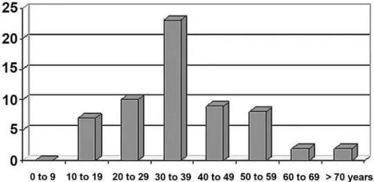
Eighty patients with urogenital tuberculosis, seen during the 1989-2005 period were retrospectively reviewed. These patients were treated in our tertiary teaching hospital that provides medical assistance free of charge to the metropolitan Sao Paulo area. Most of them have poor socio-economic conditions. There were 56 (70 %) males and 24 (30%) females with a median age of 34 years (12 to 75). Figure-1 shows the distributions according to the decades. Uro-genital tuberculosis was diagnosed by direct bacilli
LGHQWL¿FDWLRQRUFXOWXUHJURZWKLQWKHXULQHRI
%) patients; histopathology in 25 (31.3 %) patients; and a combination of strong clinical, laboratory, and
radiographic evidence of urogenital tuberculosis with negative bacilli search in the urine of 19 (23.7%) patients.
The clinical features and the organs involved
ZHUHGHVFULEHG7KHSDWLHQWVZHUHFODVVL¿HGLQVHYHQ
groups according to their patterns of initial clinical and radiographic presentation: 1) Bilateral parenchy-matous renal lesions; 2) No or minimal changes on radiographic examination; 3) Unilateral renal tuber-culosis; 4) Contracted bladder; 5) Contracted bladder with renal failure; 6) Tuberculosis on a transplanted kidney; 7) Isolated genital tuberculosis.
5(68/76
Table-1 shows details about the signs and symptoms. Storage symptoms and hematuria were the most frequent symptoms, being present in 72.5% and 56.3% of the patients, respectively. Sixteen patients
KDGVRPHIRUPRILPPXQRGH¿FLHQF\IRXUEHFDXVH
of AIDS, four with a kidney transplant and ongoing immunosuppressive therapy, four with diabetes, and four with alcohol abuse. In 35 (43.8%) patients, there was clinical or radiographic evidence of previous tuberculosis. Table-2 shows the distribution of organ involvement. After tuberculosis diagnosis, all the pa-tients received triple therapy for at least six months.
Initial clinical and radiographic assessment yielded the following patterns of urogenital tubercu-losis presentation:
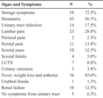
1) Bilateral parenchymatous renal lesions - In seven (8.8%) cases there were multiple bilateral parenchymatous renal lesions (Figure-2). In all cases, there were tuberculosis foci in other organs, fever, and malaise, characteristic of miliary tuberculosis. Three patients had AIDS. Case-fatality was 42.9% (three cases). The latter cases had the urogenital lesions diagnosed at postmortem examination. The other cases were diagnosed on imaging procedures. Three patients had no urologic symptom.
2) No or minimal changes on radiographic examination - In six (7.5%) cases no lesions were found (four patients) or there were minimal lesions (two patients) on radiographic examination with
XQLODWHUDO UHQDO FDOFL¿FDWLRQ RU FDO\[ GHIRUPLWLHV
(Figure-3). Tuberculosis was diagnosed when bacilli were shown in the urine. Four patients had hematuria and the other two had no urologic symptoms but the investigation was undertaken because of previous history of pulmonary tuberculosis. These patients did not require surgery and resolved well with pharma-cological treatment alone.
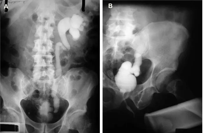
3) Unilateral renal tuberculosis - In 12 (15%) cases there was unilateral renal tuberculosis with ob-struction and dilatation of the collecting system due to stenosis (Figure-4). In one case, there was single inferior pole function loss due to infundibular stenosis, which was treated with inferior polar nephrectomy. All the other cases underwent nephrectomy due to a non-functional kidney. In all cases, the contralateral kidney was normal on radiographic examination.
4) Contracted bladder - In 37 (46.3%) pa-tients, there was contracted bladder due to tuberculo-sis. All the cases had unilateral non-function kidney with dilatation of the collecting system due to stenosis, associated with a contracted bladder, except for one case with polar renal function loss. Voiding
cystoure-WKURJUDPVKRZHGQRYHVLFRXUHWHUDOUHÀX[LQFDVHV ELODWHUDOUHÀX[LQWZRFDVHVDQGXQLODWHUDOUHÀX[WR
the contralateral functional kidney in 25. The latter
ZDVWKHPRVWIUHTXHQWUDGLRJUDSKLF¿QGLQJ)LJXUH
Table 1 – Presenting signs and symptoms in 80 patients with urogenital tuberculosis.
6LJQVDQG6\PSWRPV N %
Storage symptoms 58 72.5%
Hematuria 45 56.3%
Urinary tract infection 14 17.5%
Lumbar pain 23 28.8%
Perineal pain 2 2.5%
Scrotal pain 11 13.8%
Scrotal mass 10 12.5%
6FURWDO¿VWXOD 4 5.0%
LUTS 7 8.8%
Urinary retention 3 3.8%
Fever, weight loss and asthenia 36 45.0%
8UHWKUDO¿VWXOD 1 1.3%
Renal failure 10 12.5%
No symptoms from urinary tract 5 6.3%
LUTS = lower urinary tract symptoms.
Table 2 – Description of the affected organs.
$IIHFWHG2UJDQ N %
Kidney
Bilateral and multiple lesions Minimal lesions
Unilateral with polar loss of function Unilateral with loss of function Renal failure Transplanted kidney 72 7 2 2 47 10 4 90.0% 8.8% 2.5% 2.5% 58.8% 12.5% 5.0%
Ureter 57 71.3%
Bladder 47 58.8%
Prostate
Prostate abscess
Prostatitis with perineal pain No symptoms 6 2 2 2 7.5% 2.5% 2.5% 2.5% Epididymis Unilateral Bilateral &XWDQHRXV¿VWXOD Epididymectomy 10 8 2 4 5 12.5% 10.0% 2.5% 5.0% 6.3%
and 6). On radiographic examination, the functionally preserved contralateral kidney was normal in 23 cases and with ureterohydronephrosis in 14, one of which showing areas of cortical retraction. In all these cases
ZLWKXUHWHURK\GURQHSKURVLVYHVLFRXUHWHUDOUHÀX[WR
the functionally preserved kidney was observed. The patients underwent bladder augmentation along with total nephrectomy of the non-function kidneys and partial nephrectomy in the case with polar disease.
,QWKHFDVHVZLWKKLJKGHJUHHUHÀX[WKHXUHWHUVZHUH UHLPSODQWHG7KHSDWLHQWVGLGZHOOEXW¿YHSURJUHVVHG
to chronic renal failure.
5) Contracted bladder and renal failure - In 10 (12.5%) cases the patients had end-stage renal disease due to tuberculosis. All had contracted bladder and
XQLODWHUDOYHVLFRXUHWHUDOUHÀX[7KHSDWLHQWVXQGHU -went bilateral nephrectomy, bladder augmentation, and renal transplantation.
6) Tuberculosis on a transplanted kidney - In four (5%) cases parenchymatous renal tuberculosis was diagnosed on a transplanted kidney. The patients had creatinine elevation and tuberculosis was
diag-QRVHGWKURXJKELRSV\FDVHVRUD¿QGLQJRIEDFLOOLLQ
the urine. There was graft loss in 50% of the cases. 7) Isolated genital tuberculosis - In four (5%) cases there was genital tuberculosis without detectable
Figure 2 – Computed tomography showing multiple and bilateral kidney lesions in a patient with miliary tuberculosis.
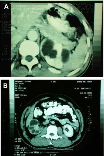
Figure 3 – Computed tomography showing calyceal dilatation in the left kidney.
Figure 4 – A) and B) Computed tomography showing unilateral renal tuberculosis with low function kidney with dilatation of the excretory system in two different patients.
A
renal lesion. Two patients presented with prostate ab-scess and two with tuberculosis of the epididymis.
Tuberculosis of the epididymis also oc-curred in another eight cases with associated renal lesion, with a total of 10 (12.5%) patients with pain and a mass over the epididymis, bilateral in two,
DQG ZLWK FXWDQHRXV ¿VWXOL]DWLRQ RI DQ HSLGLG\PLV
abscess in four. Because of extensive involvement, orchi-epididymectomy was necessary in 50% of the cases. Besides the two patients with prostate abscess, another four also had prostate tuberculosis. While two had storage symptoms and perineal pain, compatible with chronic prostatitis, two were asymptomatic and were serendipitously diagnosed by histopathology. Tuberculosis of the seminal vesicles occurred in two cases, one of which in the patient with prostate abscess
DQGDQRWKHUDVDKLVWRSDWKRORJLFDO¿QGLQJDIWHUUDGLFDO
prostatectomy.
Of the 76 patients without tuberculosis on the transplanted kidney, 15 (19.7%) developed end stage renal disease due to tuberculosis, 10 already at clinical
SUHVHQWDWLRQDQG¿YHRQIROORZXSGHVSLWHVSHFL¿F
treatment. All 57 patients with tuberculosis-related ureteral stenosis lost the corresponding kidney. Fifty-nine patients required nephrectomy and two needed polar nephrectomy.
&200(176
Of the urogenital tuberculosis cases described in the literature (range 7-39), the male-female distribu-tion was 2-1, with a mean age of 40.7 years (range 5 to 88). In only 36.6% of the cases was there history or radiographic evidence of previous tuberculosis. In cases where the renal lesions are mainly asymptom-atic, and only vesical lesions lead to symptoms (7-14), storage symptoms predominate. A total of 48.5% of males had scrotal involvement with an epididymis
PDVV HSLGLG\PLV KDUGQHVV RU ¿VWXOD RQ SK\VLFDO H[DPLQDWLRQ¿QGLQJVWKDWSRLQWWRWKHLPSRUWDQFH
of these signs. Patients in developed countries have
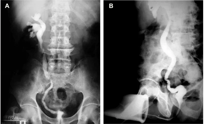
Figure 5 – A) Intravenous urography with left non-function kidney, B) Voiding cystography shows contracted bladder and right vesi-FRXUHWHUDOUHÀX[
IHZHUVSHFL¿FV\PSWRPVDQGVPDOOHUSHUFHQWDJHVRI
delayed histopathological diagnoses compared with other countries. As a result, the disease tends to be less serious, with fewer instances of renal failure, unilateral non-function kidney, ablative surgeries, and contracted bladder, with more cases
present-LQJZLWKRXWVLJQL¿FDQWOHVLRQVRIWKHXSSHUXULQDU\
tract on diagnosis. These data point to a correlation of the timing of the diagnosis with the severity of
XURJHQLWDOWXEHUFXORVLV2XU¿QGLQJVFRQWUDVWZLWK
those from developed countries, with greater rates of histopathological diagnoses, symptomatic patients at presentation, and severe destruction of the urinary tract (57.6% with contracted bladder, 19.7% with end stage renal disease, 12.5% with renal failure at initial presentation, and 71.2% with unilateral renal exclu-sion in the absence of renal failure or a transplanted kidney). Table-3 shows these features.
Since histopathology provides a delayed
XURJHQLWDO WXEHUFXORVLV GLDJQRVLV LGHQWL¿FDWLRQ RI
the tuberculosis bacillus in the urine is necessary for early diagnosis. It is achieved through direct smears (Ziehl-Neelsen stain) or through urine culture (Low-estein-Jensen media) (40,41). Direct smears provide
DIDVWHUUHVXOWZLWKKLJKVSHFL¿FLW\EXWRQO\
42.1 to 52.1% sensitivity (40,41). Culture is the di-agnostic gold standard for urogenital tuberculosis, however 3 to 6 early morning mid-stream samples are required, the sensitivity varies widely, from 10% to 90%, and the time to detection of mycobacterium growth may be 4 to 6 weeks (40-42). Faster culture has been achieved with non-radiometric automated or semi-automated liquid culture systems with a 14 to 17
GD\VUHVXOW1XFOHLFDFLGDPSOL¿FDWLRQWHVWVDV
polymerase chain reaction (PCR), for Mycobacterium
WXEHUFXORVLVLGHQWL¿FDWLRQLQWKHXULQHPD\EHFRPH Figure 6 – A) Intravenous urography with right non-function kidney, B) Voiding cystography shows contracted bladder and left vesi-FRXUHWHUDOUHÀX[
the ideal diagnostic tool, as it gives results in 24 to 48 hours and allows for the diagnosis to be made even when there are few bacilli (40,41). It has been
VKRZHGVHQVLWLYHDQGVSHFL¿FFRPSDUHG WRFXOWXUHDQGVHQVLWLYHDQGVSHFL¿F
compared to bacteriological, histological, or clinico-radiological diagnoses (41).
Primary pulmonary tuberculosis is usually subclinical and self-limited. From the pulmonary fo-cus there is bacillemia and bacilli implants in other or-gans, renal parenchymatous and prostate colonization
ensuing. After six months, spontaneous cicatrisation of the primary pulmonary tuberculosis lesion occurs and the patients enter a latent phase, with 5% reacti-vating the disease in the following two years and 5% in their lifetime. In most cases of active pulmonary or extrapulmonary disease, there is foci reactivation due to a breach of immunity brought about by malnutri-tion, diabetes mellitus, steroid use, immunosuppressor
XVHDQGLPPXQRGH¿FLHQFLHV
The present urogenital tuberculosis patients’
FODVVL¿FDWLRQLQVHYHQJURXSVZDVEDVHGRQGLVHDVH
Table 3 –Comparison between 3036 cases of urogenital tuberculosis from developed countries (USA, Europe and Japan) with 5925 cases from other countries (Russia, Latin America and Africa) and 80 cases from Brazil.
'HYHORSHG Others p Value Brazil Total
Total Men Women 3036 62.9% 37.1% 5925 65.4% 34.6% 0.02 0.02 80 70% 30% 9041 65% 35%
Median age (years) Range (years)
42.6 7 to 88
39.2 5 to 83
34 12 to 75
40.7 5 to 88
Previous tuberculosis 37.9% 38.4% 0.66 43.8% 36.6%
Signs and symptoms Storage symptoms Dysuria
Lumbar pain Hematuria
Epididymis lesion * Fever and malaise No symptoms 44.2% 33.8% 28.8% 24.5% 20.6% 23.2% 8.4% 55.2% 46.4% 42.3% 44.3% 47.4% 19.9% 0% < 0.01 < 0.01 < 0.01 < 0.01 < 0.01 0.28 < 0.01 72.5% 72.5% 28.8% 56.3% 12.5% 45.0% 6.3% 50.7% 38.2% 34.4% 35.8% 48.5% 22.1% 6.4%
Renal failure 1.4% 10.2% < 0.01 19.7% ** 5.8%
Diagnosis Urine Histopathology Clinico-radiographic 79.0% 7.8% 9.6% 55.4% 38.3% 11.3% < 0.01 < 0.01 0.36 45.0% 31.3% 23.7% 64.0% 21.8% 10.5% Kidney Unilateral non-function Normal Contracted bladder 22.8% 18.8% 4.0% 33.1% 13.2% 11.6% < 0.01 < 0.01 < 0.01 58.8% 10.0% 58.8% 27.2% 15.2% 9.3% Surgeries Ablative Nephrectomy 56.6% 35.0% 27.9% 53.6% 26.9% 26.0% < 0.01 < 0.01 0.37 76.3% 76.3% 71.3% 55.1% 27.6% 28.0%
physiopathology and made evident the correlation between symptoms and disease’s stage. Renal le-sions are initially bilateral, cortical, glomerular, and peri-capillary, typical of the hematogenous spread and concomitant with other hematogenous foci in the prostate and other organs beyond the urogenital system (15,44). These foci usually heal, with the
pa-WLHQWHQWHULQJDODWHQWSKDVH,IDQ\LPPXQRGH¿FLHQF\
ensues although, miliary tuberculosis with systemic symptoms develops (3,13),from 25 to 62% of patients with miliary tuberculosis will have a renal lesion with multiple bilateral foci (9,44). In our patients 8.8% had miliary tuberculosis with bilateral parenchymatous renal lesions and systemic symptoms, characterizing
DW\SLFDOFOLQLFDOSUHVHQWDWLRQRILPPXQHGH¿FLHQW
patients. Tuberculosis on a transplanted kidney can be also included in this multiple parenchymatous lesion presentation.
The latent period between pulmonary infec-tion with bacillemia and clinically evident urogenital tuberculosis is 22 years on average, ranging from 1 to 46 years, according to the timing of latent foci (renal, prostate, or epididymis) reactivation (12). Isolated prostate or epididymis foci reactivation may occur, characterizing genital tuberculosis without associated renal lesion. In the reactivation of renal foci, infection progresses from a single unilateral focus, with preservation of the contralateral kidney (15). This explains the greater frequency of unilateral renal tuberculosis (12,16). The contiguous involve-ment of the collecting system leads to bacilluria and descending spread of the infection to the ureter, bladder, and genital organs (44,45). Thus, a typical clinico-radiographic form exists: unilateral renal tuberculosis with preservation of the contralateral kidney and the bladder. This occurred in 15% of our cases, with obstruction and dilatation of the collecting system, with invariable renal functional loss. In fact, obstruction of the collecting system (with distal ureteral stenosis as the most frequent
¿QGLQJLVWKHPDLQFDXVHRIUHQDOIXQFWLRQDOORVV
in tuberculosis (11,46,47). The focal origin of tu-berculosis reactivation is demonstrated by two of our cases with restriction of the disease to the renal pole. If the diagnosis of the urogenital tuberculosis reactivation is at an early stage, we can detect no or minimal radiographic features such as calyceal
dilatation due to initial infundibular stricture as we observed in group 2 patients (41).
Another clinico-radiographic presentation was a contracted bladder without renal failure, occur-ring in 45% of the patients. There was unilateral non-function kidney in practically all cases, with collecting system obstruction and dilatation, primarily (66.7%)
ZLWKXQLODWHUDOUHÀX[WRWKHIXQFWLRQNLGQH\ ZLWKRXWUHÀX[DQGRQO\ZLWKELODWHUDOUHÀX[
All patients with renal failure had bladder contraction
DQGXQLODWHUDOUHÀX[RQLQYHVWLJDWLRQ7KHVH¿QGLQJV
prompted a new hypothesis for the pathophysiology of the urinary tract lesions in urogenital tuberculosis. After reactivation and progression of the unilateral renal focus, the collecting system is implicated, with unilateral descending involvement of the ipsilateral ureter and bladder ensuing (4). Ureteral stenosis with
FRUUHVSRQGLQJUHQDOIXQFWLRQORVVDQG¿EURVLVRIWKH
bladder wall are the next stages (47). At this stage, the investigations show unilateral non-function
kid-QH\ZLWKFRQWUDFWHGEODGGHUDQGDEVHQFHRIUHÀX[
Progression of the bladder infection with capacity and compliance reduction leads to distortion of the
vesico-XUHWHUDOMXQFWLRQDQGYHVLFRXUHWHUDOUHÀX[WRWKHIXQF -tional kidney, the collecting system thus functioning as a buffer for the reduced capacity of the contracted bladder, and with ascending transmission of the high intravesical pressure. Currently, the investigations show unilateral non-function kidney, contracted
blad-GHUDQGUHÀX[WRWKHIXQFWLRQNLGQH\DVVHHQLQPRVW RIRXUSDWLHQWV)LJXUHDQG1RQLGHQWL¿HGDQG QRQWUHDWHGUHÀX[FDXVHVUHQDOOHVLRQGXHWRLQIHFWLRQ
or transmission of intravesical pressure, leading to end stage renal disease. In fact, renal failure patients
SUHVHQWHGXQLODWHUDOUHÀX[XQGHUO\LQJWKHK\SRWKHVLV
that one of the kidneys loses its function because of
UHÀX[ DQG QRW GXH WR WXEHUFXORVLV LWVHOI )XUWKHU -more, patients with unilateral non-function kidney had ureterohydronephrosis and areas of retraction in the functional kidneys, as well as changes associated
ZLWKYHVLFRXUHWHUDOUHÀX[7KHIDFWWKDWWKHUHZHUHQR
typical tuberculosis lesions in these kidneys, such as ureteral or intra-renal stenosis, underlies the role of
UHÀX[QHSKURSDWK\DQGQRWWXEHUFXORVLVLWVHOILQWKH
pathogenesis of the contralateral involvement.
$IWHUWKHGLVFRYHU\RQVSHFL¿FWXEHUFXORVWDW
tuberculosis experienced dramatic changes, with reduced mortality, cure of initial lesions, reduction in the number of ablative surgeries, and increase in the number of reconstructive surgeries. In the last
GHFDGHVKRZHYHUQRVLJQL¿FDQWFKDQJHRFFXUUHGLQ
spite of technological advances (48). Since the 60`s the importance of an early diagnosis of urogenital tuberculosis has been acknowledged as the main step towards renal preservation (47). A systematic search for urogenital tuberculosis in patients with pulmonary disease yielded 10% of positive cultures (66.7% asymptomatic and 58% with normal urine exam and absence of renal lesions on pyelography) (49). In the other two series, where cultures were rou-tinely grown, a greater frequency of asymptomatic patients without lesions on intravenous pyelography was found (8,50). Although bacilluria is invariably associated with renal lesion, detection of pre-clinical bacilluria allows for an early diagnosis to be made, at a moment when the lesions are amenable to cure and the development of severe and destructive lesions can be avoided (45). Of our patients with minimum or absent lesions, two had no urologic symptoms and the others had isolated hematuria. Thus, a system-atic search for urogenital tuberculosis, regardless of symptoms, is warranted for early case detection. We propose that all patients with macroscopic hematuria or persisting microscopic hematuria or leucocituria should be submitted to Mycobacterium tuberculosis urinary culture or PCR analyses. We also propose yearly urinalysis for microscopic hematuria or leucocituria detection in patients with pulmonary tuberculosis history or immunological impairment.
+RZHYHUWKHEHWWHUGH¿QLWLRQRIKLJKHUULVNJURXSV
and optimal diagnosis strategy warrants further studies.
&21)/,&72),17(5(67
None declared.
5()(5(1&(6
1. Brasil - Ministério da Saúde, Guia de Vigilância Epidemiológica, Brasília. 1998; Cap. 5.34: pp.1-13. Tuberculose.
2. Dias Filho AC, Netto Júnior NR: Aspectos atuais da tuberculose urinária e genital masculina. Jornal Brasileiro de Urologia. 1999; 25: 453-65.
3. Leite OH: Tuberculosis. Problems in General Surgery. 2001; 18: 69-78.
4. Gow JG: Genitourinary tuberculosis. In: Walsh PC, Retik AB, Vaughan ED, Wein AJ. Campbell´s Urology. (7ª ed.), Philadelphia, WB Saunders Company. 1998; Vol. 1: pp 807-36.
5. Hijjar MA, Oliveira MJPR, Teixeira GM: A tubercu-lose no Brasil e no mundo. Boletim de Pneumologia Sanitária. 2001; 9: 9-16.
6. Schubert GE, Haltaufderheide T, Golz R: Frequency of urogenital tuberculosis in an unselected autopsy series from 1928 to 1949 and 1976 to 1989. Eur Urol. 1992; 21: 216-23.
7. Flechner SM, Gow JG: Role of nephrectomy in the treatment of non-functioning or very poorly function-ing unilateral tuberculous kidney. J Urol. 1980; 123: 822-5.
8. Ferrie BG, Rundle JS: Genito-urinary tuberculosis in Glasgow 1970 to 1979: a review of 230 patients. Scott Med J. 1985; 30: 30-4.
9. Simon HB, Weinstein AJ, Pasternak MS, Swartz MN, Kunz LJ: Genitourinary tuberculosis. Clinical features in a general hospital population. Am J Med. 1977; 63: 410-20.
10. Nzerue C, Drayton J, Oster R, Hewan-Lowe K: Geni-tourinary tuberculosis in patients with HIV infection: clinical features in an inner-city hospital population. Am J Med Sci. 2000; 320: 299-303.
11. Ramanathan R, Kumar A, Kapoor R, Bhandari M: Relief of urinary tract obstruction in tuberculosis to improve renal function. Analysis of predictive factors. Br J Urol. 1998; 81: 199-205.
12. Christensen WI: Genitourinary tuberculosis: review of 102 cases. Medicine (Baltimore). 1974; 53: 377-90. 13. Alvarez S, McCabe WR: Extrapulmonary tuberculosis
revisited: a review of experience at Boston City and other hospitals. Medicine (Baltimore). 1984; 63: 25-55.
14. Gokalp A, Gultekin EY, Ozdamar S: Genito-urinary tuberculosis: a review of 83 cases. Br J Clin Pract. 1990; 44: 599-600.
15. Narayana A: Overview of renal tuberculosis. Urology. 1982; 19: 231-7.
17. Mochalova TP, Starikov IY: Reconstructive surgery for treatment of urogenital tuberculosis: 30 years of observation. World J Surg. 1997; 21: 511-5.
18. Kao SC, Fang JT, Tsai CJ, Chen KS, Huang CC: Urinary tract tuberculosis: a 10-year experience. Changgeng Yi Xue Za Zhi. 1996; 19: 1-9.
19. Caro Muñoz M, Corvalán JR, Tenório JP, Vergara JF: Diagnosis of urogenital tuberculosis: a retrospective review (1974 - 1986). Bol Hosp San Juan de Díos. 1988; 35(4): 223-6.
20. Venegas MP, Corti OD, Foneron BA: Genitourinary TBC: experience of Hospital de Valdiva 1979-1984 years. Rev Chil Urol. 1990; 53: 131-4.
21. Núñez Vásquez N, Susaeta S R, Vogel M, Olate C: Genitourinay tuberculosis study: urologic service, Hospital San Juan de Dios 1975-1989. Rev Chil Urol. 1990; 53: 135-7.
22. Allen FJ, de Kock ML: Genito-urinary tuberculosis--experience with 52 urology inpatients. S Afr Med J. 1993; 83: 903-7.
%HQQDQL6+D¿DQL0'HEEDJK$HO0ULQL0%HQMHO
-loun S: Urogenital tuberculosis. Diagnostic aspects. J Urol (Paris). 1995; 101: 187-90.
24. Hinostroza Fuschlocher JÁ, Gorena Palominos M, Pastor Arroyo P, Rivera P, Venegas P, Orío M, Peldoza M, et al.: Genitourinary tuberculosis at Araucanía: experience at Hospital Regional de Temuco. Rev Chil Urol. 1998; 63: 113-7.
25. Mnif A, Loussaief H, Ben Hassine L, Chebil M, Ayed M. Aspects of evolving urogenital tuberculosis. 60 cases. Ann Urol (Paris). 1998; 32: 283-9.
26. Lopez de Mesa B, Cruz A, Gomes P: Review of pa-tients with genitourinary tuberculosis in San Juan de Dios Hospital. Urol Colomb. 1998; 7: 43-52.
27. Benchekroun A, Lachkar A, Soumana A, Farih MH, Belahnech Z, Marzouk M, et al.: Urogenital tubercu-losis. 80 cases. Ann Urol (Paris). 1998; 32: 89-94. 28. Chtourou M, Kbaier I, Attyaoui F, Nouira Y, Barrak
A, Sayari S, et al.: Management of genito-urinary tuberculosis. A report of 225 cases. J Urol. 1999; 161 (Suppl 4): Abstract 34.
29. Buchholz NP, Salahuddin S, Haque R: Genitourinary
WXEHUFXORVLVDSUR¿OHRILQSDWLHQWV-3DN0HG
Assoc. 2000; 50: 265-9.
30. Hemal AK, Gupta NP, Kumar R: Comparison of retroperitoneoscopic nephrectomy with open surgery for tuberculous nonfunctioning kidneys. J Urol. 2000; 164: 32-5.
HO.KDGHU./UKRU¿0+HO)DVVL-7D]L.+DFKLPL
M, Lakrissa A. Urogenital tuberculosis. Experience in 10 years. Prog Urol. 2001; 11: 62-7.
32. Gokce G, Kilicarslan H, Ayan S, Tas F, Akar R, Kaya K, et al.: Genitourinary tuberculosis: a review of 174 cases. Scand J Infect Dis. 2002; 34: 338-40.
33. Barrios F, Martinez-Peschard L, Rosas A: Surgical management of tuberculosis. BJU Int. 2002; 90 (Suppl 2): p 26.
34. Ishibashi Y, Takeda T, Nishimura R, Ohshima H: A clinical observation of genitourinary tract tuberculosis during the last decade. Hinyokika Kiyo. 1985; 31: 107-12.
35. Gow JG: Genitourinary tuberculosis: a 7-year review. Br J Urol. 1979; 51: 239-44.
36. Gow JG, Barbosa S: Genitourinary tuberculosis. A study of 1117 cases over a period of 34 years. Br J Urol. 1984; 56: 449-55.
37. Fuse H, Imazu A, Shimazaki J. A clinical observation on urogenital tuberculosis. Hinyokika Kiyo. 1984; 30: 299-304.
38. Fischer M, Flamm J: The value of surgical therapy in the treatment of urogenital tuberculosis. Urologe A. 1990; 29: 261-4.
39. Zwergel U, Wullich B, Rohde V, Zwergel T: Surgical management of urinary tuberculosis: a review of 341 patients. J Urol. 1999; 161 (Suppl 4): Abstract 33. 40. Moussa OM, Eraky I, El-Far MA, Osman HG,
Gho-neim MA: Rapid diagnosis of genitourinary tuberculo-sis by polymerase chain reaction and non-radioactive DNA hybridization. J Urol. 2000; 164: 584-8. 41. Hemal AK, Gupta NP, Rajeev TP, Kumar R, Dar L,
Seth P: Polymerase chain reaction in clinically sus-pected genitourinary tuberculosis: comparison with intravenous urography, bladder biopsy, and urine acid fast bacilli culture. Urology. 2000; 56: 570-4.
42. Dinnes J, Deeks J, Kunst H, Gibson A, Cummins E, Waugh N, et al.: A systematic review of rapid diagnos-tic tests for detection of tuberculosis infection. Health Technology Assessment. 2007; 11: 1-196.
43. Cos LR, Cockett AT: Genitourinary tuberculosis re-visited. Urology. 1982; 20: 111-7.
44. Medlar EM, Spain DM, Holliday RW: Post-mortem compared with clinical diagnosis of genito-urinary tu-berculosis in adult males. J Urol. 1949; 61: 1078-88. 45. Medlar EM, Sasano KT: Experimental renal
tubercu-losis, with special reference to excretory bacilluria. Am Rev Tuberc. 1924; 10: 370-7.
46. Barrie HJ, Kerr WK, Gale GL: The incidence and pathogenesis of tuberculous strictures of the renal pyelus. J Urol. 1967; 98: 584-9.
48. Ross JC: Renal tuberculosis. Br J Urol. 1953; 25: 277-315.
49. Bentz RR, Dimcheff DG, Nemiroff MJ, Tsang A, Weg JG: The incidence of urine cultures positive for Mycobacterium tuberculosis in a general tuberculosis patient population. Am Rev Respir Dis. 1975; 111: 647-50.
50. Butler MR, O’Flynn JD: Reactivation of genito-urinary tuberculosis: a retrospective review of 838 cases. Eur Urol. 1975; 1: 14-7.
Accepted after revision: March 17, 2008
Correspondence address: Dr. André Avarese de Figueiredo Rua Irineu Marinho 365 / 801, Bloco 3 Juiz de Fora, MG, 36021-580, Brazil Fax: + 55 11 3287-6922
E-mail: [email protected]
(',725,$/&200(176
On May 4, 1953, The New York Times pub-lished a report from The World Health Organization that stated ‘there has been an “extraordinary” drop in mortality from tuberculosis of the respiratory system since the end of World War II. The development of anti-tuberculosis medication, improved living stan-dards and Public Health measures made tuberculosis an almost forgotten disease particularly in developed countries. Unfortunately, this has not been the case. As noted in the paper tuberculosis continues to be a major world wide public health problem. Poor
socio-economic conditions, immune suppression, AIDS are factors that perpetuate tuberculosis. The authors noted that 2 to 20% of patients with pulmonary can manifest
XURJHQLWDOWXEHUFXORVLV7KHLUSDSHUµFODVVL¿HG¶*8
tuberculosis into 7 categories including one category with ‘No or minimal’ disease.
This paper renews a known caveat for the Urologist. ‘Atypical’ urologic manifestations such as renal scaring, scrotal masses, idiopathic pyuria or hematuria mandate an assessment for tuberculosis.
Dr. G. J. Wise