Detection of human adenovirus, rotavirus and enterovirus in water samples collected
on dairy farms from Tenente Portela, Northwest of Rio Grande do Sul, Brazil
Fernando Rosado Spilki
1, Roger Bordin da Luz
1, Rafael Bandeira Fabres
1,
Mayra Cristina Soliman
1, Mariana Kluge
1, Juliane Deise Fleck
1,
Manoela Tressoldi Rodrigues
1, Juliana Comerlato
1, Alexander Cenci
2, Cristine Cerva
2,
Maurício Gautério Dasso
2, Paulo Michel Roehe
2,31Laboratório de Microbiologia Molecular, Instituto de Ciências da Saúde, Universidade Feevale,
Novo Hamburgo, RS, Brazil.
2Instituto de Pesquisas Veterinárias Desidério Finamor / Fepagro Saúde Animal,
Eldorado do Sul, RS, Brazil.
3Laboratório de Virologia, Departamento de Microbiologia, Instituto de Ciências Básicas da Saúde,
Universidade Federal do Rio Grande do Sul, Porto Alegre, RS, Brazil.
Submitted: April 13, 2012; Approved: April 01, 2013.
Abstract
Viral gastroenteritis and other waterborne diseases are a major concern for health in Brazil. A number of studies were conducted about the presence of viruses on water samples from Brazilian areas. How-ever, the knowledge about the occurrence of viral contamination of drinking water sources in rural settings of the country is insufficient. On the present work, 15 samples from 5 dairy farms located at the municipality of Tenente Portela were collected and analysed for the presence of human adenovi-ruses (HAdV), as well as human enteroviadenovi-ruses (EV) and rotaviadenovi-ruses (RV). HAdV was present on 66.66% of the water samples, and have been found in all samples from artesian wells and springs, which are used as sources of drinking water for the individuals inhabiting those farms. EV and RV found only in one sample each. The detection rates of HAdV on the water from these dairy farms are alarming and point towards a situation of elevated environmental contamination by fecal microor-ganisms of human origin and poor basic sanitation conditions.
Key words:human adenovirus; water quality; dairy farms.
Introduction
Access to safe water in rural areas in Brazil is scarce; it is easily observed in the farms used for dairy milking in southern Brazil, where the production is usually conducted on small properties. Low income, poor access to technical information and improper disposal of animal waste, as well as the lack of sanitation facilities for the farmers and fami-lies, lead to a common frame of degradation of environ-mental quality in these locations (Amaralet al., 2003; de Medeiros and de Souza, 2009).
Studies involving the analysis of microbial contami-nation and chemical pollution of water in dairy farms have been conducted in different parts of the world and some
studies were made on South America (Amaralet al., 2003; Bettera et al., 2011; Derbyshire and Brown, 1978;
Schwarteet al., 2011; Weatherleyet al., 2011). In most of these studies, it is noticeable the contamination of surface and groundwater by bacteria and protozoa, but there are few studies that address the detection of enteric viruses (Ahmedet al., 2010; Schwarteet al., 2011; Verheyenet al., 2009). Enteric viruses have a number of characteristics that make them excellent markers for fecal contamination of water: i) they are extremely resistant in the environment due to its non-enveloped structure, ii) they are eliminated in large quantities in the feces of humans and animals sick or subclinical infections in iii) in most cases these viruses are
Send correspondence to F.R. Spilki. Laboratório de Microbiologia Molecular, Instituto de Ciências da Saúde, Universidade Feevale, 93352-000 Novo Hamburgo, RS, Brazil. E-mail: [email protected].
host-specific and thus allow screening of the species which is the source of fecal contamination (Fong and Lipp, 2005; Silva et al., 2011; Wolf et al., 2010; Wu et al., 2011). Among the enteric viruses three of the most studied as envi-ronmental contaminants are the adenoviruses (AdV,
Adenoviridae family, Mastadenovirus genus, double-stranded DNA), enteroviruses (EV,Picornaviralesorder,
Picornaviridaefamily,Enterovirusgenus, single-stranded RNA, positive sense) and rotaviruses (RV,Reoviridae fam-ily, Sedoreovirinae subfamily, genus Rotavirus) (Comerlato et al., 2011; Fong and Lipp, 2005; Mat-thijnssenset al., 2008; Sibleyet al., 2011). These agents are transmitted by the fecal-oral route, being associated with a number of diseases, especially gastroenteritis, either in hu-man beings or animals (Ahmadet al., 2009; Hamzaet al., 2011). In recent years, the detection of these viruses in sur-face waters, sewage and coastal waters using the previous concentration of viral particles by different methods and molecular methods for the identification of viral genomes has allowed the conclusion that there is a wide contamina-tion of water by viruses in various ecosystems (Wuet al., 2011). In rural areas, these viruses have been found con-taminating ground and surface waters and their presence may represent a risk not only the health of humans and do-mestic animals, but can also have adverse effects on the health of wildlife (Ahmedet al., 2010; Jiménez-Claveroet al., 2005; Leyet al., 2002).
In this study, water samples were collected from dif-ferent points on farms devoted to milk production in the municipality of Tenente Portela, in southern Brazil, which is inserted in a wide geographic region devoted mainly to agriculture and livestock, especially dairy. These properties have the typical characteristics of small farms attached to the chain of milk production in southern Brazil, described before. These water samples were tested by the polymerase chain reaction for the presence of human adenovirus (HadV) as an effort to determine whether the human beings are a source o fecal pollution to the water on these farms. The samples were tested also for EV and RV genomes. For HAdV and EV the primers used were capable of detecting viruses from human beings, whereas the primers for RV are pan-reactive to the group A of RV from different species. This is the first study on the contamination of water by en-teric viruses at the Northwest of the state of Rio Grande do Sul.
Materials and Methods
Sampling sites and samples
Tenente Portela is a municipality in the northwest re-gion of Rio Grande do Sul (27°22’16” S and 53°45’30” W), the southernmost state of Brazil. The estimated population of 13,719 inhabitants is decreasing through the years and the primary sector is responsible for a third of the income. From the total area of 390 km2, 19,968 ha are divided by
1,352 farms, from these 1,105 are used for dairy produc-tion. The collections were made on different water sources from 5 (five) farms on August 2009, under dry weather con-ditions. Water samples (500 mL each) were collected asep-tically from each farm. The 15 (fifteen) samples obtained were transported to the laboratory under refrigeration, and were kept at 4 °C until sample concentration.
Sample concentration
Water samples were concentrated using an adsorp-tion-elution method previously described (Katayamaet al., 2002) with minor modifications (Vecchia et al., 2012). Briefly, 0.6 g of MgCl2.6H2O were mixed with 500 mL of
water sample and pH was adjusted to 5.0 using a solution of 10% HCl. After, the resulting mixture was vacuum filtered through negatively sterile membrane (type HA, 0.45mm
pore size; 47 mm diameter). The membrane was rinsed through the washing with 87.5 mL of a 0.5 mM H2SO4
(pH 3.0) followed by elution of viral particles adsorbed to the membrane with 2.5 mL of 1 mM NaOH (pH 10.5). The pH of the filtrate was neutralized with 12.5 mL of
50 mM H2SO4 and 12.5 mL in 100X Tris-EDTA (TE)
buffer. The eluate was aliquoted and stored at -80 °C until further processing.
Viral nucleic acid extraction
The commercial kit RTP DNA / RNA Virus Mini Kit (Invitek, Germany) was used for extraction of viral nucleic acids, according to the manufacturer’s instructions, using an initial volume of 400 mL of each concentrated water
sample. The viral DNA or RNA obtained was stored at -80 °C for later processing.
Polymerase chain reaction (PCR)
In order to achieve amplification EV and RV geno-mes, a previous step of cDNA synthesis was carried out be-fore amplification. It was performed using the High Capac-ity cDNA Reverse Transcription commercial kit (Applied Biosciences, USA), using a set of random primers and RNAse Inhibitor (Applied Biosciences, USA), following manufacturer’s instructions.
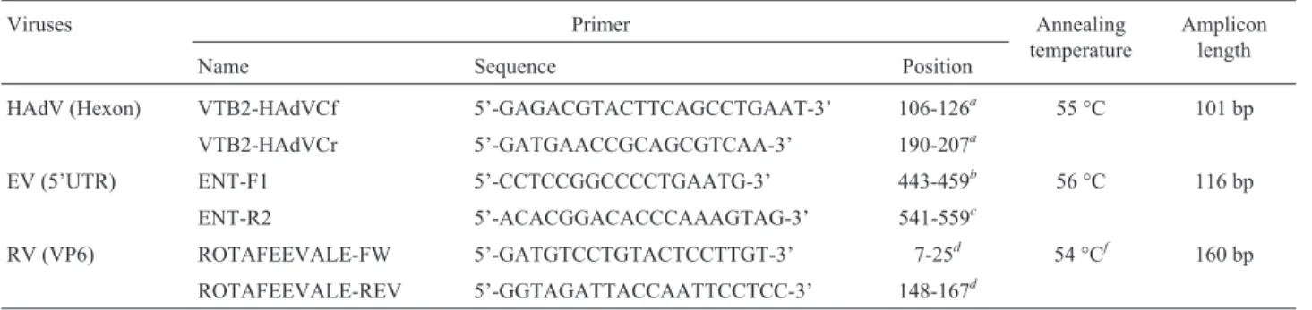
The sequences of the primers and their location in the viruses’ genomes are described on Table 1. PCR conditions were optimized and reactions were standardized as follow-ing: (a) AdV and RV: 50mL reaction mixtures consisting
25 mL of GoTaq® Green Master Mix (Promega, USA),
18mL of nuclease-free water, 1mL of each primer (20 pM)
and 5mL of nucleic acid; (b) EV: 25mL final volume
con-taining 12,5mL of 2x PCR Master Mix (LGCbio, Brazil),
7,5mL of nuclease-free water, 1mL of each primer (20 pM)
and 3mL of cDNA product; DNase/RNase free water was
kindly provided by Dr. Célia Barardi; Human-RV (isolate C-5, VP6 I-2 Genotype) was isolated from a clinical sample collected from a children with diarrhea (Vecchia et al., 2012).
Amplification of the target genomic fragments was performed using a thermal cycler (MultiGene®, Labnet In-ternational, USA). The PCR conditions were optimized for each virus group and were as follows: (a) AdV: 98 °C for 7 min, 40 cycles of 94 °C for 1 min, 55 °C for 1 min, 72 °C for 1 min; (b) EV: 98 °C for 5 min, 35 cycles of 94 °C for 1 min, 56 °C for 1 min, 72 °C for 1 min; (c) RV: 94 °C for 5 min, 40 cycles of 94 °C for 1 min, 54 °C for 1 min (which was decreased by 0.5 °C at each of the 39 subsequent cy-cles), 72 °C for 1 min. After, all reactions were left at 72 ° C for 7 min for final elongation and submitted to an infinite cycle at 4 °C.
To determine the analytical sensitivity of the assays, 10-fold serial dilutions of each EV, HAdV and RV positive controls grown on cell culture were experimentally inocu-lated onto sterile 500 mL water samples and after processed on the same manner described above for the samples. All the PCRs have analytical sensitivity enough to detect 1-10 tissue culture infective doses (TCID50) diluted on water.
These tests and results are described elsewhere (Vecchiaet al., 2012).
PCR products were stained with nontoxic fluorescent dye SYBR® SAFE DNA Gel Stain (Invitrogen, USA), ana-lyzed by electrophoresis on 2% (w/v) agarose gel and visu-alized under ultraviolet (UV) light.
Results
From the 15 samples, 10 showed HAdV genomes (66.66%), and only one sample showed contamination by EV and another by RV (Table 2). HAdV genomes were de-tected in at least one collection point from the 5 farms. Samples from only one farm resulted positive for EV and RV, and the stream contaminated by RV was also contami-nated by HAdV (farm #344). Among the eight surface
wa-ter samples collected from streams and ponds, only 3 pre-sented viral genomes, while for the six groundwater sam-ples, all were positive HAdV. A sample of tap water was analyzed and was contaminated by HAdV.
Discussion
In areas and facilities where dairy cows are milked staged between the various phases of the milking process, wastes are removed using large volumes of water. Without appropriate treatment, the sludge generated may allow the transportation of fecal microorganisms into ponds, creeks and groundwater (Pullaret al., 2011; Weatherley et al., 2011; Wilcocket al., 2011). In southern Brazil, dairy cows
Table 1- Primers and conditions used for PCR amplification of AdV, EV and RV genomes used on the present study.
Viruses Primer Annealing
temperature
Amplicon length
Name Sequence Position
HAdV (Hexon) VTB2-HAdVCf 5’-GAGACGTACTTCAGCCTGAAT-3’ 106-126a 55 °C 101 bp
VTB2-HAdVCr 5’-GATGAACCGCAGCGTCAA-3’ 190-207a
EV (5’UTR) ENT-F1 5’-CCTCCGGCCCCTGAATG-3’ 443-459b 56 °C 116 bp
ENT-R2 5’-ACACGGACACCCAAAGTAG-3’ 541-559c
RV (VP6) ROTAFEEVALE-FW 5’-GATGTCCTGTACTCCTTGT-3’ 7-25d 54 °Cf 160 bp
ROTAFEEVALE-REV 5’-GGTAGATTACCAATTCCTCC-3’ 148-167d aPrimers sequences reported by Wolfet al.(2010).
bPrimers sequences reported by Tsai
et al.(1993). cVecchia
et al.(2012), Genome position of primers based on GenBank accession number FJ859064. dVecchiaet al.(2012), Genome position of primers based on GenBank accession number HM34874.
eInitial annealing temperature, which was decreased by 0.5 °C at each of the 39 subsequent cycles (Touchdown-PCR).
Table 2- Detection of HAdV, EV and RV genomes, and coliforms quanti-fication, in water samples collected from springs, creeks, ponds and arte-sian wells on dairy farms at the municipality of Tenente Portela, Rio Grande do Sul, Brazil.
Farm Sample HAdV EV RV
#326 Artesian well #1 • ° °
Artesian well #2 • ° °
Spring • ° °
#329 Artesian well • ° °
Creek ° ° °
Pond ° ° °
#330 Spring • ° °
Tap (milking parlor) • ° °
Creek ° ° °
Pond ° ° °
#343 Spring • ° °
Creek • ° °
Pond • ° °
#344 Creek • ° •
Pond ° • °
are generally raised on a semi-intensive system, and the ex-creta deposited on pastures may be also a source of fecal pollution since contaminants may be transported into water bodies by superficial runoff (Ahmadet al., 2009; Ahmedet al., 2010). Another major problem of the farms located in this region is the poor access to treated water and absence of basic sanitation in most cases.
HAdV genomes were detected in all samples taken from wells and springs on the present study, thus indicating a high rate of contamination of the subsoil and conse-quently aquifers. This may be an effect of the poor con-struction of latrines and wells on these farms, which can permit the infiltration of the subsoil by microorganisms, and viruses may thus accumulate on the groundwater re-sources (Junget al., 2011; Pujariet al., 2012; Steyeret al., 2011; Wilcocket al., 2011). The concern is that water from artesian wells and springs is often thought to be free of con-taminants and the farmers and families living on these loca-tions have been using this as the solely source of drinking water.
The rates of detection of human HAdV on the present work are higher than those found on urban areas on the north of Brazil (Miagostovichet al., 2008), and very similar to the rates for the southeast (Piranhaet al., 2006; Santoset al., 2004) and south of Brazil (Morescoet al., 2012; Rigotto
et al., 2010). The detection rate is also very close to the found on another study conducted on pig farms, aiming the detection of porcine adenovirus (PoAdV) (Viancelliet al., 2012). Indeed, HAdV and other adenoviruses are often found as highly prevalent on environmental waters, but one may expect lower levels of detection when analyzing water from areas of low population density. Thus, it is concluded that the impact of poor sanitation conditions within these farms overpasses the small number of individuals on each local. Nevertheless, when comparing to other studies on ru-ral areas, the rates of adenoviru-ral contamination of water on the present study are very high. In a study conducted in Benin, only 12.9% of the sampling sites were positive for AdV genomes (Verheyenet al., 2009). On the other hand, the results for rotaviruses are very similar, in both studies the rates were very low for the molecular detection of RV (Verheyen et al., 2009). Other authors also found lower rates for the detection of HAdV on wastewater collected from rural areas in Australia (Ahmedet al., 2010). Lower rates of detection for AdV were reported on a previous in-vestigation conducted on dairy farms from another water-shed in Rio Grande do Sul. The detection levels also differed for the RV and EV (De Oliveiraet al., 2012). This low rate of detection was also found on water from dairy farms at the Paranhana watershed (De Oliveiraet al., 2012). Although BEV was proposed as reliable marker of fecal contamination of water by cattle manure (Comerlatoet al., 2011; Jiménez-Claveroet al., 2005; Leyet al., 2002), those samples were also submitted for molecular detection using the same protocols. However, all showed negative (data not
shown). A single sample was positive for EV on the farm #344. It is remarkable that these differences may occur in the same state, but one has to consider the possibility of in-terference from a range of factors, such as the diversity of the landscapes, the climatic factors at the time of collection, management of the animals and wastes or even the particu-lar epidemiology of these viruses in animal and human pop-ulation living at the sites of study. These findings points that there it would be difficult to find an universal viral markers of fecal contaminations, at least on rural areas.
The detection rates of HAdV in these water samples in a rural setting in southern Brazil are alarming and point towards a situation of elevated environmental contamina-tion by fecal microorganisms of human origin. Given the resistance of waterborne pathogens and its transportation on the environment, this can be a health risk to individuals inhabiting these farms and even to rural and urban areas present in the same watershed. Unfortunately, rural com-munities are often neglected by the authorities when deal-ing with investments in basic sanitation.
Acknowledgments
This work was supported by grants from the Brazilian National Council for Scientific Development (CNPq), Fun-dação de Amparo à Pesquisa do Estado do Rio Grande do Sul (FAPERGS) and Brazilian Coordination for the Im-provement of Higher Level Personnel (Capes). PMR and FRS are CNPq research fellows.
References
Ahmad F, Tourlousse DM, Stedtfeld RD, Seyrig G, Herzog AB, Bhaduri P, Hashsham SA (2009) Detection and occurence of indicator organisms and pathogens. Water Environ Res 81:959-980.
Ahmed W, Goonetilleke A, Gardner T (2010) Human and bovine adenoviruses for the detection of source-specific fecal pollu-tion in coastal waters in Australia. Water Res 44:4662-4673. Amaral LA, Rossi Jr OD, Nader Filho A, Ferreira FLA, Barros
LSS (2003) Incidence of Staphylococcus sp. in the water used by dairy farms in the State of Sao Paulo. Ocorrência de
Staphylococcussp. em água utilizada em propriedades lei-teiras do Estado de São Paulo 55:620-623.
Bettera SG, Dieser SA, Vissio C, Geuna G, Díaz C, Larriestra AJ, Odierno LM, Frigerio C (2011) Microbiological quality of the water used in a random sample from dairy farms in Córdoba, Argentina. Rev Arg Microbiol 43:111-114. Comerlato J, de Oliveira LK, Spilki FR (2011) Enterovírus como
indicadores de qualidade da água. Rev Bras Bioc 9:114-125. de Medeiros MIM and de Souza L (2009) Association of
patho-genic agents isoladed from microbiological analysis of wa-ter with the presence of clinical or subclinical mastitis in cows of dairy farms of Cerqueira Cesar region SP. Ciência e Agrotecnol 33:580-585.
En-teric viruses in water samples from Brazilian dairy farms. Agric Water Manag 111:34-39.
Derbyshire JB and Brown EG (1978) Isolation of animal viruses from farm livestock waste, soil and water. J Hyg 81:295-302.
Fong TT, Lipp EK (2005) Enteric viruses of humans and animals in aquatic environments: Health risks, detection, and poten-tial water quality assessment tools. Microbiol Molec Biol Rev 69:357-371.
Hamza IA, Jurzik L, Überla K, Wilhelm M (2011) Methods to de-tect infectious human enteric viruses in environmental water samples. International Journal of Hygiene and Environmen-tal Health 214:424-436.
Jiménez-Clavero MA, Escribano-Romero E, Mansilla C, Gómez N, Córdoba L, Roblas N, Ponz F, Ley V, Sáiz JC (2005) Sur-vey of bovine enterovirus in biological and environmental samples by a highly sensitive real-time reverse transcrip-tion-PCR. Appl Environ Microbiol 71:3536-3543. Jung JH, Yoo CH, Koo ES, Kim HM, Na Y, Jheong WH, Jeong
YS (2011) Occurrence of norovirus and other enteric viruses in untreated groundwaters of Korea. J Water Health 9:544-555.
Katayama H, Shimasaki A, Ohgaki S (2002) Development of a vi-rus concentration method and its application to detection of enterovirus and Norwalk virus from coastal seawater. Appl Environ Microbiol 68:1033-1039.
Ley V, Higgins J, Fayer R (2002) Bovine enteroviruses as indica-tors of fecal contamination. Appl Environ Microbiol 68:3455-3461.
Matthijnssens J, Ciarlet M, Rahman M, Attoui H, Bányai K, Estes MK, Gentsch JR, Iturriza-Gómara M, Kirkwood CD, Mar-tella V, Mertens PPC, Nakagomi O, Patton JT, Ruggeri FM, Saif LJ, Santos N, Steyer A, Taniguchi K, Desselberger U, Van Ranst M (2008) Recommendations for the classifica-tion of group a rotaviruses using all 11 genomic RNA seg-ments. Arch Virol 153:1621-1629.
Miagostovich MP, Ferreira FFM, Guimarães FR, Fumian TM, Diniz-Mendes L, Luz SLB, Silva LA, Leite JPG (2008) Mo-lecular detection and characterization of gastroenteritis vi-ruses occurring naturally in the stream waters of Manaus, Central Amazônia, Brazil. Appl Environ Microbiol 74:375-382.
Moresco V, Viancelli A, Nascimento MA, Souza DSM, Ramos APD, Garcia LAT, Simões CMO, Barardi CRM (2012) Mi-crobiological and physicochemical analysis of the coastal waters of southern Brazil. Marine Poll Bull 64:40-48. Piranha JM, Pacheco A, Gamba RC, Mehnert DU, Garrafa P,
Barrella KM (2006) Faecal contamination (viral and bacte-ria) detection in groundwater used for drinking purposes in São Paulo, Brazil. Geomicrobiol J 23:279-283.
Pujari PR, Padmakar C, Labhasetwar PK, Mahore P, Ganguly AK (2012) Assessment of the impact of on-site sanitation sys-tems on groundwater pollution in two diverse geological set-tings-a case study from India. Environ Monit Assess 184:251-263.
Pullar D, Allen N, Sloyan M (2011) Challenges and opportunities for sustainable livestock production in the UK. Nutr Bull 36:432-437.
Rigotto C, Victoria M, Moresco V, Kolesnikovas CK, Corrêa A, Souza DSM, Miagostovich MP, Simões CMO, Barardi CRM (2010) Assessment of adenovirus, hepatitis A virus and rotavirus presence in environmental samples in Floria-nópolis, South Brazil. J Appl Microbiol 109:1979-1987. Santos FM, Vieira MJ, Garrafa P, Monezi TA, Pellizari VH, Hársi
CM, Mehnert DU. Discrimination of adenovirus types circu-lating in urban sewage and surface polluted waters in São Paulo city, Brazil, 2004. p. 79-85.
Schwarte KA, Russell JR, Kovar JL, Morrical DG, Ensley SM, Yoon KJ, Cornick NA, Cho YI (2011) Grazing management effects on sediment, phosphorus, and pathogen loading of streams in cool-season grass pastures. J Environ Qual 40:1303-1313.
Sibley SD, Goldberg TL, Pedersen JA (2011) Detection of known and novel adenoviruses in cattle wastes via broad-spectrum primers. Appl Environ Microbiol 77:5001-5008.
Silva HD, García-Zapata MTA, Anunciação CE (2011) Why the use of adenoviruses as water quality virologic marker? Food Environ Virol 3:138-140.
Steyer A, Torkar KG, Gutiérrez-Aguirre I, Poljak-Prijatelj M (2011) High prevalence of enteric viruses in untreated indi-vidual drinking water sources and surface water in Slovenia. Intl J Hyg Environ Health 214:392-398.
Vecchia A, Fleck J, Comerlato J, Kluge M, Bergamaschi B, Da Silva J, Da Luz R, Teixeira T, Garbinatto G, Oliveira D (2012) First description of Adenovirus, Enterovirus, Rota-virus and Torque teno Rota-virus in water samples collected from the Arroio Dilúvio, Porto Alegre, Brazil. Braz J Biol 72:323-329.
Verheyen J, Timmen-Wego M, Laudien R, Boussaad I, Sen S, Koc A, Uesbeck A, Mazou F, Pfister H (2009) Detection of adenoviruses and rotaviruses in drinking water sources used in rural areas of Benin, West Africa. Food Environ Virol 75:2798-2801.
Viancelli A, Garcia LAT, Kunz A, Steinmetz R, Esteves PA, Barardi CRM Detection of circoviruses and porcine adeno-viruses in water samples collected from swine manure treat-ment systems. Res Vet Sci 98:538-543.
Weatherley AJ, Quin BF, Dassanayake KB, Rowarth JS (2011) Runoff losses from irrigated dairy pastures treated with phosphorus fertilisers of differing solubility in south-eastern Australia. Soil Res 49:633-641.
Wilcock RJ, Nash D, Schmidt J, Larned ST, Rivers MR, Feehan P (2011) Inputs of nutrients and fecal bacteria to freshwaters from irrigated agriculture: Case studies in Australia and New Zealand. Environ Manag 48:198-211.
Wolf S, Hewitt J, Greening GE (2010) Viral multiplex quantita-tive PCR assays for tracking sources of fecal contamination. Appl Environ Microbiol 76:1388-1394.
Wu J, Long SC, Das D, Dorner SM (2011) Are microbial indica-tors and pathogens correlated? A statistical analysis of 40 years of research. J Water Health 9:265-278.