R E S E A R C H
Open Access
Bacterial and protozoal agents of canine
vector-borne diseases in the blood of domestic
and stray dogs from southern Portugal
Carla Maia
1,2,3*, Bruno Almeida
3, Mónica Coimbra
4, Maria Catarina Fernandes
3, José Manuel Cristóvão
1,2,
Cláudia Ramos
1, Ângela Martins
5, Filipe Martinho
6, Pedro Silva
7, Nuno Neves
8, Mónica Nunes
2,9,
Maria Luísa Vieira
2,9, Luís Cardoso
10and Lenea Campino
1,2,11Abstract
Background:The so-called canine vector-borne diseases (CVBD) are caused by a wide range of pathogens transmitted by arthropods. In addition to their veterinary importance, many of these canine vector-borne pathogens can also affect the human population due to their zoonotic potential, a situation that requires a One Health approach. As the prevalence of vector-borne pathogens in cats from southern Portugal has been recently evaluated, the aim of the present study was to assess if the same agents were present in dogs living in the same area, and to assess positivity-associated risk factors. Methods:One thousand and ten dogs (521 domestic and 489 stray) from veterinary medical centres and animal shelters in southern Portugal were enrolled.Anaplasmaspp./Ehrlichiaspp.,Bartonellaspp.,Borrelia burgdorferisensu lato,Babesia
spp.,Hepatozoonspp. andLeishmania infantuminfections were evaluated by polymerase chain reaction (PCR) assays in blood samples.
Results:Sixty-eight (6.7%) dogs were PCR-positive to at least one of the tested CVBD agent species, genera or complex, including one dog found positive to two different genera. Nineteen (1.9%) dogs were positive toAnaplasmaspp./Ehrlichia
spp., eight (0.8%) toB. burgdorferis.l., 31 (3.1%) toHepatozoonspp. and 11 (1.1%) toL. infantum. Anaplasma platys,Ehrlichia canis,B. burgdorferis.l. andHepatozoon caniswere identified by DNA sequencing, including one animal confirmed with both
A. platysandH. canis. Furthermore,Wolbachiaspp. was amplified in blood from four dogs. None of the tested dogs was positive by PCR forBartonellaspp. orBabesiaspp.
Conclusions:The molecular identification of CVBD agents in southern Portugal, some of them with zoonotic concern, reinforces the importance to alert the veterinary community, owners and public health authorities to prevent the risk of transmission of vector-borne pathogens among dogs and to other vertebrate hosts including humans. The prevalence of the selected pathogens was lower than that previously found in cats from the same region, probably because veterinarians and owners are more aware of them in the canine population and control measures are used more often.
Keywords:Dogs, Canine vector-borne diseases, Bacteria, Protozoa, Portugal, Polymerase chain reaction
* Correspondence:carlamaia@ihmt.unl.pt
1Unidade de Parasitologia Médica, Instituto de Higiene e Medicina Tropical
(IHMT), Universidade Nova de Lisboa (UNL), Lisbon, Portugal
2Global Health and Tropical Medicine, IHMT-UNL, Lisbon, Portugal
Full list of author information is available at the end of the article
Background
Canine vector-borne diseases (CVBD) comprise a group of globally distributed and spreading illnesses that are caused by a wide range of pathogens transmitted by ar-thropods [1-4]. In addition to their veterinary importance, many of these canine vector-borne pathogens can also affect the human population due to their zoonotic poten-tial, a situation that requires a One Health approach [5,6].
Anaplasma phagocytophilum and Anaplasma platys
cause canine granulocytic anaplasmosis and infectious canine cyclic thrombocytopenia, respectively. Both agents can infect a range of domestic and wild vertebrate hosts, including dogs and humans [7-10]. A. phagocyto-philum is transmitted by ticks of the genus Ixodes and
A. platys presumably by the Rhipicephalus sanguineus
ticks. In Portugal A. platys DNA has been detected in clinically suspect dogs living in the north and south of Portugal [11,12], while the overall national seropreva-lence ofAnaplasmaspp. has ranged from 4.5% in appar-ently healthy to 9.2% in clinically suspect dogs [3].
Ehrlichia canis(transmitted byR. sanguineus) is a causa-tive agent of acute or chronic canine monocytic ehrlichi-osis. E. canis has been molecularly detected in dogs from the north [12,13] and from the south of Portugal [14]. Seroprevalence at the national level ranged from 4.1% in apparently healthy dogs to 16.4% in animals clin-ically suspected of a CVBD [3].
Seven Bartonellaspecies transmitted by several arthro-pod vectors, including fleas and Ixodes spp. ticks, have been implicated as canine pathogens [15]. To date, no dog with Bartonella spp. infection has been reported in Portugal. Spirochetes belonging to theBorrelia burgdorferi
sensu lato complex are the agents of Lyme borreliosis. In Europe,B. burgdorferi s.l. is mainly transmitted byI. rici-nus [16]. Though few infected dogs show similar clinical signs, most of them are subclinical hosts [17] and can be sentinels for this infection. In Portugal, seropositivity toB. burgdorferis.l. has ranged from 0.2% in apparently healthy dogs to 0.5% in clinical suspected animals in a country-wide investigation [3].
Canine piroplasmosis or babesiosis, mainly caused by sev-eral Babesiaspp. haemoparasites, is a protozoal tick-borne disease with worldwide distribution [18]. Babesia canis
(transmitted by Dermacentor reticulatus), Babesia vogeli
(transmitted byR. sanguineus) and theBabesia microti-like piroplasm (syn. Theileria annae) were molecularly con-firmed for the first time in Portugal in dogs from the north of the country [19,20]. Canine hepatozoonosis caused by the protozoanHepatozoon canistransmitted by the ingestion of
R. sanguineusis a common infection of dogs from the Old World [21].H. canishas already been molecularly detected in dogs from the north [13,22] and from the south of Portugal [23]. Canine leishmaniosis (CanL), a zoonotic dis-ease endemic in southern Europe is caused by the protozoan
L. infantumtransmitted byPhlebotomusspp. sand flies [24]. CanL is endemic in Portugal, with an overall national sero-prevalence of 6.3% [25].
As the prevalence of vector-borne pathogens in cats from southern Portugal was recently evaluated [26], the aim of the present study was to assess if the same agents with veterinary and zoonotic importance were present in dogs living in the same region, and to assess positivity-associated risk factors.
Methods
Animals and samples
From December 2011 to April 2014, a total of 1,010 dogs (521 domestic and 489 stray), from veterinary med-ical centres and animal shelters in southern Portugal, were studied (Table 1). Animals were from the districts of Lisbon (n = 305), Setúbal (n = 453, which include 24 dogs from the contiguous districts of Évora and Beja) and Faro (n = 252).
Domestic dogs were randomly included after owners’ informed consent. Consent for enrolment of stray dogs was obtained from the person in charge of each shelter. Out of the 489 stray animals, 457 were sheltered for adoption, and 32 others were captured and euthanized in the scope of official animal control programs.
Whole blood samples (1–2 ml) were collected by cephalic or jugular venipuncture and spotted onto filter paper for DNA extraction. Samples were dried at room temperature and kept at 4°C until tested. Whenever available, data on the region, breed, gender, age, living conditions, use of acari-cides/insecticides and clinical status (presence or absence of signs compatible with a CVBD) were registered for each dog (Table 1). Clinical signs comprised anorexia, muscular atrophy, dermatological manifestations, epistaxis, exercise intolerance, fever, gastrointestinal alterations, lameness, leth-argy, lymphadenopathy, onychogryphosis, ocular manifesta-tions, pale mucous or weight loss.
This study was ethically approved by the boards of the IHMT-UNL and of the Faculty of Veterinary Medicine (ULHT) as complying with the Portuguese legislation for the protection of animals (Law no. 92/1995).
PCR amplification and DNA sequencing
A commercial kit (Kit Citogene®, Citomed, Portugal) was used to extract DNA from blood on filter paper. Four discs of filter paper (4 mm in diameter each) were incubated with lysis buffer (150μl) and 1.5μl of proteinase K (20 mg/ml). Further DNA extraction followed the kit manufacturer’s instructions.
Positivity toAnaplasma spp./Ehrlichia spp., Bartonella
spp.,B. burgdorferis.l.,Babesiaspp.,Hepatozoonspp. and
containing 12.5 μl of NZYTaq 2x Green Master Mix (Nyztech, Portugal), 1μl of each primer (10 pmol) and 3μl of DNA template. In all amplifications a positive control containing genomic target DNA and a negative control without DNA were included. The reaction mixtures were cycled in a Thermo Electron Corporation® Px2 Termal Cy-cler (VWR, USA). PCR products were visualized under UV illumination after electrophoresis migration on a 1.5% gel agarose stained with GreenSafe Premium® (Nzytech), using a 100 bp DNA ladder as a marker.
PCR products were purified with a High Pure PCR Product Purification Kit (Roche®, Germany) according to the manufacturer’s instructions and directly sequenced (one direction) (Stabvida®, Portugal), using the same primers as those used for the DNA amplification.
Species identity of the obtained sequences was deter-mined according to the closest BLAST match (identity≥ 99% for the first 30 matches) to a GenBank® accession and deposited in DNA Data Bank of Japan (DDBJ) (http://www.DDBJ.nig.ac.jp).
Statistical analysis
Percentages of positivity to CVBD agents were compared by the Chi-square or Fisher’s exact tests. Apvalue < 0.05 was considered as statistically significant. The exact bino-mial test was used to calculate confidence intervals for the proportions, with a 95% confidence level (CI). Analyses were performed with SPSS® 21 software for Windows and with StatLib.
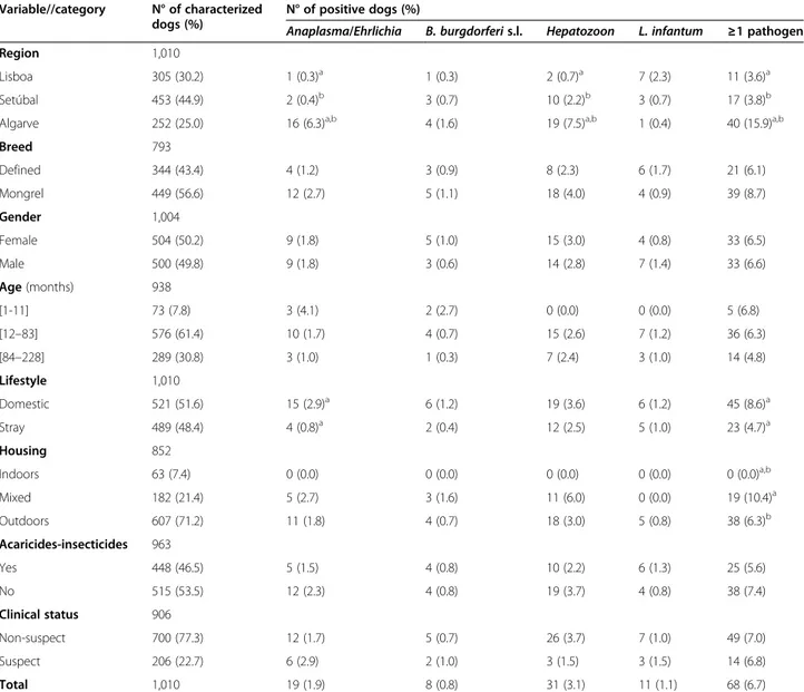
Table 1 Prevalence of vector-borne pathogen species, gender or complex as detected by PCR in 1,010 dogsfrom southern Portugal
Variable//category N° of characterized dogs (%)
N° of positive dogs (%)
Anaplasma/Ehrlichia B. burgdorferis.l. Hepatozoon L. infantum ≥1 pathogen
Region 1,010
Lisboa 305 (30.2) 1 (0.3)a 1 (0.3) 2 (0.7)a 7 (2.3) 11 (3.6)a
Setúbal 453 (44.9) 2 (0.4)b 3 (0.7) 10 (2.2)b 3 (0.7) 17 (3.8)b
Algarve 252 (25.0) 16 (6.3)a,b 4 (1.6) 19 (7.5)a,b 1 (0.4) 40 (15.9)a,b
Breed 793
Defined 344 (43.4) 4 (1.2) 3 (0.9) 8 (2.3) 6 (1.7) 21 (6.1)
Mongrel 449 (56.6) 12 (2.7) 5 (1.1) 18 (4.0) 4 (0.9) 39 (8.7)
Gender 1,004
Female 504 (50.2) 9 (1.8) 5 (1.0) 15 (3.0) 4 (0.8) 33 (6.5)
Male 500 (49.8) 9 (1.8) 3 (0.6) 14 (2.8) 7 (1.4) 33 (6.6)
Age(months) 938
[1-11] 73 (7.8) 3 (4.1) 2 (2.7) 0 (0.0) 0 (0.0) 5 (6.8)
[12–83] 576 (61.4) 10 (1.7) 4 (0.7) 15 (2.6) 7 (1.2) 36 (6.3)
[84–228] 289 (30.8) 3 (1.0) 1 (0.3) 7 (2.4) 3 (1.0) 14 (4.8)
Lifestyle 1,010
Domestic 521 (51.6) 15 (2.9)a 6 (1.2) 19 (3.6) 6 (1.2) 45 (8.6)a
Stray 489 (48.4) 4 (0.8)a 2 (0.4) 12 (2.5) 5 (1.0) 23 (4.7)a
Housing 852
Indoors 63 (7.4) 0 (0.0) 0 (0.0) 0 (0.0) 0 (0.0) 0 (0.0)a,b
Mixed 182 (21.4) 5 (2.7) 3 (1.6) 11 (6.0) 0 (0.0) 19 (10.4)a
Outdoors 607 (71.2) 11 (1.8) 4 (0.7) 18 (3.0) 5 (0.8) 38 (6.3)b
Acaricides-insecticides 963
Yes 448 (46.5) 5 (1.5) 4 (0.8) 10 (2.2) 6 (1.3) 25 (5.6)
No 515 (53.5) 12 (2.3) 4 (0.8) 19 (3.7) 4 (0.8) 38 (7.4)
Clinical status 906
Non-suspect 700 (77.3) 12 (1.7) 5 (0.7) 26 (3.7) 7 (1.0) 49 (7.0)
Suspect 206 (22.7) 6 (2.9) 2 (1.0) 3 (1.5) 3 (1.5) 14 (6.8)
Total 1,010 19 (1.9) 8 (0.8) 31 (3.1) 11 (1.1) 68 (6.7)
a,b
Results
Sixty-eight (6.7%; CI: 5.3-8.5%) dogs were PCR-positive to at least one of the tested species, genera or complex of CVBD agents (Table 1). Nineteen (1.9%; CI: 1.1-2.9%) dogs were positive to Anaplasma spp./Ehrlichiaspp., eight (0.8%; CI: 0.3-1.5%) toB. burgdorferis.l., 31 (3.1%; CI: 2.1-4.3) to Hepa-tozoonspp. and 11(1.1%; CI: 0.5-1.9) toL. infantum(Table 3).
Wolbachiaspp. DNA (amplified with the same primers used to detect Anaplasma spp./Ehrlichia spp.) was detected in four dogs, while DNA of Bartonella spp. or Babesia spp. was not amplified from any dog in the study.
As shown in Table 1, the prevalence ofAnaplasmaspp/
Ehrlichia spp. was statistically higher in domestic dogs. Positivity to these bacteria and to Hepatozoon spp. was
higher in dogs living in the Algarve region. Statistically sig-nificant differences were also found for PCR positivity to at least one of the studied agents in domestic dogs, in dogs with access to outdoors and in dogs living in the Algarve region.
Sequencing confirmedA. platysin five, E. canisin five,
B. burgdorferis.l. in six andH. canis in 18 dogs, including one animal with bothA. platysandH. canis(Table 3); and revealed Wolbachia spp. (DDBJ accessions: LC018189 to LC018192) in four dogs.
Discussion
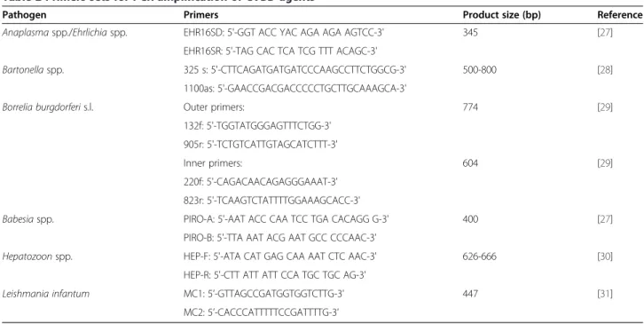
This is the most comprehensive study carried out in dogs from southern Portugal on the prevalence of infection Table 2 Primers sets for PCR amplification of CVBD agents
Pathogen Primers Product size (bp) Reference
Anaplasmaspp./Ehrlichiaspp. EHR16SD: 5'-GGT ACC YAC AGA AGA AGTCC-3' 345 [27]
EHR16SR: 5'-TAG CAC TCA TCG TTT ACAGC-3'
Bartonellaspp. 325 s: 5'-CTTCAGATGATGATCCCAAGCCTTCTGGCG-3' 500-800 [28]
1100as: 5'-GAACCGACGACCCCCTGCTTGCAAAGCA-3'
Borrelia burgdorferis.l. Outer primers: 774 [29]
132f: 5'-TGGTATGGGAGTTTCTGG-3' 905r: 5'-TCTGTCATTGTAGCATCTTT-3'
Inner primers: 604 [29]
220f: 5'-CAGACAACAGAGGGAAAT-3' 823r: 5'-TCAAGTCTATTTTGGAAAGCACC-3'
Babesiaspp. PIRO-A: 5'-AAT ACC CAA TCC TGA CACAGG G-3' 400 [27]
PIRO-B: 5'-TTA AAT ACG AAT GCC CCCAAC-3'
Hepatozoonspp. HEP-F: 5'-ATA CAT GAG CAA AAT CTC AAC-3' 626-666 [30]
HEP-R: 5'-CTT ATT ATT CCA TGC TGC AG-3'
Leishmania infantum MC1: 5’-GTTAGCCGATGGTGGTCTTG-3’ 447 [31]
MC2: 5’-CACCCATTTTTCCGATTTTG-3’
Table 3 Single and mixed PCR-positivity to species, genera and/or complex of CVBD agents in 1,010 dogs from southern Portugal
Agents No. positive dogs (%) DDBJ accessions
Single infections 67 (6.6)
Anaplasmaspp./Ehrlichiaspp. 18 (1.8)
[Anaplasma platys] [4 (0.4)] [LC018179 to LC018182]
[Ehrlichia canis] [5 (0.5)] [LC018184 to LC018188]
Borrelia burgdorferis.l. 8 (0.8) [LC018211 to LC018216]
Hepatozoonspp. 30 (3.0)
[Hepatozoon canis] [17 (1.7)] [LC018193 to LC018209]
Leishmania infantum 11 (1.1)
Co-infections 1 (0.1)
Anaplasmaspp./Ehrlichiaspp. andHepatozoonspp. 1 (0.1)
[A. platysandH. canis] [1 (0.1)] [LC018183 and LC018210]
with CVBD agents as it included domestic and stray animals with and without clinical signs compatible with a vector-borne disease. DNA from these pathogens taken all together was less frequently detected in dogs (6.7%;p< 0.001) than in cats (29.9%; 194/649) from the same region [26]. Further-more, only one (0.1%) dog was found co-infected (with two pathogens), whereas 29 (4.5%) cats were positive to two agents and four (0.6%) cats to three agents [26].
In this studyA. platyshas been molecularly confirmed to infect dogs from the south of the country, corroborating previous detection of this bacterium in dogs [11,23] and in
R. sanguineus[32] from the same region. The prevalence of positivity to Anaplasma/Ehrlichia in this work (1.9%) was lower than the 4.0% obtained in Spain [33] and the 3.7-6.0% in Italy [34], which might be related with the targeted popu-lation. In fact, in the works of Tabar et al. [33] and Trotta et al. [34], all the positive dogs were sick animals with clinical signs compatible with vector-borne diseases and admitted for medical treatment, while in the present work most of the enrolled animals were apparently healthy. Inter-estingly, in our study most of the animals harbouring
Anaplasma/Ehrlichia DNA were from Faro, overlapping the Algarve region, the southern most district of continen-tal Portugal, which seem to follow the trend revealed by Cardoso et al. [3] that the prevalence of antibodies against
Anaplasma spp. and E. canis in dogs from southern Portugal was significantly higher than in dogs from the northern and central regions of the country.
In the present work,B. burgdorferis.l. DNA was ampli-fied from 0.8% of the screened animals, providing the first molecular evidence of naturally occurringB. burgdorferis. l. infection in dogs from Portugal. The exposure of dogs to these spirochetes was previously demonstrated by specific serology in the Algarve [35] and in the Alentejo and Lisbon regions [3]. Furthermore,B. burgdorferis.l. genos-pecies, Borrelia lusitaniae was isolated from humans [36-38] and DNA of B. burgdorferi s.l. was detected in ticks [32,39] and cats from the south of the country [26]. Nevertheless, information on the clinical signs associated withBorreliainfections in dogs and their role as sentinels is still limited [6].
H. caniswas the most prevalent pathogen detected in all the assessed dogs, with a significantly higher prevalence in animals living in the Algarve. In fact,H. canishas recently been identified in dogs [23], in R. sanguineus collected from dogs living in this region [32], and also in foxes from the south [40], showing that the protozoan is widespread in this area of the country. Although in this study only three out of the 31 infected dogs presented clinical signs, subclinical infections should not be neglected as they may progress to a severe disease and warrant treatment [41]. Concurrent infections ofH. caniswith other canine patho-gens are common [21]; however, in the present work only one animal apparently healthy was co-infected with A.
platys and the protozoan. Although this individual dog had no clinical signs of a CVBD, co-infections may po-tentiate disease pathogenesis, altering clinical manifesta-tions associated with single infecmanifesta-tions [42].
The overall prevalence of L. infantum infection in the present study (1.1%) was much lower (p< 0.001) than the 34.9% obtained in 152 dogs from Lisbon [43]. The lower detection of Leishmania DNA might be due to the: (i) dynamics of infection over time, which may de-pend on the abundance and distribution of the proven vector species in conjunction with the number of in-fected vertebrate hosts [44], and (ii) insufficient data re-garding the duration of parasitaemia in infected dogs. In fact, and taking into account a seroprevalence of 18.2% recently obtained in 170 dogs from the Algarve region [45], PCR with blood should be used to complement serological results and not only by itself to detect Leish-mania infection, as it can lead to false negative results, especially in subclinically infected dogs [46].
PCR-positivity to one or more genera/complex of CVBD agents was found to be associated with domestic dogs, with animals living in the Algarve and with an out-door or mixed (i.e. with outout-door access) housing. In fact, most of the domestic dogs harbouring DNA of the stud-ied pathogens lived in rural areas from the Algarve re-gion and used to spend most of their time exclusively outdoors, thus increasing their exposure to arthropod vectors and the agents they might transmit.
The role of domestic dogs as reservoirs of Bartonella
spp. is less clear than for cats, and the former are probably accidental hosts. Nevertheless, they are excellent sentinels for human infections because a similar disease spectrum develops in dogs [47]. Serologic and molecular evidence of
Bartonella henselae and Bartonella clarridgeiae exist for cats from the south of Portugal [26,48]. Thus, the non-detection of Bartonella DNA in the present study might be related with differences in immune responses, host preference of particular vectors or innate resistance in dogs to these bacteria. On the other hand, the definitive diagnosis ofBartonellainfection is challenging due to the fastidious nature and intracellular tropism of these bac-teria for erythrocytes and endothelial cells [49]. According to Perez et al. [50], enrichment culture and subculture, followed by PCR amplification, enhances molecular diag-nostic sensitivity in dogs. Thus, it is possible that the PCR done directly from blood samples might have missed some positive cases; nevertheless, the prevalence of infection at the population level, if any, must be very low.
expected as its vector, D. reticulatus, is more abundant in the north of the country. However, the absence ofB. vogeli
andB. microti-like DNA is more difficult to explain, since both have already been detected in southern Portugal, the former in cats [26], dogs [23] and ticks [32], and the latter in foxes [51]. According to a recent questionnaire-based survey on the distribution of canine babesiosis in western Europe, the annual incidence of this parasitosis in southern Spain, which is geographically close to the area surveyed in this study, was estimated to be 0.0-0.7% [52]. Furthermore, a 58% prevalence of antibodies anti-Babesia spp. was re-ported among 331 dogs from kennels/shelters in southern Portugal [53]. The absence ofBabesiaspp. infection in the present study might be related with differences in the gen-etic background/immune system or between vector-dog in-teractions. Further studies are needed to better understand the epidemiological importance of these findings.
Conclusion
The identification of CVBD agents in southern Portugal, some of them with zoonotic concern, reinforces the im-portance to alert the veterinary community, owners and public health authorities to prevent the risk of transmission of vector-borne pathogens among dogs and to other verte-brate hosts including humans. Interestingly, the prevalence of the selected pathogens was much lower than that previ-ously found in cats from the same region [26], probably be-cause veterinarians and owners are much aware of them in the canine population and prophylatic measures such as in-secticides and acaricides are used.
Competing interests
The authors declare that they have no competing interests.
Authors’contributions
CM planned, designed and supervised the study, and wrote the manuscript; BA, CR, and MCF collected samples and clinical data, and performed DNA extraction and molecular analyses; AM, FM, JMC, MC, NN and PS collected samples and clinical data; MN performedB. burgdorferis.l. nested-PCR; LuC performed data analysis and revised the manuscript; MLV and LeC reviewed the manuscript. All authors read and approved the final manuscript.
Acknowledgements
This work was supported by Centro de Malária e outras Doenças Tropicais, IHMT-UNL, Portugal, and EU grant FP7-261504 EDENext, and is catalogued by the EDENext Steering Committee as EDENext302 (http://www.edenext.eu). The contents of this publication are the sole responsibility of the authors and do not necessarily reflect the views of the European Commission. The authors thank the cooperation of veterinarians, auxiliary staff, dog owners and shelters that contributed with collection of samples, and also acknowledge Prof. G. Baneth for providing DNA ofAnaplasmaspp./Ehrlichiaspp.,Babesiaspp. and
Hepatozoonspp. CM (SFRH/BPD/44082/2008) and MN (SFRH/BD/78325/2011) hold scholarships from Fundação para a Ciência e a Tecnologia, Ministério da Educação e Ciência, Portugal.
Publication of the CVBD10 thematic series has been sponsored by Bayer HealthCare–Animal Health division.
Author details
1Unidade de Parasitologia Médica, Instituto de Higiene e Medicina Tropical
(IHMT), Universidade Nova de Lisboa (UNL), Lisbon, Portugal.2Global Health
and Tropical Medicine, IHMT-UNL, Lisbon, Portugal.3Faculdade de Medicina
Veterinária, Universidade Lusófona de Humanidades e Tecnologias, Lisbon,
Portugal.4Clínica Veterinária Porto Seguro, Olhão, Portugal.5Hospital
Veterinário da Arrábida, Azeitão, Portugal.6Nyctea Lda., Lisbon, Portugal. 7Vetévora, Évora, Portugal.8Clube Animal, Beja, Portugal.9Unidade de
Microbiologia Médica, IHMT-UNL, Lisbon, Portugal.10Department of
Veterinary Sciences, School of Agrarian and Veterinary Sciences, University of Trás-os-Montes e Alto Douro, Vila Real, Portugal.11Departamento de Ciências
Biomédicas e Medicina, Universidade do Algarve, Faro, Portugal.
Received: 15 January 2015 Accepted: 23 February 2015
References
1. Otranto D, Dantas-Torres F, Breitschwerdt EB. Managing canine vector borne diseases of zoonotic concern: part one. Trends Parasitol. 2009;25:157–63. 2. Baneth G, Bourdeau P, Bourdoiseau G, Bowman D, Breitschwerdt E, Capelli
G, et al. Vector-borne diseases–constant challenge for practicing veterinarians: recommendations from the CVBD World Forum. Parasit Vectors. 2012;5:55.
3. Cardoso L, Mendão C, Madeira de Carvalho L. Prevalence ofDirofilaria immitis,Ehrlichia canis,Borrelia burgdorferisensu lato,Anaplasmaspp. and
Leishmania infantumin apparently healthy and CVBD-suspect dogs in Portugal–a national serological stud. Parasit Vectors. 2012;5:62. 4. Miró G, Montoya A, Roura X, Gálvez R, Sainz A. Seropositivity rates for
agents of canine vector-borne diseases in Spain: a multicentre study. Parasit Vectors. 2013;6:117.
5. Day MJ. One health: the importance of companion animal vector-borne diseases. Parasit Vectors. 2011;4:49.
6. Mencke N. Future challenges for parasitology: vector control and 'One health' in Europe: the veterinary medicinal view on CVBDs such as tick borreliosis, rickettsiosis and canine leishmaniosis. Vet Parasitol. 2013;195:256–71.
7. Doudier B, Olano J, Parola P, Brouqui P. Factors contributing to emergence ofEhrlichiaandAnaplasmaspp. as human pathogens. Vet Parasitol. 2010;167:149–54.
8. Stuen S, Granquist EG, Silaghi C.Anaplasma phagocytophilum–a widespread multi-host pathogen with highly adaptive strategies. Front Cell Infect Microbiol. 2013;3:31.
9. Arraga-Alvarado CM, Qurollo BA, Parra OC, Berrueta MA, Hegarty BC, Breitschwerdt EB. Molecular evidence ofAnaplasma platysinfection in two women from Venezuela. Am J Trop Med Hyg. 2014;91:1161–5.
10. Jahfari S, Coipan EC, Fonville M, van Leeuwen AD, Hengeveld P, Heylen D, et al. Circulation of fourAnaplasma phagocytophilumecotypes in Europe. Parasit Vectors. 2014;7:365.
11. Santos AS, Alexandre N, Sousa R, Núncio MS, Bacellar F, Dumler JS. Serological and molecular survey ofAnaplasmaspecies infection in dogs with suspected tickborne disease in Portugal. Vet Rec. 2009;164:168–71. 12. Cardoso L, Tuna J, Vieira L, Yisaschar-Mekuzas Y, Baneth G. Molecular
detection ofAnaplasma platysandEhrlichia canisin dogs from the North of Portugal. Vet J. 2010;183:232–3.
13. Yisaschar-Mekuzas Y, Jaffe CL, Pastor J, Cardoso L, Baneth G. Identification of
Babesiaspecies infecting dogs using reverse line blot hybridization for six canine piroplasms, and evaluation of co-infection by other vector-borne pathogens. Vet Parasitol. 2013;191:367–73.
14. Alexandre N, Santos AS, Núncio MS, Sousa RD, Boinas F, Bacellar F. Detection ofEhrlichia canisby polymerase chain reaction in dogs from Portugal. Vet J. 2009;181:343–4.
15. Diniz PP, Billeter SA, Otranto D, De Caprariis D, Petanides T, Mylonakis ME, et al. Molecular documentation ofBartonellainfection in dogs in Greece and Italy. J Clin Microbiol. 2009;47:1565–7.
16. Rizzoli A, Hauffe H, Carpi G, Vourc HG, Neteler M, Rosa R. Lyme borreliosis in Europe. Euro Surveill. 2011;16:19906.
17. Hovius KE, Stark LA, Bleumink-Pluym NM, van de Pol I, Verbeek-de Kruif N, Rijpkema SG, et al. Presence and distribution ofBorrelia burgdorferisensu lato species in internal organs and skin of naturally infected symptomatic and asymptomatic dogs, as detected by polymerase chain reaction. Vet Q. 1999;21:54–8.
18. Solano-Gallego L, Baneth G. Babesiosis in dogs and cats–expanding parasitological and clinical spectra. Vet Parasitol. 2011;181:48–60. 19. Cardoso L, Costa A, Tuna J, Vieira L, Eyal O, Yisaschar-Mekuzas Y, et al.
20. Simões PB, Cardoso L, Araújo M, Yisaschar-Mekuzas Y, Baneth G. Babesiosis due to the canineBabesia microti-like small piroplasm in dogs-first report from Portugal and possible vertical transmission. Parasit Vectors. 2011;4:50. 21. Baneth G. Perspectives on canine and feline hepatozoonosis. Vet Parasitol.
2011;181:3–11.
22. Cardoso L, Yisaschar-Mekuzas Y, Rodrigues FT, Costa A, Machado J, Diz-Lopes D, et al. Canine babesiosis in northern Portugal and molecular characterization of vector-borne co-infections. Parasit Vectors. 2010;3:27. 23. René-Martellet M, Lebert I, Chêne J, Massot R, Leon M, Leal A, et al.
Diagnosis and incidence risk of clinical canine monocytic ehrlichiosis under field conditions in Southern Europe. Parasit Vectors. 2015;8:3.
24. Campino L, Maia C. The role of reservoirs: canine leishmaniasis. In: Ponte-Sucre A, Padron-Nieves M, Diaz E, editors. Drug resistance in Leishmania parasites–consequences, molecular mechanism and possible treatments. Vienna: Springer Verlag; 2013. p. 45–64.
25. Cortes S, Vaz Y, Neves R, Maia C, Cardoso L, Campino L. Risk factors for canine leishmaniasis in an endemic Mediterranean region. Vet Parasitol. 2012;189:189–96.
26. Maia C, Ramos C, Coimbra M, Bastos F, Martins A, Pinto P, et al. Bacterial and protozoal agents of feline vector-borne diseases in domestic and stray catsfrom southern Portugal. Parasit Vectors. 2014;7:115.
27. Harrus S, Perlman-Avrahami A, Mumcuoglu K, Morick D, Eyal O, Baneth G. Molecular detection ofEhrlichia canis,Anaplasma bovis,Anaplasma platys,
CandidatusMidichloria mitochondrii andBabesia canis vogeliin ticks from Israel. Clin Microbiol Infect. 2011;17:459–63.
28. Diniz P, Maggi R, Schwartz D, Cadenas M, Bradley J, Hegarty B, et al. Canine bartonellosis: serological and molecular prevalence in Brazil and evidence of co-infection withBartonella henselaeandBartonella vinsoniisubsp.berkhoffii. Vet Res. 2007;38:697–710.
29. Wodecka B, Leońska A, Skotarczak B. A comparative analysis of molecular markers for the detection and identification ofBorreliaspirochaetes in
Ixodes ricinus. J Med Microbiol. 2010;59(Pt 3):309–14.
30. Inokuma H, Okuda M, Ohno K, Shimoda K, Onishi T. Analysis of the 18S rRNA gene sequence of aHepatozoondetected in two Japanese dogs. Vet Parasitol. 2002;106:265–71.
31. Cortes S, Rolão N, Ramada J, Campino L. PCR as a rapid and sensitive tool in the diagnosis of human and canine leishmaniasis usingLeishmania donovanis.l.–specific kinetoplastid primers. Trans R Soc Trop Med Hyg. 2004;98:12–7.
32. Maia C, Ferreira A, Nunes M, Vieira ML, Campino L, Cardoso L. Molecular detection of bacterial and parasitic pathogens in hard ticks from Portugal. Ticks Tick Borne Dis. 2014;5:409–14.
33. Tabar MD, Francino O, Altet L, Sánchez A, Ferrer L, Roura X. PCR survey of vectorborne pathogens in dogs living in and around Barcelona, an area endemic for leishmaniasis. Vet Rec. 2009;164:112–6.
34. Trotta M, Fogliazza A, Furlanello T, Solano-Gallego L. A molecular and serological study of exposure to tick-borne pathogens in sick dogs from Italy. Clin Microbiol Infect. 2009;15:62–3.
35. Alexandre NML. Estudo clínico e epidemiológico da febre botonosa, ehrlichiose canina e borreliose de Lyme numa população de canídeos domésticos do Algarve. MSc dissertation. Lisbon: Technical University of Lisbon, Faculty of Veterinary Medicine; 2006.
36. Collares-Pereira M, Couceiro S, Franca I, Kurtenbach K, Schäfer SM, Vitorino L, et al. First isolation ofBorrelia lusitaniaefrom a human patient. J Clin Microbiol. 2004;42:1316–8.
37. de Carvalho IL, Fonseca JE, Marques JG, Ullmann A, Hojgaard A, Zeidner N, et al. Vasculitis-like syndrome associated withBorrelia lusitaniaeinfection. Clin Rheumatol. 2008;27:1587–91.
38. de Carvalho IL, Zeidner N, Ullmann A, Hojgaard A, Amaro F, Zé-Zé L, et al. Molecular characterization of a new isolate ofBorrelia lusitaniaederived fromApodemus sylvaticusin Portugal. Vector Borne Zoonotic Dis. 2010;10:531–4.
39. Baptista S, Quaresma A, Aires T, Kurtenbach K, Santos-Reis M, Nicholson M, et al. Lyme borreliosis spirochetes in questing ticks from mainland Portugal. Int J Med Microbiol. 2004;293:109–16.
40. Cardoso L, Cortes HC, Eyal O, Reis A, Lopes AP, Vila-Viçosa MJ, et al. Molecular and histopathological detection ofHepatozoon canisin red foxes (Vulpes vulpes) from Portugal. Parasit Vectors. 2014;7:113.
41. Baneth G, Weigler B. Retrospective case–control study of hepatozoonosis in dogs in Israel. J Vet Intern Med. 1997;11:365–70.
42. De Tommasi AS, Otranto D, Dantas-Torres F, Capelli G, Breitschwerdt EB, de Caprariis D. Are vector-borne pathogen co-infections complicating the clinical presentation in dogs? Parasit Vectors. 2013;6:97.
43. Maia C, Gomes J, Cristóvão J, Nunes M, Martins A, Rebêlo E, et al. Feline
Leishmaniainfection in a canine leishmaniasis endemic region, Portugal. Vet Parasitol. 2010;174:336–40.
44. Maia C, Dionísio L, Afonso MO, Neto L, Cristóvão JM, Campino L.Leishmania
infection and host-blood feeding preferences of phlebotomine sandflies and canine leishmaniasis in an endemic European area, the Algarve Region in Portugal. Mem Inst Oswaldo Cruz. 2013;108:481–7.
45. Maia C, Coimbra M, Ramos C, Cristóvão JM, Cardoso L, Campino L. Serological investigation of Leishmania infantum, Dirofilaria immitis and Angiostrongylus vasorum in dogs from southern Portugal. Parasit Vectors (in press).
46. Maia C, Campino L. Methods for diagnosis of canine leishmaniasis and immune response to infection. Vet Parasitol. 2008;158:274–87.
47. Chomel B, Boulouis H, Maruyama S, Breitschwerdt E.Bartonellaspp. in pets and effect on human health. Emerg Infect Dis. 2006;12:389–94.
48. Alves AS, Milhano N, Santos-Silva M, Santos AS, Vilhena M, de Sousa R. Evidence ofBartonellaspp.,Rickettsiaspp. andAnaplasma phagocytophilum
in domestic, shelter and stray cat blood and fleas, Portugal. Clin Microbiol Infect. 2009;15:1–3.
49. Breitschwerdt EB, Maggi RG, Chomel BB, Lappin MR. Bartonellosis: an emerging infectious disease of zoonotic importance to animals and human beings. J Vet Emerg Crit Care (San Antonio). 2010;20:8–30.
50. Perez C, Maggi RG, Diniz PP, Breitschwerdt EB. Molecular and serological diagnosis ofBartonellainfection in 61 dogs from the United States. J Vet Intern Med. 2011;25:805–10.
51. Cardoso L, Cortes HC, Reis A, Rodrigues P, Simões M, Lopes AP, et al. Prevalence ofBabesia microti-like infection in red foxes (Vulpes vulpes) from Portugal. Vet Parasitol. 2013;196:90–5.
52. Halos L, Lebert I, Abrial D, Danlois F, Garzik K, Rodes D, et al. Questionnaire-based survey on the distribution and incidence of canine babesiosis in countries of Western Europe. Parasite. 2014;21:13.
53. Menn B, Lorentz S, Naucke TJ. Imported and travelling dogs as carriers of canine vector-borne pathogens in Germany. Parasit Vectors. 2010;3:34.
Submit your next manuscript to BioMed Central and take full advantage of:
• Convenient online submission
• Thorough peer review
• No space constraints or color figure charges
• Immediate publication on acceptance
• Inclusion in PubMed, CAS, Scopus and Google Scholar
• Research which is freely available for redistribution