Native
Wolbachia
from
Aedes albopictus
Blocks Chikungunya Virus Infection
In Cellulo
Vincent Raquin1¤
*, Claire Valiente Moro1, Yoann Saucereau1, Florence-Hélène Tran1,
Patrick Potier1, Patrick Mavingui1,2*
1Université de Lyon, UMR 5557 Ecologie Microbienne, CNRS, USC1190 INRA, VetAgro Sup, Université Lyon 1, Villeurbanne, France,2Université de La Réunion, UMR PIMIT, INSERM U1187, CNRS 9192, IRD 249, Plateforme de Recherche CYROI, Saint-Denis, La Réunion, France
¤ Current address: Insect-Virus Interactions group, Genomes & Genetics Department, Institut Pasteur, Paris, France
*[email protected](VR);[email protected](PM)
Abstract
Wolbachia, a widespread endosymbiont of terrestrial arthropods, can protect its host against viral and parasitic infections, a phenotype called "pathogen blocking". However, in some casesWolbachiamay have no effect or even enhance pathogen infection, depending on the host-Wolbachia-pathogen combination. The tiger mosquitoAedes albopictusis natu-rally infected by two strains ofWolbachia,wAlbA andwAlbB, and is a competent vector for different arboviruses such as dengue virus (DENV) and Chikungunya virus (CHIKV). Inter-estingly, it was shown in some cases thatAe.albopictusnativeWolbachiastrains are able to inhibit DENV transmission by limiting viral replication in salivary glands, but no such impact was measured on CHIKV replicationin vivo. To better understand theWolbachia/ CHIKV/Ae.albopictusinteraction, we generated a cellular model usingAe.albopictus
derived C6/36 cells that we infected with thewAlbB strain. Our results indicate that CHIKV infection is negatively impacted at both RNA replication and virus assembly/secretion steps in presence ofwAlbB. Using FISH, we observed CHIKV andwAlbB in the same mosquito cells, indicating that the virus is still able to enter the cell in the presence of the bacterium. Further work is needed to decipher molecular pathways involved inWolbachia-CHIKV inter-action at the cellular level, but this cellular model can be a useful tool to study the mecha-nism behind virus blocking phenotype induced byWolbachia. More broadly, this underlines that despiteWolbachiaantiviral potential other complex interactions occurin vivoto deter-mine mosquito vector competence inAe.albopictus.
Introduction
Human infectious diseases caused by vector-borne pathogens have an increasing incidence worldwide, accounting for 17% of the estimated burden of infectious diseases as referred by World's Health Organization [1]. Notably, arthropod-borne viruses (arboviruses) are emerging or re-emerging viruses transmitted to vertebrate hosts by the bite of infected arthropod vectors, a11111
OPEN ACCESS
Citation:Raquin V, Valiente Moro C, Saucereau Y, Tran F-H, Potier P, Mavingui P (2015) Native
WolbachiafromAedes albopictusBlocks
Chikungunya Virus InfectionIn Cellulo. PLoS ONE 10 (4): e0125066. doi:10.1371/journal.pone.0125066
Editor:Kostas Bourtzis, International Atomic Energy Agency, AUSTRIA
Received:January 4, 2015
Accepted:March 20, 2015
Published:April 29, 2015
Copyright:© 2015 Raquin et al. This is an open access article distributed under the terms of the
Creative Commons Attribution License, which permits unrestricted use, distribution, and reproduction in any medium, provided the original author and source are credited.
Data Availability Statement:All relevant data are within the paper and its Supporting Information files.
2087/6-mainly mosquitoes. Among them, Chikungunya is a mosquito-borne viral infection caused by an alphavirus from theTogaviridaefamily. Chikungunya virus (CHIKV) is transmitted to humans byAedes(Stegomyia) spp mosquitoes, primarilyAedes aegypti. Since 2004, CHIKV started a global spread with severe outbreaks in the Indian Ocean region, the Indian subconti-nent and Central Africa, all associated with a single amino-acid change in the virus E1 glyco-protein that allowed an enhanced transmission by a secondary mosquito species,Aedes albopictus[2–4]. Autochthonous transmissions in Europe were also reported from Italy, with 217 confirmed cases in 2007 [5] and from France with two confirmed cases in 2010 [6]. Con-secutively to a major chikungunya outbreak started in the French Antilles in 2013, autochtho-nous cases were reported in the United States [7] and more recently in France [8], bringing the threat of multiple outbreaks caused by virus-carrying travellers; in both temperate areas,Ae.
albopictuswas the vector responsible for CHIKV transmission.
The speciesAe.albopictus, also known as tiger mosquito, is native from Southern and East-ern Asia but recently spread worldwide [9]. The rapid extension ofAe.albopictuscombined with its ecological plasticity and vector competence for diverse arboviruses make the tiger mos-quito a significant threat for public health [10]. In absence of effective vaccines or prophylaxis against most of arboviruses included CHIKV, current efforts are mainly based on controlling vector populations with insecticides. However, the development of mosquito resistance, as well as environmental contamination and side effects on non-target organisms has called chemical-based control methods into question [11]. Consequently, alternative and innovative vector control strategies emerged, and one of the most promising is based on the use of symbiotic bac-teria [12]. In this framework, the endosymbiontWolbachiahas been the most studied candi-date including arboviruses and parasites transmission control [13–15].
Wolbachiais an obligate intracellular bacterium that infects around 40% of arthropods [16], and manipulates their reproduction to facilitate its own spread among populations [17]. When thewMel strain ofWolbachia, originated fromDrosophila, was transinfected intoAe.aegypti
embryos, mosquitoes presented limited vector competence for a large panel of pathogens including dengue virus (DENV) [18,19], CHIKV [19], yellow fever (YFV) [20], West-Nile virus (WNV) [21] andPlasmodiumparasite [14,19]. However, it appears thatWolbachia -tran-sinfected mosquitoes are markedly associated with a viral inhibition phenotype compared to naturally infected populations, which most of time exhibit no inhibition or even an enhancing of the infection [22]. In the field,Ae.aegyptilacks this association withWolbachiawhileAe.
albopictusmosquitoes naturally carry two strains, namelywAlbA andwAlbB [23,24]. The nativeWolbachiafromAe.albopictuswas associated with a decrease of DENV transmission in mosquitoes from La Réunion island [25]. However, this phenotype was shown to be dependent on the mosquito population considered as no inhibition was observed in population from Houston, Texas [18]. Intriguingly, no significant impact ofWolbachiawas observed on CHIKV transmission inAe.albopictuspopulation from La Réunion [26]. This suggests that the
Wolbachiainhibition phenotype also depends on the viral strain considered. Together, these observations clearly indicate that the tripartite interaction betweenWolbachia, arboviruses and their mosquito host is complex and varies according to the nature of the interacting partners.
The molecular and cellular mechanisms ofWolbachia-mediated inhibition of arboviruses are poorly known, but current hypotheses suggest a competition for host cell resources, sup-ported by the bacterial density-dependent interference and the intra-host competition for amino acids and cholesterol [27,28]. Insect immune pathways activated uponWolbachia infec-tion have been also suggested to mediate the blocking phenotype, like autophagy [29], oxidative stress [30] or miRNA pathway [31]. It appears thatWolbachia-mediated activation of the Toll and Imd immune pathways was unlikely to trigger antiviral interference, as suggested by a recent study inDrosophila[32]. In addition, as being an obligate intracellular bacterium,
1), part of the 2012-2013 BiodivERsA call for research proposals.
studies onWolbachiaare difficult using standard techniques. Interestingly,Wolbachia-infected cell lines were used as a tool to study the mechanisms involved inWolbachia-pathogen interac-tion [29,30,33–35]. To facilitate the understanding of theWolbachia/CHIKV/Ae.albopictus
interaction, we built a cellular model by culturing thewAlbB strainin vitrointo theAe. albopic-tusCHIKV-permissive cell line C6/36. Using this simplifiedin vitromodel, we measured the viral dynamic in the presence or absence ofWolbachia, and tried to decipher at which step of the viral cycleWolbachiainterferes with CHIKV infection. More broadly, this work provides a suitable tool to studyWolbachia-arbovirus interaction at the cellular level.
Material and Methods
Establishment of
Wolbachia-infected mosquito cell line
The C6/36 cells, derived fromAe.albopictuslarvae and originally non-infected byWolbachia, were used for culturingwAlbB strain. This bacterial strain originated from naturally infected Aa23 cells isolated fromAe.albopictuseggs [36]. Both cell types were cultured at 28°C in growth medium consisting of equal volumes of Mitsuhashi/Maramorosh (Bioconcept, Swit-zerland) and Schneider’s insect medium (Sigma, France) supplemented with 10% (v/v) of heat-inactivated foetal bovine serum (PAA, USA) and penicillin/streptomycin (50 U/50μg/ mL; Gibco, Invitrogen, France). Briefly, three 25 cm2flasks of confluent Aa23 cells were scrapped, pelleted for 10 min at 300×g and crushed by vortexing 10 min with 5-mm diameter sterile borosilicate beads (Biospec, OK, USA). Cell lysates were centrifuged for 5 min at 300×g, and supernatants were filtered through a 5-μM syringe filter (Millipore) to eliminate cellular debris. Fresh filtrate (500μL) containing bacteria was inoculated onto 80% confluent monolayer of C6/36 cells, in shell vial tube (Sterilin, UK). After centrifugation 5 min at 2000×g, cells were incubated overnight at 28°C then the coverslip bearing cells was trans-ferred into a 25 cm2flask with fresh culture medium and incubation period extended to reach 80% confluence. After this first round of infection, cells were harvested, resuspended in
500μL of fresh medium and used for a second infection procedure. TheWolbachiainfection
in cells was characterized using electron microscopy, FluorescentIn SituHybridization (FISH) and quantitative PCR (qPCR). For each assay, we used as control tetracycline-treated cells (TET) to remove bacteria without modifying the host cell genetic background. This was achieved by adding 10μg/mL of tetracycline hydrochloride (Sigma, France) in culture media ofWolbachia-infected (wAlbB) cells for 5 passages, and then cells were maintained in culture without tetracycline until use. The original C6/36 uninfected (CTRL), TET andwAlbB infected cells were continuously passaged in 25 cm2flasks by scrapping and seeding a new flask with 1:5 of the cell suspension in 5 mL of fresh medium, every 4 days.
Electron microscopy
Virus
The CHIKV 06.21 strain derived from newborn serum sample with neonatal encephalopathy, was collected in La Reunion Island in 2005 [37]. This isolate was highly passaged in C6/36. Viral stocks were produced on C6/36 cells in 25-cm2flasks, at Multiplicity Of Infection (MOI) of 0.01. After 3 days at 28°C, supernatants from infected cells were recovered and virus titration was performed using plaque assay on Vero E6 (green monkey kidney) cells [38]. To measure the impact of tetracycline treatment on viral dynamics, CHIKV RNA titer was compared between CTRL and TET cells using quantitative RT-PCR (RT-qPCR), at two different MOI of 0.1 and 3. To that end, cells were transferred into 12-well plates at 1×10⁶cells per well and allowed to attach for 24 h, at 28°C. Infection with CHIKV 06.21 was performed in 2% FBS medium, using virus-free medium as control. After 1 h, 1.5 mL of fresh media with 10% FBS was added. Cells and supernatants were harvested at 2, 4, 6, 8, 10, 24, 48, 72, 96 and 168 hours post-infection. Residual cells were removed from supernatant by centrifugation for 3 min at full-speed and samples were stored at -80°C until titration. Adherent cells were rinsed twice in PBS and scrapped, pelleted by centrifugation and kept at -80°C prior to RNA isolation. Experi-ment was conducted with two independent samples. To assess the role ofWolbachiaduring CHIKV infection, we compared virus titer between TET andwAlbB bearing cells. The day prior infection, cells from three to six independent flasks were transferred in 12-well plates at 1×10⁶cells per well while another fraction was inoculated in shell vial tubes at 5×10⁵cells per tube for FISH staining. CHIKV 06.21 infection was performed as mentioned above, at MOI of 0.1 and 3, with cells and supernatant harvested at 1, 3, 5 and 7 days post-infection. Samples were stored at -80°C until use.
DNA and RNA isolation
Genomic DNA isolation was performed usingDNeasy blood and tissueskit (Qiagen, France) following manufacturer's recommendations. After lysis in 180μL of ATL buffer, samples were incubated for 2 h a 37°C with lysozyme (Euromedex, France) at a final concentration of 2 mg/ mL. Residual co-extracted RNA was eliminated by adding 100 mg/mL of RNase A, for 2 min at room temperature. The isolated DNA was eluted in 30μL of DNase-free water. Total RNA was isolated using theRNeasy Mini Kit(Qiagen, France) as recommended by supplier. Cell pellets were crushed in 350μL RLT lysis buffer using RNase-free piston pellet (Kontes, USA), and RNA was eluted in 37μL of RNase-free water. RNA solution was treated with DNase using the AmbionTURBO-DNA freekit (Ambion, USA) in 50μL final volume following the manufac-turer’s instructions. DNA and RNA were quantified using a UV-mc2spectrophotometer and diluted to 5 ng/μL, then frozen at -20°C (DNA) or -80°C (RNA) until use.
Quantitative
Wolbachia
PCR analysis
The relative density ofWolbachiaper cell was monitored by qPCR usingWolbachiaSurface Protein (wsp) gene for the bacterium andactingene for the host cell. Standard curves were drawn on 10-fold serial dilutions from 1×10⁸to 1×101copies/μL of the DNA plasmid pQuan-tAlb16Scontainingwspandactingene fragments [23]. Each 20μL reaction contained 10 ng
(2μL) of template DNA, 10μL Fast-SYBR-Green Master Mix (Roche, Suisse), 200 mM (wsp)
CHIKV RT-qPCR analysis
The CHIKV RNA copy number was quantified by RT-qPCR targeting the envelopeE2gene. Viral RNA copies were assessed using a standard curve of 10-fold serial dilution of a synthetic CHIKV RNA transcript [26]. One-step RT-qPCR was performed using EXPRESS One-Step SYBR GreenER Kit (Invitrogen, France) in a volume of 20μL containing 10 ng (2μL) of RNA template, 10μL EXPRESS SYBR GreenER SuperMix Universal, 200 nM of sense Chik/E2/ 9018/+ and anti-sense Chik/E2/9235/−primers (Table 1) and 0.5μL EXPRESS Superscript
Mix. Amplification was performed on a LC480 LightCycler (Roche, France) and consisted of 15 min at 50°C followed by 95°C for 2 min, then 40 cycles of 95°C for 15 s and 63°C for 1 min. All PCR reactions were performed in triplicate and RNA from CHIKV-uninfected C6/36 cells was used as negative control.
Fluorescent focus assay (FFA)
Virus infectious titer was quantified using an indirect immunofluorescent detection of infec-tious foci on C6/36 monolayer [39]. Cells were seeded in 96-well plates at a density of 3×10⁶ cells/well and incubated for 36 h at 28°C to produce confluent monolayers. Ten-fold serial dilu-tions of sample supernatants were inoculated in a final volume of 50μL/well. After 1 h incuba-tion at 28°C to allow viral adsorpincuba-tion, with gently rocking every 15 min to spread viral
inoculum, an overlay consisting of 5% FBS, 1.6% of carboxymethyl cellulose (CMC, VWR) was added in a final volume of 200μL per well. Plates were incubated 3 days at 28°C then 150μL of freshly prepared 4% formaldehyde solution in PBS was added without removing the overlay. Cell monolayers were fixed for 20 min at RT, washed three times in PBS, then incubate for 30 min at RT in PBS-0.1% Triton X-100. Plates were stained for 1 h at 37°C with a 1:1000 dilution of hyper-ascetic immune fluid specific to CHIKV 06.21 in PBS-0.1% Bovine Serum Albumin (BSA, Sigma, France). After 3 washes in PBS, cells were incubated for 1 h at 37°C with an anti-mouse Alexa488-conjugated antibody (Molecular probes, Invitrogen, France) diluted at 1:200 in PBS-0.1% BSA followed by three washes in PBS and a final wash in distilled water. Cell monolayers were observed using an EVOS inverted fluorescence microscope (Life Technolo-gies, France) with a FITC-filter, under 10X objective. The total number of fluorescent foci was counted from 5 to 50 at the appropriate dilution, and virus titer was calculated as fluorescent focus unit per mL. The titer represents a mean of two independent samples.
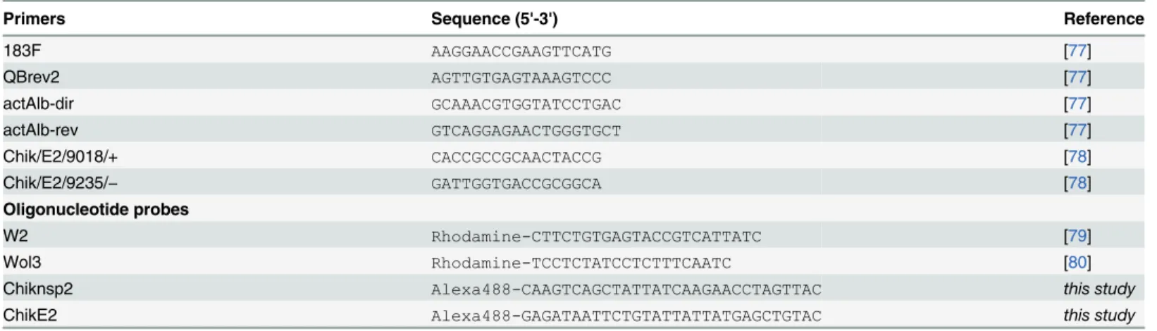
Table 1. List of primers and probes used in this study.
Primers Sequence (5'-3') Reference
183F AAGGAACCGAAGTTCATG [77]
QBrev2 AGTTGTGAGTAAAGTCCC [77]
actAlb-dir GCAAACGTGGTATCCTGAC [77]
actAlb-rev GTCAGGAGAACTGGGTGCT [77]
Chik/E2/9018/+ CACCGCCGCAACTACCG [78]
Chik/E2/9235/− GATTGGTGACCGCGGCA [78]
Oligonucleotide probes
W2 Rhodamine-CTTCTGTGAGTACCGTCATTATC [79]
Wol3 Rhodamine-TCCTCTATCCTCTTTCAATC [80]
Chiknsp2 Alexa488-CAAGTCAGCTATTATCAAGAACCTAGTTAC this study
ChikE2 Alexa488-GAGATAATTCTGTATTATTATGAGCTGTAC this study
Fluorescent
In Situ
Hybridization
After two washes in PBS, cells were fixed on the coverslip for 10 min in freshly prepared 4% formaldehyde in PBS. Hybridization was conducted overnight at 37°C in 1 mL of hybridization buffer [formamide 50%, SSC (saline-sodium citrate) 5X, 200 mg dextran sulfate per mL and
250μg poly(A) per mL, 250μg salmon sperm DNA per mL, 250μg tRNA per mL, DTT
(1,4-dithiothreitol) 0.1 mg/L, Denhartdt’s solution 0.5X] containing 200 ng ofWolbachia probes W2 and Wol3 labelled in their 5'-end with Rhodamine Red-X and CHIKV probe labelled in 5'-end with Alexa488 fluor (Table 1). After hybridization, samples were washed twice in 1X SSC-10 mmol/L DTT and then twice in 0.5X SSC-10 mmol/L DTT at 55°C for 15 min each. Cells were then rinsed in PBS, mounted on a glass slide with 3μL of DAPI (4’ ,6-dia-midino-2-phenylindole, dihydrochloride) solution (1μg/mL of dye) in glycerol/PBS (1:1). Samples were viewed under a fluorescence microscope (AXIO Imager.ZI; Zeiss, France). To estimate the proportion of cells infected byWolbachia, five different microscope fields were analyzed with at least 50 cells per field [40].
Statistics
The continuous response variables (viral and bacterial titers) were log10-transformed. They
were analysed using a multifactorial linear model, with a normal error distribution and an identity link function that included the effect of the time and MOI as ordinal variables, treat-ment as discrete variable and their interactions. All the statistical analysis was performed using R environment (version 3.1.0).
Results
Characterization of
wAlbB infection in mosquito cells
Previous studies mentioned that thewAlbB strain could be maintained in C6/36 [41,42]. Despite this,wAlbB dynamics of infection in C6/36 remains unknown. ThewAlbB cells were purified from Aa23 cells, as they were already adapted to cell line culture. The C6/36 cells tend to grow in adhesive cell clusters, forming patchy monolayers independently ofWolbachia
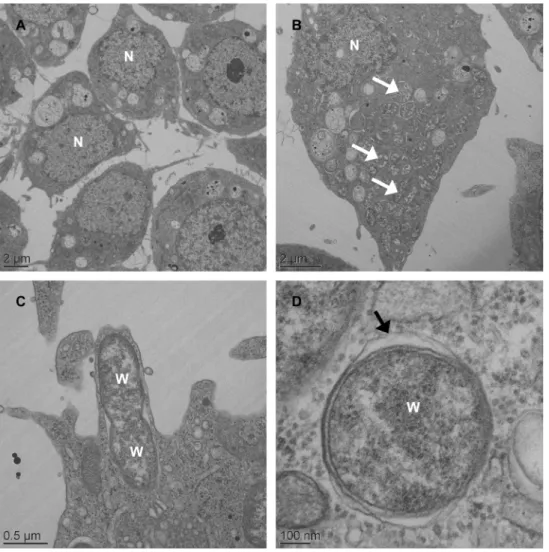
infection (S1 Fig). Two attempts were necessary to obtainWolbachiainfected cells, designated C6/36_wAlbB, with awspsignal in PCR persisting in cells after several passages (not shown). Electron microscopy of C6/36_wAlbB cells (P.30) revealed the presence ofWolbachiaas round-shaped particles of varying size inside the cytoplasm, surrounded by a host cell mem-brane where the bacteria seem to divide (Fig 1). As expected, noWolbachiawas seen outside a cell, while some bacteria could be released after the lysis of their host cell. In C6/36_TET cells, i.e. cells cured fromWolbachiaby tetracycline treatment, no difference in cell aspect was noted compared toWolbachia-infected cells, despite the absence ofWolbachiainfection. The C6/ 36_wAlbB cells were maintained in continuous culture for 40 passages, corresponding to approximately 5 months. Quantitative PCR analysis showed that the density ofWolbachiawas highly dynamic according to the passages (Fig 2), with the lowest density of 0.9wsp/actinratio at P.7 to 67.6wsp/actinratio at P.17 for the highest. After P.17,Wolbachia's density decreased to remain around 10wsp/actinratio from P.36 to P.40. The C6/36_TET cells were negative for
Reduced CHIKV infection by
wAlbB
in vitro
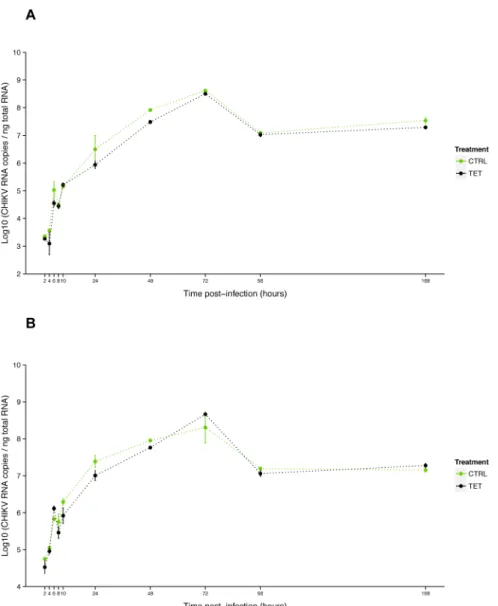
As no viral inhibition was measured for CHIKV 06.21 in orally infectedAe.albopictus mosqui-toes [26], we tested the interaction ofwAlbB and CHIKV 06.21 in C6/36. First, we assessed that CHIKV replication was not affected by anti-Wolbachiatetracycline treatment, as viral RNA titer was not significantly different between C6/36_TET and C6/36_CTRL cells at MOIs of 0.1 (P= 0.45) and 3 (P= 0.68) (Fig 4). The viral RNA titer increased from 2 h to 72 h post-infection (pi), with a short eclipse phase between 8 h and 10 h pi, then decreased until 96 h to reach a plateau until day 7 pi. The viral replication was dramatically reduced in C6/36_wAlbB compared to C6/36_TET cells as measured by RT-qPCR after infection at MOI 0.1 (Fig 5A). The RNA titer significantly decreased in C6/36_wAlbB cells by at least ten-fold across all time-points. Interestingly,Wolbachia-mediated inhibition depended on the time of infection ( Wol-bachiatime interaction,P<2E-16), suggesting that presence ofWolbachiacould delay virus replication as previously mentioned [43]. Although viral RNA titer decreased, inhibition was
Fig 1. Electron microscopy ofWolbachiainAedes albopictusC6/36 cells.Low-magnification transmission electron micrograph of C6/36_TET cells with no bacterial signal in host cell cytoplasm (A) whereasWolbachia(white arrowhead) are seen throughout the cytoplasm of C6/36_wAlbB cells (B).
Wolbachiapresumably is undergoing the process of cell division (C). High-magnification micrograph of
Wolbachiain cytoplasm of the host cell showing a membranous structure surrounding the bacterium (black arrowhead) (D).
Fig 2. Dynamics ofwAlbB infection in C6/36 cells.Ratio ofWolbachia wspcopies per hostactincopies during continuous cell culture, measured by qPCR on total genomic DNA (error bars represent the standard deviation of the mean of three independent samples).
doi:10.1371/journal.pone.0125066.g002
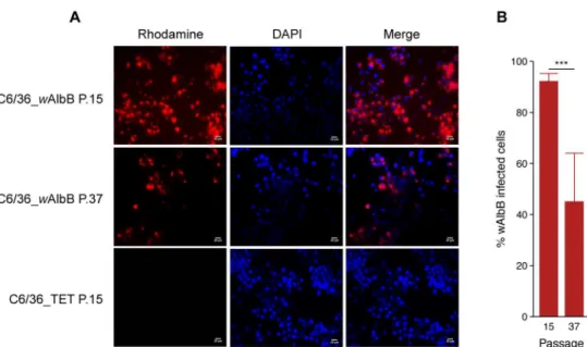
Fig 3. Proportion ofWolbachia-infected cells detected by Fluorescencein situHybridization. Rhodamine-labelled oligonucleotide probe designed onWolbachia16S rRNA gene (red) detected the bacteria in the cytoplasm of the host cell at passages P.15 and P.37 (A). Nuclei of the host cells are shown in blue after DAPI labelling (bars = 20μm). Percentage of cells with aWolbachia-positive signal in FISH at P.15 and P.37 (B) (Error bars represent the standard deviation of the mean of 50 independent microscope fields from three independent samples).
not complete with at least 4.81 log10CHIKV RNA copies per ng total RNA in C6/36_wAlbB
cells at day 1 pi, whereWolbachiaantiviral effect seemed to be the strongest. CHIKV inhibition bywAlbB was also measured at the RNA infectious particles level using FFA assay on cell supernatants (Fig 5B). A major decrease of viral infectious titer was detected in C6/36_wAlbB compared to C6/36_TET cells, depending on the time post-infection (Wolbachiatime
interac-tion,P= 0.00177). As for viral RNA, this suggests thatWolbachia-mediated inhibition of viral infectious particles production decreases with the time of infection, even if the time effect is lower than for viral RNA decrease. ThewAlbB density was monitored in both CHIKV infected (CHIKV+) and uninfected (CHIKV-) cells using qPCR (Fig 6). The bacterial load did not vary according to viral infection (P= 0.228) but time had a significant effect (P<2E-16). The Wolba-chiatiter increased with time, ranging from 13.3 to 25.7wsp/actinratio at day 1 and 7 pi, respectively.
Fig 4. Effect of tetracycline treatment on CHIKV growth in C6/36.Kinetics of CHIKV RNA titer at MOI 0.1 (A) and 3 (B) measured by RT-qPCR on total cellular RNA isolated from C6/36 cells (non infected by
Wolbachia) treated with tetracycline (TET) or not (CTRL). Error bars represent the standard deviation of the mean of two independent samples.
CHIKV infection of
wAlbB-colonized cells
The FISH technique was shown to be an efficient method to detect viruses in mosquito cells [44]. This is the first time such a technique was used to detect CHIKV. The oligonucleotide-probes designed can also detect other alphaviruses, namely Sindbis virus and Ross River virus (not shown). The results showed that CHIKV could be labelled in the cytoplasm of infected cells whereas no CHIKV signal was detected in uninfected cells (Fig 7). Moreover, viral RNA was also detected in cells previously infected withWolbachia, indicating that at least in some cells the virus is able to penetrate in spite of the presence of the bacterium. However, the co-localization of bothWolbachiaand CHIKV was not detected in many cells, and the use of
Fig 5. Effect ofWolbachiaon CHIKV replication and infectiosity.Kinetics at MOI 0.1 of CHIKV RNA titer measured by RT-qPCR on total cellular RNA (A) and CHIKV infectious titer in supernatant measured by FFA (B) in presence ofWolbachia(wAlbB) or in cells cured from the bacteria by tetracycline treatment (TET). Error bars represent the standard deviation of the mean of three independent samples.
FISH technique did not allowed us to tell if the presence of both micro-organisms in the same host cell was correlated with the load of either bacterium or virus.
Discussion
Ae.albopictusis naturally infected byWolbachiaand remains an important vector of CHIKV [45,46] and in a lesser extent of DENV [47,48]. Intriguingly, the pattern ofWolbachia -arbovi-rus interaction inAe.albopictusremains unclear. Previous studies showed that transinfection ofAe.albopictuswith thewMel strain ofWolbachiais likely to induce DENV and CHIKV inhi-bition [49,50]. However,Ae.albopictusis naturally co-infected withWolbachia wAlbA and
wAlbB strains but no blocking phenotype was measured against DENV and CHIKV in popula-tions from Houston [18,51] and La Réunion [26], respectively. Conversely, a decrease of DENV titer was observed in the saliva of symbiotic females in theAe.albopictuspopulation from La Réunion [25]. This suggests thatWolbachia's potential to interact with viral replication in its native mosquito host depends on the combination of bacterial strain, vector and virus fac-tors thus making the study of this multipartite interaction very complex. Therefore, simplified models are needed to exploreWolbachia-pathogen interaction in mosquito. AsWolbachiais an obligate intracellular bacterium, insect cell lines have been widely used for culturing the
Fig 6. Dynamics ofwAlbB in C6/36 during CHIKV infection.Ratio ofWolbachia wspcopies per hostactincopies during CHIKV infection at MOI 0.1, measured by qPCR on genomic DNA. Error bars represent the standard deviation of the mean of three independent samples.
Fig 7. Detection and localization ofwAlbB and CHIKVin celluloby FISH.Detection ofWolbachia16S rRNA gene (red) and CHIKVEnvRNA (green) using oligonucleotide probes labelled with Rhodamine and Alexa488, respectively.Wolbachiasignal is detected in C6/36_wAlbB but not in tetracycline treated cells (C6/36_TET). CHIKV signal is detected only in CHIKV infected modality, in the absence or in the presence ofWolbachiawhere it co-localize with the bacteria in the cytoplasm of C6/36_wAlbB cells. Nuclei of host cells are shown in blue after DAPI labelling (bars = 10μm).
bacterium with special emphasis onAe.albopictusderived cells [29,36,41,42,52–55]. It is worth noting that previous studies showed a high discrepancy inWolbachiadensity in cell culture [42,55,56], possibly due to cells passaging method that could result in reduction or loss of bac-terial infection [55]. The initial burst of infection followed by a rapid decrease ofwAlbB density in C6/36 could be interpreted as an adaptation of the bacterium to cell culture as previous work noticed a shift inWolbachiaphenotype in its natural host after long-term passage in mos-quito cell line [52]. It would be worth to study the mechanisms underpinningWolbachia estab-lishment in mosquito cell line and explore possible correlation with antiviral activity.
Moreover, mosquito cell lines are generally permissive to arbovirus infection, providing a use-ful tool to studyWolbachia-arbovirus interaction at a finer scale [21,35,51]. In adult mosquito, during the Extrinsic Incubation Period (EIP), the virus infects essentially somatic tissues including midgut and salivary glands [57] which are both infected byWolbachiainAe. albopic-tus[23]. C6/36 cells, which originated from uninfected somatic tissue, appear to be an appro-priate model in complement to Aa23 cells to studyWolbachia-arbovirus interaction in anAe.
albopictusbackground.
Previous studies suggested that DENV inhibition seems to depend onWolbachiadensity [51,58]. We showed that in C6/36,wAlbB density is highly dynamic but remains low compared to Aa23 with a maximum at 72.5wsp/actincopies against 1,888.3wsp/actin, respectively [51]. However, we observed a significant CHIKV interference in C6/36_wAlbB at a relative Wolba-chiadensity of 13.7 to 25.6wsp/actin, although inhibition was not complete. These results sug-gest thatWolbachia-mediated antiviral activity can occurin vitroeven at low bacterial density. Interestingly, Lu and colleagues extrapolated from their observations in Aa23_wAlbB cells that a relative density ofwAlbB of 0.3, 5.3, and 12.3wsp/actinin midgut, salivary gland and fat body ofAe.albopictus, respectively was too low to interfere with DENV infectionin vivo[51]. The lower abundance ofwAlbB inAe.albopictusorgans compared to C6/36 cells [23] is in line with this observation, and with the absence of CHIKV inhibition measuredin vivoinAe. albo-pictus. Conversely, the viral load did not seem to counteract with virus blocking byWolbachia
as demonstrated in C6/36_wMelPop-CLA cells infected with DENV [35]. Using theAe.aegypti
cell line Aag-2 to culturewMelPop-CLA, it was recently shown thatWolbachia-induced antivi-ral activity occurred as soon as the RNA replication step for DENV, but only at the step of virion assembly/secretion for WNV [21]. These results emphasize the importance of measuring both RNA and infectious particles to assessWolbachia-antiviral activity, and suggest that dis-tinct antiviral cellular mechanisms are involved duringWolbachia-virus interaction. In our model, CHIKV replication is inhibited bywAlbB in C6/36 cells, in a time-dependent manner with the lowest viral RNA load measured at 24 h pi. We also observed a decrease of infectious particles titer in supernatant as early as 24 h pi, indicating that viral blocking could occur at both stages of the viral cycle. This also suggests that CHIKV blocking bywAlbB could occur at the early stage of viral infection. Considering this, FISH was used to label bothWolbachiaand CHIKV during co-infection of C6/36 cells. The FISH experiment showed thatWolbachiaand CHIKV could be localized in the same host cell, indicating thatwAlbB did not seem to inhibit CHIKV infection by preventing viral entry, at least in some cells. This hypothesis is reinforced byin vivoconfocal microscopy whereWolbachiawas co-localized with DENV inAe.albopictus
salivary glands [25] as well inAe.aegyptitissues, where detection by FISH supported a cellular exclusion of DENV by thewMel strain ofWolbachia[19]. However, even ifWolbachia-virus co-infected cells or tissues are detected inAe.albopictusbothin vitroandin vivo, their magni-tude cannot exclude that viruses preferentially infectWolbachia-free compartment.
main one being the small interfering RNA (siRNA) pathway [59]. It was recently shown that C6/36 lacks a functional siRNA mechanism [60], suggesting that siRNA pathway is not involved inwAlbB-mediated CHIKV interference.Wolbachiawas shown to manipulate another RNA interference pathway, the micro-RNA (miRNA) pathway, to facilitate its own spread in the mosquito, and this mechanism could be involved in DENV interference [61,62]. It has been proposed thatWolbachia-induced antiviral phenotype relies through the activation of mosquito innate immune system, including Imd and Toll pathways [19]. However, a recent study usingDrosophilamutant’s deficient for Toll and Imd genes conclude that neither is required for the bacteria to inhibit DENV [32]. In the meantime, it has been suggested that
Wolbachiaand the virus could engage a direct competition for host cell resources, as under-lined by the importance of host cholesterol levels forDrosophilaC virus blocking inD. melano-gaster[27]. We demonstrated in previous work thatwMel manipulates iron metabolism inAe.
albopictusRML-12 cells through bacterioferritin expression [63], another potential explanation for its antiviral activity as iron load is involved in the modulation of innate immunity [64]. Fur-ther unexplored hypothesis is autophagy, a mechanism that has been shown recently to regu-lateWolbachiadensity across different arthropod hosts including mosquito cells [29]. The autophagy pathway is required by CHIKV to replicate [65], and this cellular function could be involved inWolbachiaantiviral interference.
Overall, insect cell lines may represent a promising tool to facilitate the understanding of
Wolbachia-pathogen interaction notably through electron microscopic observations of cell structural changes, and transcriptomic or proteomic studies which could allow to identify host infection regulatory pathways influenced byWolbachia[34,66–68]. The potential direct activ-ity ofWolbachiaderived compounds against pathogens remains unknown but need further exploration, especially in the light of recent results suggesting the direct anti-DENV activity of aChromobacteriumsp (Csp_P) isolated fromA.aegyptimidgut [69]. Our results showed a sig-nificant antiviral effect ofwAlbB against CHIKVin cellulothat was not measuredin vivoat the mosquito organ level, even if CHIKV RNA load was constraint in a smaller range in symbiotic females organs [26]. This emphasizes the need to better understandWolbachiasymbiosis in its native hostAe.albopictus, and its impact on vector competence [70]. Mosquito vector compe-tence for arboviruses depends on multiple factors such as mosquito genotype, virus genotype and their interaction [71] but also temperature [72,73] or mosquito microbiota [74]. Recent studies showed that pathogen blocking byWolbachiawas influenced by temperature [75] and that bacteria from the genusAsaiacan inhibit vertical transmission ofWolbachiainAn. gam-biae[76]. Together, these results underline the importance of exploringWolbachia-pathogen interaction, especially in a context whereWolbachia-infected mosquitoes represent a promis-ing strategy to control vector-borne diseases.
Supporting Information
S1 Fig. C6/36_wAlbB cells in transmission-light microscopy.Pictures in light microscopy of C6/36 cells infected byWolbachia(C6/36_wAlbB) or tetracycline-treated (C6/36_TET) during their growth in F25 cm2flasks, between two passages (bars = 20μm).
(TIF)
Acknowledgments
platform of SFR BioSciences Gerland Lyon Sud (UMS3444/US8) as well as the DTAMB plat-form of the FR41 Bio-Environment and Health (University Lyon 1) and the Centre Technolo-gique des Microstructures (University Lyon 1). This work was carried out within the
framework of Groupement de Recherche International (GDRI) Biodiversity and Infectious Diseases and Biodiversité et Développement Durable.
Author Contributions
Conceived and designed the experiments: VR CVM PM. Performed the experiments: VR CVM YS FHT. Analyzed the data: VR CVM PM. Contributed reagents/materials/analysis tools: PP PM. Wrote the paper: VR CVM PM.
References
1. WHO | About vector-borne diseases. In: WHO [Internet]. [cited 15 Nov 2014]. Available:http://www. who.int/campaigns/world-health-day/2014/vector-borne-diseases/en/
2. Tsetsarkin KA, Vanlandingham DL, McGee CE, Higgs S. A single mutation in chikungunya virus affects vector specificity and epidemic potential. PLoS Pathog. 2007; 3: e201. doi:10.1371/journal.ppat. 0030201PMID:18069894
3. Tsetsarkin KA, Weaver SC. Sequential adaptive mutations enhance efficient vector switching by Chi-kungunya virus and its epidemic emergence. PLoS Pathog. 2011; 7: e1002412. doi:10.1371/journal. ppat.1002412PMID:22174678
4. De Lamballerie X, Leroy E, Charrel RN, Ttsetsarkin K, Higgs S, Gould EA. Chikungunya virus adapts to tiger mosquito via evolutionary convergence: a sign of things to come? Virol J. 2008; 5: 33. doi:10. 1186/1743-422X-5-33PMID:18304328
5. Angelini R, Finarelli AC, Angelini P, Po C, Petropulacos K, Silvi G, et al. Chikungunya in north-eastern Italy: a summing up of the outbreak. Euro Surveill Bull Eur Sur Mal Transm Eur Commun Dis Bull. 2007; 12: E071122.2.
6. La Ruche G, Souarès Y, Armengaud A, Peloux-Petiot F, Delaunay P, Desprès P, et al. First two autoch-thonous dengue virus infections in metropolitan France, September 2010. Euro Surveill Bull Eur Sur Mal Transm Eur Commun Dis Bull. 2010; 15: 19676.
7. Leparc-Goffart I, Nougairede A, Cassadou S, Prat C, de Lamballerie X. Chikungunya in the Americas. Lancet. 2014; 383: 514. doi:10.1016/S0140-6736(14)60185-9PMID:24506907
8. WHO | Chikungunya–France. In: WHO [Internet]. [cited 15 Nov 2014]. Available:http://www.who.int/ csr/don/23-october-2014-chikungunya/en/
9. Bonizzoni M, Gasperi G, Chen X, James AA. The invasive mosquito speciesAedes albopictus: current knowledge and future perspectives. Trends Parasitol. 2013; 29: 460–468. doi:10.1016/j.pt.2013.07. 003PMID:23916878
10. Benedict MQ, Levine RS, Hawley WA, Lounibos LP. Spread of the tiger: global risk of invasion by the mosquitoAedes albopictus. Vector Borne Zoonotic Dis Larchmt N. 2007; 7: 76–85. doi:10.1089/vbz. 2006.0562PMID:17417960
11. David J-P, Coissac E, Melodelima C, Poupardin R, Riaz MA, Chandor-Proust A, et al. Transcriptome response to pollutants and insecticides in the dengue vectorAedes aegyptiusing next-generation sequencing technology. BMC Genomics. 2010; 11: 216. doi:10.1186/1471-2164-11-216PMID: 20356352
12. Christodoulou M. Biological vector control of mosquito-borne diseases. Lancet Infect Dis. 2011; 11: 84– 85. PMID:21351390
13. Slatko BE, Luck AN, Dobson SL, Foster JM.Wolbachiaendosymbionts and human disease control. Mol Biochem Parasitol. 2014; 195: 88–95. doi:10.1016/j.molbiopara.2014.07.004PMID:25046729 14. Bian G, Joshi D, Dong Y, Lu P, Zhou G, Pan X, et al.WolbachiaInvadesAnopheles stephensi
Popula-tions and Induces Refractoriness toPlasmodiumInfection. Science. 2013; 340: 748–751. doi:10.1126/ science.1236192PMID:23661760
15. Frentiu FD, Zakir T, Walker T, Popovici J, Pyke AT, van den Hurk A, et al. Limited dengue virus replica-tion in field-collectedAedes aegyptimosquitoes infected withWolbachia. PLoS Negl Trop Dis. 2014; 8: e2688. doi:10.1371/journal.pntd.0002688PMID:24587459
17. Werren JH, Baldo L, Clark ME.Wolbachia: master manipulators of invertebrate biology. Nat Rev Micro-biol. 2008; 6: 741–751. doi:10.1038/nrmicro1969PMID:18794912
18. Bian G, Xu Y, Lu P, Xie Y, Xi Z. The endosymbiotic bacteriumWolbachiainduces resistance to dengue virus inAedes aegypti. PLoS Pathog. 2010; 6: e1000833. doi:10.1371/journal.ppat.1000833PMID: 20368968
19. Moreira LA, Iturbe-Ormaetxe I, Jeffery JA, Lu G, Pyke AT, Hedges LM, et al. AWolbachiasymbiont in
Aedes aegyptilimits infection with Dengue, Chikungunya, andPlasmodium. Cell. 2009; 139: 1268– 1278. doi:10.1016/j.cell.2009.11.042PMID:20064373
20. Van den Hurk AF, Hall-Mendelin S, Pyke AT, Frentiu FD, McElroy K, Day A, et al. Impact ofWolbachia
on infection with chikungunya and yellow fever viruses in the mosquito vectorAedes aegypti. PLoS Negl Trop Dis. 2012; 6: e1892. doi:10.1371/journal.pntd.0001892PMID:23133693
21. Hussain M, Lu G, Torres S, Edmonds JH, Kay BH, Khromykh AA, et al. Effect ofWolbachiaon replica-tion of West Nile virus in a mosquito cell line and adult mosquitoes. J Virol. 2013; 87: 851–858. doi:10. 1128/JVI.01837-12PMID:23115298
22. Zélé F, Nicot A, Berthomieu A, Weill M, Duron O, Rivero A.Wolbachiaincreases susceptibility to Plas-modiuminfection in a natural system. Proc Biol Sci. 2014; 281: 20132837. doi:10.1098/rspb.2013. 2837PMID:24500167
23. Zouache K, Voronin D, Tran-Van V, Mousson L, Failloux A-B, Mavingui P. PersistentWolbachiaand cultivable bacteria infection in the reproductive and somatic tissues of the mosquito vectorAedes albo-pictus. PloS One. 2009; 4: e6388. doi:10.1371/journal.pone.0006388PMID:19633721
24. Zouache K, Raharimalala FN, Raquin V, Tran-Van V, Raveloson LHR, Ravelonandro P, et al. Bacterial diversity of field-caught mosquitoes,Aedes albopictusandAedes aegypti, from different geographic regions of Madagascar. FEMS Microbiol Ecol. 2011; 75: 377–389. doi:10.1111/j.1574-6941.2010. 01012.xPMID:21175696
25. Mousson L, Zouache K, Arias-Goeta C, Raquin V, Mavingui P, Failloux A-B. The NativeWolbachia
Symbionts Limit Transmission of Dengue Virus inAedes albopictus. PLoS Negl Trop Dis. 2012; 6: e1989. doi:10.1371/journal.pntd.0001989PMID:23301109
26. Mousson L, Martin E, Zouache K, Madec Y, Mavingui P, Failloux AB.Wolbachiamodulates Chikungu-nya replication inAedes albopictus. Mol Ecol. 2010; 19: 1953–1964. doi:10.1111/j.1365-294X.2010. 04606.xPMID:20345686
27. Caragata EP, Rancès E, Hedges LM, Gofton AW, Johnson KN, O’Neill SL, et al. Dietary cholesterol modulates pathogen blocking byWolbachia. PLoS Pathog. 2013; 9: e1003459. doi:10.1371/journal. ppat.1003459PMID:23825950
28. Caragata EP, Rancès E, O’Neill SL, McGraw EA. Competition for amino acids betweenWolbachiaand the mosquito host,Aedes aegypti. Microb Ecol. 2014; 67: 205–218. doi:10.1007/s00248-013-0339-4 PMID:24337107
29. Voronin D, Cook DAN, Steven A, Taylor MJ. Autophagy regulatesWolbachiapopulations across diverse symbiotic associations. Proc Natl Acad Sci U S A. 2012; 109: E1638–1646. doi:10.1073/pnas. 1203519109PMID:22645363
30. Brennan LJ, Keddie BA, Braig HR, Harris HL. The endosymbiontWolbachiapipientis induces the expression of host antioxidant proteins in anAedes albopictuscell line. PloS One. 2008; 3: e2083. doi: 10.1371/journal.pone.0002083PMID:18461124
31. Hussain M, Frentiu FD, Moreira LA, O’Neill SL, Asgari S.Wolbachiauses host microRNAs to manipu-late host gene expression and facilitate colonization of the dengue vectorAedes aegypti. Proc Natl Acad Sci U S A. 2011; 108: 9250–9255. doi:10.1073/pnas.1105469108PMID:21576469
32. Rancès E, Ye YH, Woolfit M, McGraw EA, O’Neill SL. The relative importance of innate immune priming inWolbachia-mediated dengue interference. PLoS Pathog. 2012; 8: e1002548. doi:10.1371/journal. ppat.1002548PMID:22383881
33. Hussain M, O’Neill SL, Asgari S.Wolbachiainterferes with the intracellular distribution of Argonaute 1 in the dengue vectorAedes aegyptiby manipulating the host microRNAs. RNA Biol. 2013; 10: 1868– 1875. doi:10.4161/rna.27392PMID:24351659
34. Hughes GL, Ren X, Ramirez JL, Sakamoto JM, Bailey JA, Jedlicka AE, et al.Wolbachiainfections in
Anopheles gambiaecells: transcriptomic characterization of a novel host-symbiont interaction. PLoS Pathog. 2011; 7: e1001296. doi:10.1371/journal.ppat.1001296PMID:21379333
35. Frentiu FD, Robinson J, Young PR, McGraw EA, O’Neill SL.Wolbachia-mediated resistance to dengue virus infection and death at the cellular level. PloS One. 2010; 5: e13398. doi:10.1371/journal.pone. 0013398PMID:20976219
37. Schuffenecker I, Iteman I, Michault A, Murri S, Frangeul L, Vaney M-C, et al. Genome microevolution of chikungunya viruses causing the Indian Ocean outbreak. PLoS Med. 2006; 3: e263. doi:10.1371/ journal.pmed.0030263PMID:16700631
38. Dubrulle M, Mousson L, Moutailler S, Vazeille M, Failloux A-B. Chikungunya virus andAedes mosqui-toes: saliva is infectious as soon as two days after oral infection. PloS One. 2009; 4: e5895. doi:10. 1371/journal.pone.0005895PMID:19521520
39. Payne AF, Binduga-Gajewska I, Kauffman EB, Kramer LD. Quantitation of flaviviruses by fluorescent focus assay. J Virol Methods. 2006; 134: 183–189. doi:10.1016/j.jviromet.2006.01.003PMID: 16510196
40. Venard CM-P, Crain PR, Dobson SL. SYTO11 staining vs FISH staining: a comparison of two methods to stainWolbachia pipientisin cell cultures. Lett Appl Microbiol. 2011; 52: 168–176. doi:10.1111/j. 1472-765X.2010.02986.xPMID:21214605
41. Fenollar F, La Scola B, Inokuma H, Dumler JS, Taylor MJ, Raoult D. Culture and phenotypic characteri-zation of aWolbachia pipientisisolate. J Clin Microbiol. 2003; 41: 5434–5441. PMID:14662922 42. Voronin D, Tran-Van V, Potier P, Mavingui P. Transinfection and growth discrepancy ofDrosophila
WolbachiastrainwMel in cell lines of the mosquitoAedes albopictus. J Appl Microbiol. 2010; 108: 2133–2141. doi:10.1111/j.1365-2672.2009.04621.xPMID:19951376
43. Osborne SE, Leong YS, O’Neill SL, Johnson KN. Variation in antiviral protection mediated by different
Wolbachiastrains inDrosophila simulans. PLoS Pathog. 2009; 5: e1000656. doi:10.1371/journal.ppat. 1000656PMID:19911047
44. Raquin V, Wannagat M, Zouache K, Legras-Lachuer C, Moro CV, Mavingui P. Detection of dengue group viruses by fluorescencein situhybridization. Parasit Vectors. 2012; 5: 243. doi: 10.1186/1756-3305-5-243PMID:23110979
45. Bonilauri P, Bellini R, Calzolari M, Angelini R, Venturi L, Fallacara F, et al. Chikungunya virus inAedes albopictus, Italy. Emerg Infect Dis. 2008; 14: 852–854. doi:10.3201/eid1405.071144PMID:18439383 46. Tsetsarkin KA, Chen R, Yun R, Rossi SL, Plante KS, Guerbois M, et al. Multi-peaked adaptive
land-scape for chikungunya virus evolution predicts continued fitness optimization inAedes albopictus mos-quitoes. Nat Commun. 2014; 5: 4084. doi:10.1038/ncomms5084PMID:24933611
47. Delatte H, Paupy C, Dehecq JS, Thiria J, Failloux AB, Fontenille D. [Aedes albopictus, vector of chikun-gunya and dengue viruses in Reunion Island: biology and control]. Parasite Paris Fr. 2008; 15: 3–13. 48. Ponlawat A, Scott JG, Harrington LC. Insecticide susceptibility ofAedes aegyptiandAedes albopictus
across Thailand. J Med Entomol. 2005; 42: 821–825. PMID:16363166
49. Blagrove MSC, Arias-Goeta C, Di Genua C, Failloux A-B, Sinkins SP. AWolbachia wMel transinfection inAedes albopictusis not detrimental to host fitness and inhibits Chikungunya virus. PLoS Negl Trop Dis. 2013; 7: e2152. doi:10.1371/journal.pntd.0002152PMID:23556030
50. Blagrove MSC, Arias-Goeta C, Failloux A-B, Sinkins SP.WolbachiastrainwMel induces cytoplasmic incompatibility and blocks dengue transmission inAedes albopictus. Proc Natl Acad Sci U S A. 2012; 109: 255–260. doi:10.1073/pnas.1112021108PMID:22123944
51. Lu P, Bian G, Pan X, Xi Z.WolbachiaInduces Density-Dependent Inhibition to Dengue Virus in Mos-quito Cells. PLoS Negl Trop Dis. 2012; 6: e1754. doi:10.1371/journal.pntd.0001754PMID:22848774 52. McMeniman CJ, Lane AM, Fong AWC, Voronin DA, Iturbe-Ormaetxe I, Yamada R, et al. Host adapta-tion of aWolbachiastrain after long-term serial passage in mosquito cell lines. Appl Environ Microbiol. 2008; 74: 6963–6969. doi:10.1128/AEM.01038-08PMID:18836024
53. Fallon AM, Witthuhn BA. Proteasome activity in a naïve mosquito cell line infected withWolbachia pipientis wAlbB. In Vitro Cell Dev Biol Anim. 2009; 45: 460–466. doi:10.1007/s11626-009-9193-6 PMID:19296184
54. Fallon AM, Baldridge GD, Higgins LA, Witthuhn BA.Wolbachiafrom the planthopperLaodelphax stria-tellusestablishes a robust, persistent, streptomycin-resistant infection in clonal mosquito cells. In Vitro Cell Dev Biol Anim. 2013; 49: 66–73. doi:10.1007/s11626-012-9571-3PMID:23271364
55. Khoo CCH, Venard CMP, Fu Y, Mercer DR, Dobson SL. Infection, growth and maintenance of Wolba-chia pipientisin clonal and non-clonalAedes albopictuscell cultures. Bull Entomol Res. 2013; 103: 251–260. doi:10.1017/S0007485312000648PMID:23113940
56. Noda H, Miyoshi T, Koizumi Y. In vitro cultivation ofWolbachiain insect and mammalian cell lines. In Vitro Cell Dev Biol Anim. 2002; 38: 423–427. doi:10.1290/1071-2690(2002)038<0423:IVCOWI>2.0.
CO;2PMID:12534342
58. Osborne SE, Iturbe-Ormaetxe I, Brownlie JC, O’Neill SL, Johnson KN. Antiviral Protection and the Importance ofWolbachiaDensity and Tissue Tropism inDrosophila simulans. Appl Environ Microbiol. 2012; 78: 6922–6929. doi:10.1128/AEM.01727-12PMID:22843518
59. Kingsolver MB, Huang Z, Hardy RW. Insect antiviral innate immunity: pathways, effectors, and connec-tions. J Mol Biol. 2013; 425: 4921–4936. doi:10.1016/j.jmb.2013.10.006PMID:24120681
60. Brackney DE, Scott JC, Sagawa F, Woodward JE, Miller NA, Schilkey FD, et al. C6/36Aedes albopic-tuscells have a dysfunctional antiviral RNA interference response. PLoS Negl Trop Dis. 2010; 4: e856. doi:10.1371/journal.pntd.0000856PMID:21049065
61. Zhang G, Hussain M, O’Neill SL, Asgari S.Wolbachiauses a host microRNA to regulate transcripts of a methyltransferase, contributing to dengue virus inhibition inAedes aegypti. Proc Natl Acad Sci U S A. 2013; 110: 10276–10281. doi:10.1073/pnas.1303603110PMID:23733960
62. Zhang G, Hussain M, Asgari S. Regulation of arginine methyltransferase 3 by aWolbachia-induced microRNA inAedes aegyptiand its effect onWolbachiaand dengue virus replication. Insect Biochem Mol Biol. 2014; 53: 81–88. doi:10.1016/j.ibmb.2014.08.003PMID:25158106
63. Kremer N, Voronin D, Charif D, Mavingui P, Mollereau B, Vavre F.Wolbachiainterferes with ferritin expression and iron metabolism in insects. PLoS Pathog. 2009; 5: e1000630. doi:10.1371/journal. ppat.1000630PMID:19851452
64. Schaible UE, Kaufmann SHE. Iron and microbial infection. Nat Rev Microbiol. 2004; 2: 946–953. doi: 10.1038/nrmicro1046PMID:15550940
65. Krejbich-Trotot P, Gay B, Li-Pat-Yuen G, Hoarau J-J, Jaffar-Bandjee M-C, Briant L, et al. Chikungunya triggers an autophagic process which promotes viral replication. Virol J. 2011; 8: 432. doi:10.1186/ 1743-422X-8-432PMID:21902836
66. Baldridge GD, Baldridge AS, Witthuhn BA, Higgins L, Markowski TW, Fallon AM. Proteomic profiling of a robustWolbachiainfection in anAedes albopictusmosquito cell line. Mol Microbiol. 2014; 94: 537– 556. doi:10.1111/mmi.12768PMID:25155417
67. Darby AC, Christina Gill A, Armstrong SD, Hartley CS, Xia D, Wastling JM, et al. Integrated transcrip-tomic and proteomic analysis of the global response ofWolbachiato doxycycline-induced stress. ISME J. 2014; 8: 925–937. doi:10.1038/ismej.2013.192PMID:24152719
68. Kambris Z, Blagborough AM, Pinto SB, Blagrove MSC, Godfray HCJ, Sinden RE, et al.Wolbachia stim-ulates immune gene expression and inhibitsPlasmodiumdevelopment inAnopheles gambiae. PLoS Pathog. 2010; 6: e1001143. doi:10.1371/journal.ppat.1001143PMID:20949079
69. Ramirez JL, Short SM, Bahia AC, Saraiva RG, Dong Y, Kang S, et al.Chromobacterium Csp_P Reduces Malaria and Dengue Infection in Vector Mosquitoes and Has Entomopathogenic and In Vitro Anti-pathogen Activities. PLoS Pathog. 2014; 10: e1004398. doi:10.1371/journal.ppat.1004398PMID: 25340821
70. Bourtzis K, Dobson SL, Xi Z, Rasgon JL, Calvitti M, Moreira LA, et al. Harnessing mosquito-Wolbachia
symbiosis for vector and disease control. Acta Trop. 2014; 132 Suppl: S150–163. doi:10.1016/j. actatropica.2013.11.004PMID:24252486
71. Fansiri T, Fontaine A, Diancourt L, Caro V, Thaisomboonsuk B, Richardson JH, et al. Genetic mapping of specific interactions betweenAedes aegyptimosquitoes and dengue viruses. PLoS Genet. 2013; 9: e1003621. doi:10.1371/journal.pgen.1003621PMID:23935524
72. Richards SL, Anderson SL, Lord CC, Tabachnick WJ. Effects of virus dose and extrinsic incubation temperature on vector competence ofCulex nigripalpus(Diptera:Culicidae) for St. Louis encephalitis virus. J Med Entomol. 2012; 49: 1502–1506. PMID:23270182
73. Carrington LB, Seifert SN, Armijos MV, Lambrechts L, Scott TW. Reduction ofAedes aegyptivector competence for dengue virus under large temperature fluctuations. Am J Trop Med Hyg. 2013; 88: 689–697. doi:10.4269/ajtmh.12-0488PMID:23438766
74. Jupatanakul N, Sim S, Dimopoulos G. The Insect Microbiome Modulates Vector Competence for Arbo-viruses. Viruses. 2014; 6: 4294–4313. doi:10.3390/v6114294PMID:25393895
75. Murdock CC, Blanford S, Hughes GL, Rasgon JL, Thomas MB. Temperature alters Plasmodium block-ing byWolbachia. Sci Rep. 2014; 4: 3932. doi:10.1038/srep03932PMID:24488176
76. Hughes GL, Dodson BL, Johnson RM, Murdock CC, Tsujimoto H, Suzuki Y, et al. Native microbiome impedes vertical transmission ofWolbachiainAnopheles mosquitoes. Proc Natl Acad Sci U S A. 2014; 111: 12498–12503. doi:10.1073/pnas.1408888111PMID:25114252
78. Vazeille M, Moutailler S, Coudrier D, Rousseaux C, Khun H, Huerre M, et al. Two Chikungunya isolates from the outbreak of La Reunion (Indian Ocean) exhibit different patterns of infection in the mosquito,
Aedes albopictus. PloS One. 2007; 2: e1168. doi:10.1371/journal.pone.0001168PMID:18000540 79. Sanguin H, Herrera A, Oger-Desfeux C, Dechesne A, Simonet P, Navarro E, et al. Development and
validation of a prototype 16S rRNA-based taxonomic microarray forAlphaproteobacteria. Environ Microbiol. 2006; 8: 289–307. doi:10.1111/j.1462-2920.2005.00895.xPMID:16423016