K. I. R. Teixeira1, P. V. Araújo1, R. D. Sinisterra2, M. E. Cortés1*
1
Faculdade de Odontologia, Departamento de Odontologia Restauradora, Universidade Federal de Minas Gerais, Belo Horizonte,
MG, Brasil; 2 Departamento de Química, Instituto de Ciências Exatas, Universidade Federal de Minas Gerais, Belo Horizonte, MG, Brasil.
Submitted: September 08, 2010; Returned to authors for corrections: February 03, 2011; Approved: June 07, 2012.
ABSTRACT
Chlorhexidine (Cx) augmented with beta-cyclodextrin (β-cd) inclusion compounds, termed Cx:β-cd
complexes, have been developed for use as antiseptic agents. The aim of this study was to examine the
interactions of Cx:β-cd complexes, prepared at different molecular ratios, with sterol and yeast membranes.
The Minimal Inhibitory Concentration (MIC) against the yeast Candida albicans (C.a.) was determined for
each complex; the MICs were found to range from 0.5 to 2 µg/mL. To confirm the MIC data, quantitative
analysis of viable cells was performed using trypan blue staining. Mechanistic characterization of the
interactions that the Cx:β-cd complexes have with the yeast membrane and assessment of membrane
morphology following exposure to Cx:β-cd complexes were performed using Sterol Quantification Method
analysis (SQM) and scanning electron microscopy (SEM). SQM revealed that sterol extraction increased
with increasing β-cd concentrations (1.71 × 10³; 1.4 × 10³; 3.45 × 10³, and 3.74 × 10³ CFU for 1:1, 1:2, 1:3, and 1:4, respectively), likely as a consequence of membrane ergosterol solubilization. SEM images
demonstrated that cell membrane damage is a visible and significant mechanism that contributes to the
antimicrobial effects of Cx:β-cd complexes. Cell disorganization increased significantly as the proportion of β-cyclodextrin present in the complex increased. Morphology of cells exposed to complexes with 1:3 and 1:4 molar ratios of Cx:β-cd were observed to have large aggregates mixed with yeast remains, representing
more membrane disruption than that observed in cells treated with Cx alone. In conclusion, nanoaggregates
of Cx:β-cd complexes block yeast growth via ergosterol extraction, permeabilizing the membrane by
creating cluster-like structures within the cell membrane, possibly due to high amounts of hydrogen bonding.
Key words: Chlorhexidine, β-cyclodextrin, membrane-drug interactions
INTRODUCTION
Chlorhexidine (Cx) is an effective chemical antiseptic that
has a bactericidal effect at high concentrations (2-5%) and a
bacteriostatic effect at low concentrations (0.6%). The
antimicrobial activity of Cx is thought to be due to bacterial
membrane disruption, which leads to increased cell
permeability and leakage of intracellular ions (Na+, K)(17, 26,
36, 37). Agents containing high concentrations of Cx have
been used in periodontic and endodontic clinical practice. Cx
has also been safely used at very low concentrations in several
commercial products for dental hygiene, including
mouthwashes, gels, and sprays. However the transitory effects
of Cx have been shown to have deleterious effects on cells in
vitro (15, 17, 40).
Cyclodextrins have been used as tools to manipulate the
lipid composition of biological and model membranes (29).
Their hydrophilic outer surface and hydrophobic inner core
enable them to form inclusion complexes by trapping small
amphiphilic molecules in their core. Beta-cyclodextrins (β-cds)
are particularly efficient sterol acceptors, apparently because
the size of their inner hydrophobic cavity matches the size of
the sterol molecule (18, 28, 30, 34). Indeed, Cholesterol can be
removed from cells by extraction into small unilamellar
vesicles of β-cds (5, 6, 7).
Hence, in order to reduce the toxicity caused by high
concentrations of Cx, chlorhexidine:beta-cyclodextrin (Cx:β
-cd) complexes have been developed for use as antiseptic
agents. These complexes offer advantages in effectiveness,
long-term activity, and antimicrobial efficacy at low
concentrations (10). Their key component, β-cd, creates
regions of hydrophobic cavity that surround Cx. Gels
containing Cx:β-cd complexes were shown to exhibit higher
antimicrobial activity against oral microorganismal pathogens
than non-augmented Cx in a long-term in vitro study (10).
However, previous studies characterizing Cx activity provide
little information regarding the mechanisms underlying
microorganism-drug interactions on a nanometer scale.
Understanding the structure and properties of microbial
surfaces at the nanometer level is of great importance for the
development of novel antimicrobial compounds.
Candida species, including Candida albicans (C.a.), are
commensal organisms that proliferate when the body is out of
homeostasis, leading to the development of disease. Although
it is almost universally described as dimorphic, C.a.’s cell
morphology actually lies on a pleomorphic continuum, ranging
from ovoid yeast cells to filamentous hyphae (1, 2, 12, 13, 27,
42). This yeast has developed specific mechanisms of virulence
that allow it to colonize epithelial cells of the host for the
purpose of invading deeper layers or influencing the body's
defenses. Interestingly, exposure of C.a. to 0.1% Cx has been
shown to result in cell wall changes, leading to a loss of
intracellular components and aggregation of nuclear proteins
(2, 13, 14,16 19, 21, 22, 23).
Loftsson and Brewster (24) have shown that molecular
envelopment of Cx with β-cd increases the gradient
concentration at the membrane site. This increase most likely
occurs due to Cx:β-cd developing a stronger interaction the cell
membrane than that which forms between Cx and the
membrane. Therefore, complexes of Cx with β-cd inclusion
compounds may represent more effective microbicidal agents
than pure Cx. Although data regarding the interactions between
Cx inclusion complexes and biological membranes are limited,
these complexes have attracted interest in many fields, as they
could be used to release biologically-active drugs through
membrane contact in diverse tissues (35).
The aim of this study was to investigate the anti-fungal
activity of Cx inclusion complexes comprised of different
Cx:β-cd molar ratios and to characterize the interactions of
these complexes with yeast membranes using a C.a. strain as
an experimental model.
MATERIALS AND METHODS
Chemicals and yeast stocks
Chlorhydrate of Cx was obtained from Ecadil® (SP, Brazil), and β-cd from Cerestar®, Co. (Milwaukee, WI, USA). Fluconazole was obtained from Pfizer® Pharmaceutics Group (New York, NY, USA). Sabouraud dextrose agar (SDA) and
broth (SDB) were purchased from Biobras S.A® (MG, Brazil).
C.a. (ATCC 28804) was obtained from the American Type
and solvents were of analytical grade.
Preparation of supramolecular complexes
Cx:β-cd inclusion complexes were prepared by
freeze-drying aqueous solutions of β-cd and Cx at different relative
molar ratios (Cx:β-cd: 1:1, 1:2, 1:3, and 1:4) as described
elsewhere (9).
Antimicrobial assay
Yeast cultures were grown and maintained in Sabouraud
dextrose broth (SDB) and Sabouraud dextrose agar. Prior to the
assay, C.a. cells were cultured for 24 h at 34 °C under aerobic
conditions in SDB. Yeast cultures were then further diluted to
obtain a density of 1.5 × 10⁸ CFU/mL for inoculums. Minimal Inhibitory Concentration (MIC90) is the lowest concentration of drug that inhibits more than 90% of a microorganism
population and MIC90 is the lowest concentration of drug that inhibits 90% of the microorganism population. MIC90 was determined using the macrodilution method (8). Briefly,
aliquots containing 500 µL of 128 µg/mL Cx or 1:1; 1:2; 1:3,
1:4 Cx:β-cd solutions were serially diluted in 3.4 mL of SDB.
Normalized Cx or Cx:β-cd dilutions were then inoculated with
100 µL of C.a. inoculums. Cx alone and fluconazole (FLZ)
were used as standard antimicrobial positive controls and β-cd
was used as a negative control (8).
Sample absorbance was measured using a standard
spectrophotometer (Spectrumlamb 22PC®) set to 580 nm, and sample data were analyzed and compared to controls. Six
replicates were analyzed for each condition, and MIC values
were recorded and evaluated using the non-parametric
Kruskal-Wallis test. P-values less than 0.05 were considered significant.
Yeast viability assay
The concentration of cells in a 0.5 µg/mL suspension was
determined using a standard hemocytometer. The yeast
suspension was diluted 100-fold with sterile water. Cells were
then diluted in an equal volume of 0.4% trypan blue to assess
the number of viable cells. Total cell counts were also
performed with a hemocytometer. Calculations involving cell
populations were based on viable cell counts (33).
Sterol Quantification Method (SQM)
Total intracellular sterols were extracted using the SQM
described by Arthington-Skaggs et al. (3) with slight
modifications. Briefly, a single C.a. colony from an SBD agar
plate incubated overnight was used to inoculate SBD broth
containing 0, 1, 2, 4, or 8 μg/mL of Cx or Cx:β-cd inclusion
compounds (1:1, 1:2, 1:3, 1:4). The cultures were incubated
with agitation for 16 h at 34 °C. Stationary-phase cells were
harvested by centrifugation at 2700 rpm in a centrifuge for 5
min and washed once with sterile distilled water. The net wet
weight of the cell pellet was determined. To each pellet was
added 3 mL of a 25% alcoholic potassium hydroxide solution,
followed by vortexing for 1 min. Cell suspensions were
incubated at 85 °C in a water bath for 1 h. Following this
incubation, the tubes were allowed to cool to room
temperature. Sterols were then extracted by the addition of a
mixture of 1 mL sterile distilled water and 3 mL n-heptane,
followed by vigorous vortexing for 3 min. The heptane layer
was transferred to a clean tube and stored at 20 °C for 24 h.
Prior to analysis, a 20-µl aliquot of sterol extract was diluted
five times in 100% ethanol and analyzed spectroscopically over
a range of wavelengths from 240 to 300 nm. The presence of
ergosterol and the late sterol intermediate 24(28)DHE in the
extracted sample resulted in a characteristic four-peaked curve.
A dose-dependent decrease in the height of the absorbance
peaks was evident, corresponding to decreased ergosterol
concentration. Ergosterol content was calculated as a
percentage of the wet weight of the cells using the following
equations:
% ergosterol % + 24(28) DHE = [(A281.5/ 290) F] / pellet weight
% 24(28) DHE = [(A230/518) x F] / pellet weight
Where F is the factor for dilution in ethanol and 290 and
518 are the E values (%/cm) determined for crystalline
ergosterol and 24(28)DHE, respectively.
Scanning electron microscopy (SEM)
SEM was used to assess cellular membrane integrity and
morphologic changes caused by the drug treatments. Yeast
suspensions (1 mL) treated with inclusion complexes were
centrifuged at 3000 rpm for 5 min, and the resulting pellets
were pre-fixed at 4 °C for 3 h in 2.5% glutaraldehyde in 0.1 M
cacodylate buffer (pH 7.2). After fixation, 5-µl aliquots of this
dilution were added to a solid substrate made of mica. A
contact tapping mode fluid cell was sealed against a freshly
cleaved muscovite mica substrate using a silicone O-ring.
Samples were dried for 7 d at 37 °C. Samples were then coated
with gold in a Penning sputter system in a high-vacuum
chamber and observed using a field emission scanning electron
microscope (model Zeiss-DSM 950). Micrographs were
obtained using secondary electrons.
RESULTS
Antifungal activity
The MIC90 data are summarized in Table 1. Cx alone exhibited robust fungicidal activity against C.a., with a MIC90 value of 2 μg/mL. The MIC90 value of the 1:4 Cx:β-cd complex, 0.5 μg/mL, differed significantly from that of Cx .
Similar patterns were found with the 1:2 and 1:3 Cx:β-cd
complexes, which exhibited MIC values of ~1 μg/mL.
Results from cell counts of viable cells obtained using a
hemocytometer were in agreement with the MIC90 values (Table 1). For cells treated with 0.5 µg/mL Cx, the number of
viable cells after treatment was 3.55 × 10³. Increasing concentrations of β-cd in Cx:β-cd complexes (1:1, 1:2, 1:3, and
1:4) led to concomitant decreases in cell viability. . Indeed, as
can be seen in Table 1, the 1:4 complex resulted in a 4-fold
reduction in C.a growth (0.5 µg/ mL), relative to Cx alone.
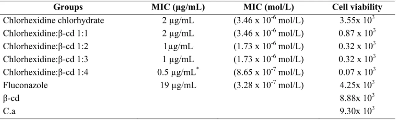
Table 1. Inhibition concentration of Cx and Cx:β-cd inclusion compounds (1:1; 1:2; 1:3; 1:4) against Candida albicans and viable
cell counting.
Groups MIC (µg/mL) MIC (mol/L) Cell viability
Chlorhexidine chlorhydrate 2 µg/mL (3.46 x 10⁻⁶ mol/L) 3.55x 10³ Chlorhexidine:β-cd 1:1 2 µg/mL (3.46 x 10⁻⁶ mol/L) 0.87 x 10³
Chlorhexidine:β-cd 1:2 1µg/mL (1.73 x 10⁻⁶ mol/L) 0.32 x 10³
Chlorhexidine:β-cd 1:3 1 µg/mL (1.73 x 10⁻⁶ mol/L) 0.32 x 10³ Chlorhexidine:β-cd 1:4 0.5 µg/mL* (8.65 x 10⁻⁷ mol/L) 0.07 x 10³
Fluconazole 19 µg/mL (3.28 x 10⁻⁷ mol/L) 4.25x 10³
β-cd 8.88x 10³
C.a 9.30x 10³
P<0.01 significance level
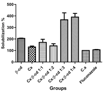
Effects of Cx:β-cd complexes on sterols as shown by SQM
analysis
Yeast was treated for 24 h with the complexes analyzed at
concentrations equivalent to the MIC90 values. Spectrophotometric analysis (wavelength range, 240–330 nm)
showed that treatment with Cx alone or with the inclusion
compounds, particularly Cx:β-cd 1:3 and 1:4, led to significant
reductions in ergosterol levels (Fig. 1). Membrane leakage
increased progressively as cells were exposed to greater
concentrations of β-cd (1:1, 1:2 vs. 1:3 or 1:4, P <0.05).
Furthermore, the 1:3 and 1:4 exposed cells showed significant
increases in ergosterol levels in the SQM test, 100% (3.15 to
-cd
Cx
-cd 1:1 Cx:
-cd 1 :2
Cx:
-cd 1:3 Cx:
-cd 1:4 Cx:
C.a
Fluc onaz
ole 0
100 200 300 400 500
Groups
S
o
lu
b
il
iz
a
ti
o
n
%
Figure 1. Percentage of solubilization of Candida albicans
membrane treated with Chlorhydrate of Chlorhexidine,
Chlorhexidine:β-cyclodextrin 1:1, 1:2, 1:3, 1:4; β-cyclodextrin
and Fluconazole.
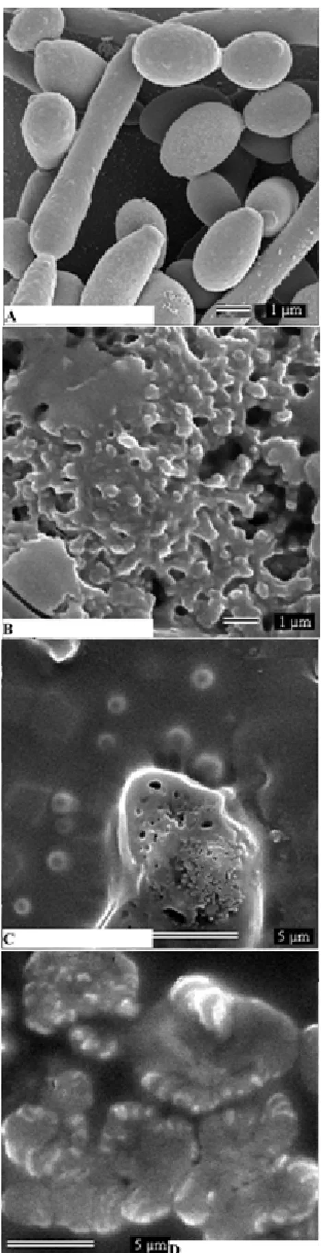
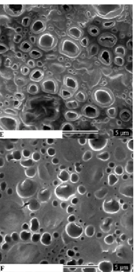
SEM analysis of Cx:β-cd complex interactions with
membranes
SEM revealed crystalline structures in some regions of the
membranes that were exposed to Cx (see micrograph in Fig.
2A). The β-cd alone control molecules (Fig. 2B) produced a
semi-crystalline structure. However, the addition of β-cd
molecules appeared to disrupt the crystalline character of the
Cx structures. That is, with increasing ratios of β-cd in the
Cx:β-cd complexes from 1:1 to 1:4 (Fig. 2C-F) —and therefore
increasing concentration of β-cd being delivered to the cellular
membranes—the membrane-associated Cx molecules appeared
to lose their regular crystalline appearance, becoming
increasingly amorphous (Fig. 3C-D).
C.a. cells treated with Cx and Cx:β-cd complexes were
also analyzed by SEM; untreated C.a. cells served as controls
to assess normal yeast membrane morphology (Fig. 3A). In the
Cx alone treatment group, Cx molecules surrounded the yeast
cells, binding the membranes only at singular points (Fig. 3B).
Structures resembling Cx were visualized around the yeast
cellular membrane, generally near one or two break points in
the membrane. Initial morphological changes in Cx-treated
cells included the appearance of indentations on the surface of
some cells as well as the presence of micelle-like structures and
membrane residues near the cells (Fig. 3C-D).
The membranes of C.a. cells treated with Cx:β-cd
complexes (1:1, 1:2, 1:3, and 1:4 ratios) showed slight
differences in the size and shape of the liquid-ordered domain
relative to membranes exposed to Cx alone (Fig. 3C-F), and
showed various leakage points in the membrane structure that
were not seen in Cx-exposed membranes (Fig. 3B). Exposure
of C.a. to Cx:β-cd complexes at a concentration of 1 μg/mL led
to dramatic changes in membrane structure. In these samples,
the C.a. membrane domains and inclusion compounds
coalesced into larger, less rounded domains (Fig. 3E-F).
Additionally, the cells appeared to lose their characteristic
shape and to allow leakage of intracellular substances (Fig. 3
Figure 2. SEM micrographs of Chlorhexidine Chlorhydrate (A),
Figure 3. SEM micrographs of Candida albicans untreated (A)
and C.a cells treated with Chlorhydrate Chlorhexidine (B),
Chlorhexidine:β-cyclodextrin 1:1(C), 1:2 (D), 1:3 (E), 1:4 (F).
DISCUSSION
In this study, enveloping Cx in β-cd inclusion compounds
was shown to significantly alter the inhibitory effect of Cx
exposure on C.a., and greater molar ratios of β-cd were
associated with enhanced antifungal activity. The heightened
antifungal activity of the Cx:β-cd complexes, especially the
1:4 complex, can be explained by the observed greater
adhesion efficiency of these complexes with the membrane
versus free Cx or β-cd. Indeed, as can be seen in Table 1, the
1:4 complex resulted in a 4-fold reduction in C.a growth (0.5
µg/ mL), relative to Cx alone.
Importantly, our cell counts of viable cells and MIC value
data were in agreement with one another. The heightened
antifungal activity that is produced by enveloping Cx in β-cd
inclusion molecules should allow Cx to be efficacious at lower
concentrations. The antifungal activity on C.a. of Cx alone
observed here was consistent with previous research, which
reported that Cx inhibited C.a at a concentration of 2.19 µg/
mL (16).
Our SQM findings showing that the 1:4 ratio Cx:β-cd
complex produced greater ergosterol extraction than the other
lower ratio complexes indicate that the complex was more
potent at the 1:4 ratio than at the lower ratio solutions analyzed.
On the other hand, the toxicity of antibiotics, such as Cx,
toward human cells may be reduced in the presence of β-cd
inclusion compound molecules since the antibiotic molecules
must then compete with phospholipids in order to interact with
cholesterol and form pores (25, 32).
Our SEM images showed that exposure of cells to Cx:β-cd
complexes with increasing amounts of β-cd led to significant
increases in C.a. membrane leakage. These observations
indicate that addition of β-cd can increase leakage of ergosterol
from C.a., a phenomenon which may lead to depletion of
cellular amino acids and render the C.a. membranes unstable
(32, 41). This leak-inducing effect is likely due to the intrinsic
chemical affinity of β-cd molecules, wherein hydrophobic
interactions and electrostatic contributions favor binding of β
-cd with cellular membrane molecules (23, 25, 38, 39).
In general, membrane surfaces are strongly affected by the
ability of the aqueous structures to form hydrogen bonds (24,
38). Hydrophilic membrane structure-disrupting cyclodextrins
can enhance drug delivery by increasing the concentration
gradient of a drug and favoring more rapid drug delivery to the
membrane surface.
In this study, we found that sterol extraction increased with
rising concentrations of β-cd in the Cx:β-cd complex solutions.
The cholesterol content of the host cell membrane plays a
particularly important role in the potential of agents to perturb
membranes. The eukaryotic membrane cannot be
though drugs can bind lipoprotein rafts in the eukaryotic
membrane, and then be taken up via endocytosis (3, 4). The
cholesterol-free composition of the prokaryotic membrane
makes prokaryotes susceptible to permeabilization. Hence, data
describing the ability of molecules to extract sterol provide
important information for understanding the function and
mechanism of action of positively-charged antimicrobial agents
and how they interact with eukaryotic and prokaryotic cell
membranes.
Modifications of the plasma membrane lipid composition
can affect the capacity of cells to fuse with model membranes.
Hence, since membrane lipid composition has the potential to
modulate membrane fusogenic capacity, it may also affect
susceptibility to pathogens (31).
In conclusion, the experiments reported here demonstrated
that β-cd has an important effect on the interaction between Cx
and C.a. cells. We showed for the first time that β-cd facilitates
the interaction between Cx and the yeast cell membrane by
enhancing the molecular affinity of Cx for the membrane,
wherein Cx:β-cd complexes form nanoaggregate clusters
within the yeast cell membrane. We further showed that these
Cx:β-cd nanoaggregates inhibit yeast growth by extracting
ergosterol and thereby permeabilizing the yeast cell membrane.
Finally, this permeabilization effect appears to enhance the
antifungal efficacy of Cx.
ACKNOWLEDGEMENTS
We are grateful for the financial support from CAPES,
FAPEMIG, and INCT/Nanobiofar that funded this research.
REFERENCES
1. Anibal, P.C.; Sardi, J.C.O.; Peixoto, I.T.A.; Moraes, J.T.C; Hofling, J.F. (2010). Conventional and alternative antifungal therapies to oral candidiasis. Braz. J. Microbiol. 41 (4), 824-831.
2. Anil, S.; Ellepola, A.N.B; Samaranayake, L.P. (2001). The impact of chlorhexidine gluconate on the relative cell surface hydrofobicity of oral
Candida albicans. Oral Dis. 7 (2), 119-122.
3. Arthington-Skaggs, B.A.; Jradi, H.; Desai, T.; Morrison, C.J. (1999). Quantitation of ergosterol content: Novel method for determination of fluconazole susceptibility of Candida albicans. J. Clin. Microbiol. 37 (10), 3332-3337.
4. Barman, H.; Walch, M.; Latinovic-Golic, S.; Dumrese, C.; Dolder, M.; Groscurth, P.; Ziegler,U. (2006). Cholesterol In negatively charged lipid bilayers modulates the effect of the antimicrobial protein granulysin. J. Membr. Biol. 212 (1), 29-39.
5. Bittman, R.; Clejan, S.; Hui, SW. (1990).Increased rates of lipid exchange between Mycoplasma capricolum membranes and vesicles in relation to the propensity of forming nonbilayer lipid structures. J. Biol. Chem. 265(25):15110-7.
6. Clejan, S.; Bittman, R. (1984a). Kinetics of cholesterol and phospholipid exchange between Mycoplasma gallisepticum cells and lipid vesicles. Alterations in membrane cholesterol and protein content. J. Biol. Chem.
259(1):441–448.
7. Clejan, S.; Bittman R. (1984). Decreases in rates of lipid exchange between Mycoplasma gallisepticum cells and unilamellar vesicles by incorporation of sphingomyelin. J. Biol. Chem. 259(17):10823–10826. 8. CLSI (Clinical and Laboratory Standard Institute). (2005). Reference
method for broth dilution Antifungal susceptibility testing of yeast: Approved standards. Document CLSI M27-A2, CLSI, Wayne, Pennsylvania.
9. Cortés, M.E.; Sinisterra, R.D.; Ávila-Campos, M.J.; Tortamano, N.; Rocha, R.G. (2001). The chlorhexidine:beta-cyclodextrin inclusion compound: preparation, characterization and microbiological evaluation.
J. Inclus. Phenom. Macrocyc. Chem. 40 (15), 297-302.
10. Denadai, A.M.L.; Teixeira, K.I.R.; Santoro, M.M.; Pimenta, A.M.C; Cortés, M.E.; Sinisterra, R.D. (2007). Supramolecular self-assembly of
β-cyclodextrin: an effective Carrier of the antimicrobial agent chlorhexidine. Carbohydr. Res. 342 (15), 2286-2296.
11. Dennison, S.R.; Morton, L.H.G.; Frederick, H.; Phoenix, D.A. (2008). The impact of membrane lipid composition on antimicrobial function of an α-helical peptide. Chem. Phys. Lipids. 151 (2), 92-102.
12. Eggimann, P.; Garbino, J.; Pittet, D. (2003). Management of Candida species infections in critically ill patients. Lancet Infect. Dis. 3 (12), 772-785.
13. Ellepola, A.N.B; Samaranayake, L.P. (2001). Adjunctive use of a chlorhexidine in oral candidoses: review. Oral Dis. 7 (1), 11-17. 14. Gaetti-Jardim Júnior, E.; Nakano, V.; Wahasughi, T.C.; Cabral, F.C.;
Gamba, R.; Avila-Campos, M.J. (2008). Occurrence of yeasts, enterococci and other enteric bacteria in subgingival biofilm of HIV-positive patients with chronic gingivitis and necrotizing periodontitis.
Braz. J. Microbiol. 39 (2), 257-261.
16. Giuliana, G.; Pizzo, G.; Milici, M.; Giangreco, R. (1999). In vitro activities of antimicrobial agents against Candida species. Oral Surg. Oral Med. Oral Pathol. Oral Radiol. Endod. 87 (1), 44-49.
17. Hidalgo, E.; Dominguez, C. (2001). Mechanisms underlying chlorhexidine-induced citotoxicity. Toxicol. In Vitro. 15(4-5), 271-276. 18. Irie, T.; Fukunaga, K.; Pitha, J. (1992). Hydroxypropylcyclodextrins in
parenteral use. I: lipid dissolution and effects on lipid transfers in vivo. J. Pharm. Sci.81 (6): 521-523.
19. Kadir, T.; Gümrüm, B.; Uygun-Can, B. (2007). Phospholipase activity of
Candida albicans isolates from patients with denture stomatitis: the influence of chlorhexidine gluconate on phospholipase production. Arch. Oral Biol. 52 (7), 691-696.
20. Komljenovic, I.; Marquardt, D.; Harroun, T.A.; Sternin, E. (2010). Location of chlorhexidine in DMPC model membranes: a neutron diffraction study. Chem. Phys. Lipids. 163 (6), 480-487.
21. Lemos, J. A.; Costa, C.R.; Araújo, C.R.; Hasimoto e Souza, L.K.; Silva, M.R.R. (2009). Susceptibility testing of Candida albicans isolated from oropharyngeal mucosa of HIV+ patients to fluconazole, amphotericin B
and Caspofungin: killing kinetics of caspofungin and amphotericin B against fluconazole resistant and susceptible isolates. Braz. J. Microbiol.
40 (1), 163-169.
22. Lima, K. C.; Neves, A.A.; Beiruth, J.B.; Magalhães, F.A.C.; Uzeda, M. (2001). Levels of infection and colonization of some oral bacteria after use of naf, chlorhexidine and a combined chlorhexidine with naf mouthrinses. Braz. J. Microbiol. 32 (2), 158-161.
23. Lin, D.M.; Kalachandra, S.; Yaliyaparambil, J.; Offenbacher, S. (2003). A polymeric device for delivery of antimicrobial and anti-fungal drugs in the oral environment: effect of temperature and medium on the rate of drug release. Dent. Mater. 19 (7), 589-596.
24. Loftsson, T.; Brewster, M.E. (2008). Physicochemical properties of water and its effect on drug delivery: a commentary. Int. J. Pharm. 354 (1-2), 248-54.
25. Miñones-Jr, J.; Pais, S.; Miñones, J.; Conde, O.; Latka, P.D. (2009). Interactions between membrane and phospholipids in model mammalian and fungi cellular membranes- a Langmuir monolayer study. Biophys. Chem. 140 (1-3), 69-77.
26. Nunez, L.; Moretton, J. (2007). Disinfectant- resistant bacteria in Buenos Aires city hospital wastewater. Braz. J. Microbiol. 38 (4), 644-648. 27. Odds, F.C. (2000). Pathogenic fungi in the 21st century. Trends
Microbiol. 8 (5), 200-201.
28. Ohtani, H.; Wilson, R.J.; Chiang, S.; Mate C.M. (1988). Scanning Tunneling Microscopy Observations of Benzene Molecules on the Rh(111)-(3 × 3) (C6H6 + 2CO). Phys. Rev. Lett. 60, 2398–2401.
29. Ohvo, H.; Olsio, C.; Slotte, J. P. (1997). Effects of sphingomyelin and
phosphatidylcholine degradation on cyclodextrin-mediated cholesterol efflux from cultured fibroblasts. Biochim. Biophys. Acta. 1349:131–141. 30. Ohvo, H.; Slotte, J. P. (1996). Cyclodextrin-mediated removal of sterols
from monolayers: effects of sterol structure and phospholipids on desorption rate. Biochemistry. 35:8018–8024.
31. Pankov,R.; Markovska, T.; Antonov, P.; Ivanova, L.; Momchilova, A. (2006). The plasma membrane lipid composition affects fusion between cells and model membranes. Chem. Biol. Interact. 164 (3), 167-173. 32. Parks, L.W.; Casey, W.M. (1995). Physiological implications of sterol
biosynthesis in yeast. Annu. Rev. Microbiol. 49, 95-116.
33. Pfaller, M.A.; Burmeister, L.; Bartlett, M.S.; Rinaldi, M.G. (1988). Multicenter evaluation of four methods of yeast inoculum preparation. J. Clin. Microbiol. 26 (8), 1437-1441.
34. Pitha, J.; Irie, T.; Sklar, P.B.; Nye, J.S. (1988). Drug solubilizers to aid pharmacologists: amorphous cyclodextrin derivatives. Life Sci.
43(6):493–502.
35. Prabahar an, M.; Jayakuma, R. (2009). Chitosan-graft beta-cyclodextrin scaffolds with controlled drug release capability for tissue engineering applications. Int. J. Biol. Macromol. 44 (4), 320-325.
36. Pusateri, C.R.; Monaco, E.A.; Edgerton, M. (2009). Sensitivity of
Candida albicans biofilm cells grown on denture acrylic to antifungal proteins and chlorhexidine. Arch. Oral Biol. 54 (6), 588-594.
37. Redmerski, R.; Bulla, J.R.; Moreno, T.; Garcia, L.B.; Cardoso, C.L. (2007). Disinfection of gutta-percha cones with chlorhexidine. Braz. J. Microbiol. 38 (4), 649-655.
38. Shaw, J.E.; Epand, R.F.; Hsu, J.C.Y.; Mo, G.C.H.; Epand, R.M.; Yip, C.M. (2008). Cationic peptide-induced remodeling of model membranes: Direct visualization by in situ atomic force microscopy. J. Struct. Biol.
162 (1), 121-38.
39. Tanida, T.; Okamoto, T.; Ueta, E.; Yamamoto, T.; Osaki, T. (2006). Antimicrobial peptides enhance the candidacidal activity of antifungal drugs by promoting the efflux of ATP from Candida cells. J. Antimicrob. Chemother. 57 (1), 94-103.
40. Teixeira, K.I.R.; Cortés, M.E. (2005). Estado actual de la indicación de antimicrobianos para la medicación intracanal. Acta Odontol. Venez.
43(2), 177-180.
41. Thati, B.; Noble, A.; Rowan, R.; Criaven, B.S.; Walsh, M.; McCann, M.; Egan, D.; Kavanagh, K. (2007). Mechanism of action of coumarin and silver (I) -coumarin complexes against the pathogenic yeast Candida albicans. Toxicol. in vitro. 21 (5), 801-808.
42. Traboulsi, R.S.; Mukherjee, P.K.; Ghannoum, M.A. (2008). In vitro
activity of inexpensive topical alternatives against Candida spp isolated from the oral cavity of HIV infected patients. Int. J. Antimicrob. Agents.
31(3), 272-276.